Gonioscopy: Candidacy, Procedure Steps, and Recovery Timeline

Gonioscopy examines the eye’s drainage angle and is commonly used in glaucoma evaluation. The test is usually brief, done with numbing drops, and does not require an incision.
Key Takeaways
- Gonioscopy examines the eye’s drainage angle and is commonly used in glaucoma evaluation.
- The test is usually brief, done with numbing drops, and does not require an incision.
- Most people return to normal activities right away, though mild blur from drops may last a short time.
- Gonioscopy can help detect narrow or closed angles, pigment changes, scar tissue, or abnormal new blood vessels.
- The results may guide follow-up care, medicines, laser procedures, or glaucoma surgery if needed.
Gonioscopy is a quick in-office eye examination used to look at the drainage angle where the cornea and iris meet. It helps an ophthalmologist assess glaucoma risk, identify angle closure, and decide whether monitoring, medication, laser treatment, or surgery may be needed.
Overview: what gonioscopy is and why it is done
Gonioscopy is a specialized eye examination that allows an ophthalmologist to see the drainage angle of the eye. This is the area where the clear front surface of the eye, called the cornea, meets the colored part of the eye, called the iris. The angle contains structures that help fluid drain from the eye, so looking at it can provide important information about eye pressure and glaucoma risk.
The exam is often recommended when a doctor needs to determine whether the angle is open, narrow, or closed. This distinction matters because different forms of glaucoma affect the angle in different ways. Gonioscopy may also help explain raised eye pressure, sudden eye pain, or other findings seen during a routine eye exam.
Unlike standard imaging or visual acuity tests, gonioscopy gives a direct view of angle structures that cannot usually be seen by looking straight through the pupil. It is considered a key part of a complete glaucoma assessment and is commonly performed in the clinic as part of a broader eye examination.
Who may need gonioscopy
Gonioscopy is not only for people who already have glaucoma. It may be advised for anyone whose eye doctor suspects a narrow drainage angle, angle-closure glaucoma, or another condition affecting how fluid leaves the eye. It is also helpful when eye pressure is elevated without a clear explanation.
Common reasons a clinician may recommend gonioscopy include:
- Suspicion of open-angle or angle-closure glaucoma
- Higher-than-normal intraocular pressure
- A family history of glaucoma or other risk factors
- Eye pain, halos around lights, redness, or sudden blurred vision
- Abnormal findings on slit-lamp examination or optic nerve assessment
- Monitoring after eye trauma, inflammation, or prior laser treatment
Some people are more likely to have narrow angles, including those with smaller eyes, farsightedness, increasing age, or certain anatomical features of the iris and lens. Gonioscopy can also be useful when the doctor wants to assess pigment dispersion, scar tissue, abnormal blood vessel growth, or changes after surgery. The test helps guide treatment decisions rather than serving as a treatment itself.
How gonioscopy works
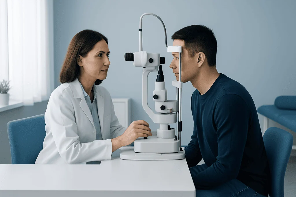
The eye’s drainage angle is difficult to view directly because of the way light bends as it passes through the cornea. Gonioscopy solves this by placing a small mirrored contact lens on the eye after numbing drops are used. The lens changes the path of light so the doctor can inspect the angle through a slit lamp microscope.
During the exam, the ophthalmologist looks for whether the angle is open, narrow, or closed and whether the trabecular meshwork and related structures are visible. They also check for pigment deposits, adhesions, signs of prior inflammation, trauma-related changes, or abnormal vessels. These findings help show how well fluid may be draining and whether pressure problems could develop or worsen.
In some cases, the doctor gently applies pressure with the lens to distinguish a temporary blockage from a more fixed closure caused by scar-like adhesions. This can clarify whether laser treatment such as glaucoma treatment may be helpful. The information gained from gonioscopy is often interpreted together with eye pressure measurements, optic nerve evaluation, and visual field testing.
Step-by-step: what happens during the procedure
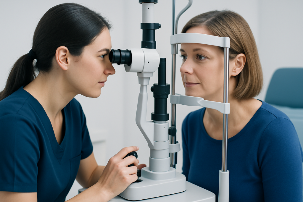
Gonioscopy is usually performed in an ophthalmology clinic and often takes only a few minutes. Before the test begins, the patient is seated at a slit lamp, which is the same microscope used for many eye examinations. The doctor or assistant places anesthetic drops in the eye so the lens can be applied comfortably.
A clear gel may be placed on the gonioscopy lens, depending on the type of lens used. The patient is then asked to keep the head steady against the chin and forehead supports while looking in a specific direction. The doctor gently places the lens on the eye and uses the microscope light to examine the angle from different positions.
The patient may notice light pressure or awareness of the lens, but the exam is not usually painful. Because the doctor may ask for small changes in gaze or briefly adjust the lens position, it can help to blink when instructed and remain as relaxed as possible. The lens is removed once the angle has been fully inspected, and the eye may be rinsed if needed.
If the doctor is evaluating a narrow angle or planning next steps, gonioscopy may be done alongside pressure checks, optic nerve imaging, or a dilated exam. In selected patients, the findings may support consideration of laser-based approaches such as laser eye surgery when appropriate for the underlying diagnosis and after a full specialist assessment.
Recovery timeline, benefits, and possible risks
Recovery after gonioscopy is usually immediate. Most people can return to usual activities right away, especially if no dilating drops were used. Some may have mild temporary blur from the numbing drops, gel, or bright examination light, but this generally settles quickly.
The main benefit of gonioscopy is that it gives direct, clinically useful information about the drainage angle that other routine tests may miss. It can help confirm whether glaucoma is likely to be open-angle or angle-closure, identify who may be at risk of sudden angle closure, and detect angle damage from inflammation, trauma, or new blood vessel formation. This can make treatment more precise and timely.
Risks are generally minimal. A small number of patients may experience brief eye irritation, watering, or light sensitivity after the exam. In uncommon situations, the eye surface may feel mildly scratched for a short time, especially in people with dry eyes or corneal sensitivity. Serious complications from routine gonioscopy are rare when the examination is performed by a trained eye specialist.
If the eye remains painful, very red, or significantly blurry after the visit, medical advice is important because symptoms may not be due to the gonioscopy itself and could reflect the underlying eye condition. Follow-up depends on the results: some patients only need monitoring, while others may need medication, laser therapy, or surgical review.
Understanding the results and next steps
After gonioscopy, the ophthalmologist explains whether the drainage angle appears open, narrow, or closed. An open angle does not rule out glaucoma, but it helps classify the type and informs the next tests or treatment plan. A narrow or closed angle may require closer observation or preventive treatment to reduce the chance of pressure rising quickly.
Results can also show whether there are peripheral anterior synechiae, which are adhesions that may keep the angle from opening properly. Other findings may include heavy pigmentation, recession after trauma, or abnormal blood vessels seen in secondary glaucoma. Each pattern points to different causes and can change how treatment is approached.
Depending on the findings, the doctor may recommend observation, pressure-lowering eye drops, laser treatment, or glaucoma surgery. If cataract anatomy is contributing to angle crowding, further evaluation for cataract surgery may sometimes be part of the discussion. The aim is to protect vision by addressing the source of impaired fluid drainage and monitoring the optic nerve over time.
For international patients who need further assessment, Acibadem International’s multidisciplinary eye specialists and JCI-accredited hospitals diagnose and treat glaucoma-related conditions with coordinated follow-up plans.
When to seek medical care
Medical care should be sought promptly if there are symptoms that could suggest sudden angle closure or a rapid rise in eye pressure. These may include severe eye pain, headache, nausea, redness, blurred vision, or seeing halos around lights. Such symptoms need urgent ophthalmic assessment.
Even without urgent symptoms, a person should arrange an eye evaluation if they have elevated eye pressure, a strong family history of glaucoma, previous eye injury, or have been told they may have narrow angles. Regular follow-up matters because glaucoma can progress without obvious early symptoms.
After gonioscopy, it is sensible to contact the doctor if discomfort is more than mild, if vision does not clear as expected, or if redness worsens instead of improving. Timely review helps confirm whether symptoms are simply temporary irritation or a sign that the underlying eye condition needs faster treatment.
Frequently asked questions
Is gonioscopy painful?
Gonioscopy is usually not painful because numbing eye drops are used before the lens touches the eye. Some people feel mild pressure or temporary awareness of the lens, but the test is generally well tolerated.
How long does a gonioscopy exam take?
The examination itself often takes only a few minutes, although the full appointment may be longer if other glaucoma tests are done the same day. Timing can vary depending on whether the doctor needs to examine both eyes or perform additional measurements.
Can a person drive after gonioscopy?
Many people can drive afterward if only gonioscopy was performed and vision feels normal. If dilating drops or other tests were also used, temporary blur or light sensitivity may make it better to arrange transportation.
What does gonioscopy diagnose?
Gonioscopy helps assess the drainage angle and supports the diagnosis of open-angle glaucoma, narrow angles, angle-closure glaucoma, and some secondary glaucomas. It can also reveal pigment, scar tissue, trauma-related changes, or abnormal new blood vessels in the angle.
Is gonioscopy the same as measuring eye pressure?
No. Eye pressure measurement and gonioscopy are different tests that provide complementary information. Pressure testing shows the current intraocular pressure, while gonioscopy shows the anatomy of the drainage angle that may explain pressure problems.
How often is gonioscopy needed?
The frequency depends on the person’s eye anatomy, glaucoma risk, and previous findings. Some people may need it only when first assessed, while others with narrow angles or glaucoma may need repeat exams during follow-up.
References
- American Academy of Ophthalmology
- National Eye Institute
- American Optometric Association
- Glaucoma Research Foundation
This article is for general information only and is not a substitute for professional medical advice. Please consult a qualified doctor about your individual situation.
Explore treatments in Turkey — costs, top hospitals & a free quote
JCI-accredited · board-certified surgeons · reply within 24h
Add us as a Preferred Source to see more of our trusted health content across Google Search, AI Overviews and Discover.