

Hip Labral Tear Xray: Preparation, Procedure and Results

A standard hip X-ray cannot usually show the labrum, which is soft cartilage around the hip socket. X-rays can identify bone abnormalities, arthritis, fractures, and signs of femoroacetabular impingement that may be linked with labral tears.
Key Takeaways
- A standard hip X-ray cannot usually show the labrum, which is soft cartilage around the hip socket.
- X-rays can identify bone abnormalities, arthritis, fractures, and signs of femoroacetabular impingement that may be linked with labral tears.
- MRI, often with contrast injected into the joint, is commonly used when a labral tear is suspected.
- Hip X-rays are quick, painless, and use a low dose of radiation; preparation is usually minimal.
- Urgent assessment is important after a serious injury, inability to bear weight, fever, or a painful swollen hip.
A hip labral tear X-ray is often an early imaging test for persistent hip or groin pain. Although an X-ray cannot directly show a torn labrum, it can reveal bone shape changes, arthritis, injury, or other conditions that may contribute to symptoms and guide the next diagnostic step.
Hip Labral Tear X-Ray: What It Can and Cannot Show
A hip labral tear X-ray is commonly used as part of the assessment of hip or groin pain. The labrum is a ring of strong cartilage that lines the rim of the hip socket, helping deepen the joint and support smooth movement. Because it is soft tissue, the labrum itself is not usually visible on a standard X-ray.
Rather than confirming a tear directly, an X-ray helps clinicians look for changes in the bones of the hip and pelvis. These may include arthritis, a fracture, joint-space narrowing, calcium deposits, or bone shapes associated with femoroacetabular impingement (FAI). FAI can place repeated pressure on the labrum and may contribute to a tear.
If symptoms and examination findings suggest a labral injury, the next test may be an MRI. In some cases, an MR arthrogram, where contrast material is placed into the hip joint before MRI scanning, provides more detailed images of the labrum. A clinician considers the full picture, including symptoms, physical examination, activity level, and imaging results, before making a diagnosis.
How Hip X-Ray Imaging Works
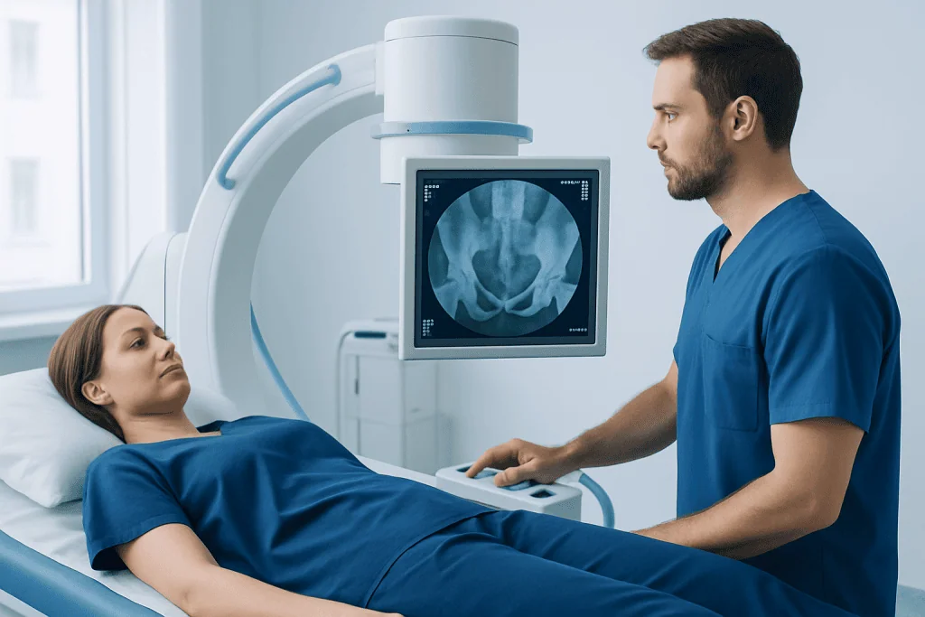
An X-ray uses a small amount of ionizing radiation to create images of dense structures, especially bone. The X-ray beam passes through the body at different rates, allowing bones and joint alignment to appear on the resulting images. It does not provide the same level of detail for cartilage, tendons, muscles, or the labrum.
For hip symptoms, images are often taken from more than one angle. These may include a front-facing image of the pelvis and a side or angled image of the affected hip. Different views help the radiologist and orthopedic specialist assess the contour of the femoral head and neck, the hip socket, joint alignment, and any bony prominence that may affect hip movement.
X-rays are particularly useful for excluding or identifying other explanations for pain. For example, they can show osteoarthritis, a stress injury in some circumstances, bone lesions, prior surgical changes, or a dislocation following trauma. They also provide a useful baseline if further imaging or treatment is needed.
Preparation and Candidacy for a Hip X-Ray

A hip X-ray is appropriate for many people with ongoing hip, buttock, upper-thigh, or groin pain, especially when symptoms began after an injury or interfere with walking, sport, work, or daily activities. It may also be requested when a clinician suspects hip arthritis, impingement, a fracture, or another bone or joint condition.
Preparation is generally simple. Patients should tell the imaging team if they may be pregnant, as X-rays involve radiation. The team can discuss whether the scan is necessary, whether alternative imaging may be appropriate, and what precautions are used to limit radiation exposure. Patients should also mention previous hip surgery, implants, or any recent imaging studies.
Jewellery, belts, clothing fasteners, and objects in pockets may interfere with images and may need to be removed. Comfortable clothing is helpful, but the imaging center will provide instructions based on the views required. Eating, drinking, and usual medicines are generally not restricted for a standard hip X-ray unless a clinician has given different instructions.
Do you need to take pants off for hip X-ray?
Whether pants need to be removed depends on the clothing and the imaging center’s protocol. Metal zippers, buttons, belts, thick seams, and decorative items can obscure the hip area, so patients may be asked to change into a gown or wear clothing without metal. Privacy is respected, and the radiographer explains what needs to be removed before the examination begins.
What Happens During the Procedure and When Results Are Ready
A hip X-ray is usually performed by a radiographer in an imaging department. The patient may stand, lie on an X-ray table, or be positioned in more than one way depending on the requested views and their comfort. The radiographer carefully positions the pelvis and leg, sometimes asking the patient to turn the foot inward or outward slightly.
It is important to remain still for a few seconds while each image is taken, as movement can blur the picture. The radiographer normally steps behind a protective screen while taking the image but can see and communicate with the patient throughout. The scan itself is painless and generally takes only a short time.
Afterward, normal activities can usually be resumed immediately. A radiologist interprets the images and sends a report to the referring clinician, who discusses the findings in the context of the patient’s symptoms. Timing varies between facilities, but results are commonly reviewed within a few days unless urgent assessment is needed.
Will a torn hip labrum show up on an X-ray?
No, a torn hip labrum usually does not show up directly on an X-ray because the labrum is cartilage rather than bone. However, the X-ray may show structural features that increase the likelihood of a labral problem, such as FAI or arthritis. When a tear remains a concern, MRI or MR arthrography may be recommended to assess the labrum and other soft tissues more closely.
Interpreting Results, Benefits and Limitations
A normal hip X-ray does not rule out a labral tear. It means that no clear bone or joint abnormality was identified on the images, or that any changes seen do not explain all symptoms. A person can have a normal X-ray and still need further assessment when pain, catching, clicking, stiffness, or reduced hip function continues.
An abnormal result may show FAI-related bone shape changes, osteoarthritis, reduced joint space, a fracture, or another finding that requires follow-up. Some findings are common and may not be the direct source of pain. For this reason, imaging results should not be interpreted alone; the clinician compares them with the history and physical examination.
The main benefits of X-ray are that it is accessible, quick, noninvasive, and useful for evaluating bone. Radiation exposure is low, but it is not zero, which is why imaging is requested only when it is expected to help clinical decision-making. The test cannot provide a complete evaluation of the labrum, cartilage, tendons, or muscles.
If a labral tear is confirmed, treatment may range from activity changes and targeted physiotherapy to injections or surgery in selected cases. Hip arthroscopy may be considered when symptoms persist despite appropriate non-surgical care and clinical assessment indicates that repair or treatment inside the joint could be beneficial.
Pain, Treatment Planning and Recovery After Surgery
How bad is hip labral tear pain?
Hip labral tear pain varies widely. Some people have mild discomfort only during sport or deep hip movements, while others experience persistent pain in the groin, side of the hip, buttock, or upper thigh. Clicking, catching, a sense of instability, stiffness, and pain with sitting, twisting, squatting, or walking can occur, but these symptoms are not specific to a labral tear.
Initial care often focuses on reducing movements that aggravate symptoms, maintaining safe activity, and completing a clinician-guided rehabilitation program. Pain relief options, injections, and surgical assessment may be discussed according to the cause of the tear, associated hip structure changes, symptom severity, and personal goals. Not every imaging-detected labral tear requires surgery.
Recovery after hip arthroscopy differs according to the procedure performed and whether related conditions, such as impingement, are treated at the same time. Patients may use crutches for a period and begin supervised rehabilitation early to restore motion, strength, balance, and gradual return to daily activities. The orthopedic team provides an individualized recovery plan.
How to poop after hip labrum surgery?
Constipation can occur after hip labrum surgery because of anesthesia, reduced movement, changes in eating and drinking, and some prescription pain medicines. Following the surgeon’s instructions, drinking enough fluids if permitted, eating fiber-containing foods, and taking gentle walks as advised may help support bowel regularity. A clinician may also recommend a stool softener or laxative when appropriate.
Patients should contact their care team if they have severe abdominal pain, vomiting, marked bloating, or constipation that does not improve with the recommended plan. They should not strain heavily or change prescribed medicines without medical advice, particularly during the early recovery period.
When to Seek Medical Care
Medical evaluation is advisable for hip or groin pain that lasts more than a few weeks, recurs with activity, causes limping, disrupts sleep, or limits work, exercise, or everyday movement. A clinician can assess whether symptoms may relate to the hip joint, surrounding muscles and tendons, the lower back, or another condition.
Prompt medical care is important after a fall, collision, or other injury when a person cannot bear weight, has severe pain, notices a visible deformity, or develops numbness or weakness in the leg. Urgent assessment is also needed for a hot, swollen, very painful hip, particularly if accompanied by fever or feeling unwell, as these features may indicate a condition requiring rapid treatment.
For patients travelling for care, Acibadem International’s multidisciplinary specialists and JCI-accredited hospitals can assess hip conditions and coordinate imaging, orthopedic treatment, and rehabilitation for international patients. A specialist can explain whether X-ray, MRI, conservative care, or a surgical opinion is the most appropriate next step.
Frequently asked questions
Can an X-ray diagnose a hip labral tear?
An X-ray cannot usually diagnose a hip labral tear directly because it does not show the labrum in sufficient detail. It can identify bone and joint changes that may contribute to symptoms or suggest the need for MRI or MR arthrography.
How long does a hip X-ray take?
The image-taking portion of a hip X-ray is usually brief, often only a few minutes. Allow additional time for registration, changing clothes if needed, and positioning for different views.
Is a hip X-ray painful?
The X-ray itself is painless. Some positions may be uncomfortable when the hip is already painful or stiff, but the radiographer can adapt positioning where possible and should be told about significant discomfort.
Why might a doctor order an MRI after a normal hip X-ray?
A normal X-ray may exclude many bone problems but cannot fully assess the labrum, cartilage, tendons, or muscles. MRI can provide more detailed soft-tissue images when symptoms and examination findings still suggest an injury within or around the hip joint.
Can a hip labral tear heal without surgery?
Symptoms from a hip labral tear may improve with activity modification, physiotherapy, and other non-surgical treatments, although the tear itself may not fully heal. The best approach depends on symptoms, hip anatomy, associated conditions, and the person's daily and sporting goals.
What symptoms can be confused with a hip labral tear?
Hip pain can also arise from osteoarthritis, tendon problems, bursitis, muscle strains, stress injuries, hernias, or pain referred from the lower back. A clinical examination and appropriate imaging help distinguish between these possibilities.
References
- American Academy of Orthopaedic Surgeons
- American College of Radiology
- Radiological Society of North America
- National Institute of Arthritis and Musculoskeletal and Skin Diseases
This article is for general information only and is not a substitute for professional medical advice. Please consult a qualified doctor about your individual situation.
Joint and spine care in Turkey — expert assessment & treatment
JCI-accredited · board-certified surgeons · reply within 24h
Add us as a Preferred Source to see more of our trusted health content across Google Search, AI Overviews and Discover.

More from the Health Library



Related Specialists

Prof. Dr. Mustafa Karaçelik
Cardiovascular Surgery
Dr. Bayarmaa Khishigsuren
Medical Oncology
Dr. Bahar Anayurdu
Radiology
Dr. Yusuf İzzet Güngörmüş
Ophthalmology