Images of Athlete’s Foot: An Evidence-Based Guide for Patients

Athlete's foot often appears as peeling, scaling, redness, or soggy white skin between the toes. The condition can also affect the sole, heel, or sides of the foot and may cause itching, burning, or fissures.
Key Takeaways
- Athlete's foot often appears as peeling, scaling, redness, or soggy white skin between the toes.
- The condition can also affect the sole, heel, or sides of the foot and may cause itching, burning, or fissures.
- Photos are useful for comparison, but athlete's foot can resemble eczema, contact dermatitis, or psoriasis.
- Keeping feet clean and dry, changing socks, and treating shoes can help reduce recurrence.
- Medical assessment is important if symptoms are painful, widespread, recurrent, or do not improve with standard care.
Images of athlete's foot commonly show peeling, scaling, redness, and cracks, especially between the toes or along the sole. While pictures can help patients recognize patterns, athlete's foot can look similar to eczema, psoriasis, or irritation, so diagnosis should be confirmed by a qualified clinician when symptoms are persistent or severe.
What images of athlete's foot typically show
Images of athlete’s foot usually show a fungal rash called tinea pedis. In many patients, the most recognizable pattern is peeling, scaling, and redness between the fourth and fifth toes, sometimes with soft, white, soggy-looking skin. Other photos may show dry flaking on the sole, cracking at the heel, or a more inflamed rash with small blisters.
These images can be helpful because athlete’s foot does not look exactly the same in every person. On lighter skin tones, the rash may appear pink or red. On darker skin tones, it may look brown, gray, purple, or simply lighter or darker than the surrounding skin. Some cases appear dry and powdery, while others look moist and irritated.
Although online pictures can guide recognition, they cannot confirm the cause. Several skin conditions can mimic fungal infection, including eczema, contact dermatitis, and psoriasis. A patient who is unsure, or who has diabetes, poor circulation, or a weakened immune system, should seek medical advice rather than relying on images alone.
How athlete's foot can look in different areas of the foot
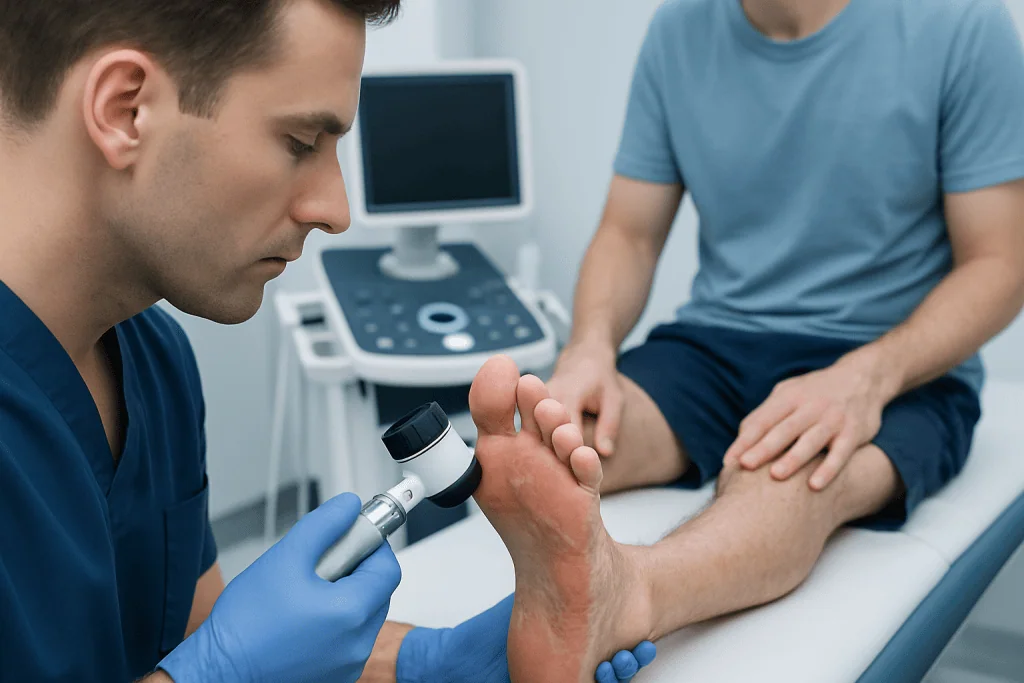
Athlete’s foot has several common visual patterns. The classic toe-web type affects the spaces between the toes. Pictures often show peeling edges, redness, and fissures, with skin that may look macerated or waterlogged. This form can also cause a noticeable odor if moisture is trapped for long periods.
Another pattern is the “moccasin” type, which involves the sole, heel, and sides of the foot. Images often show diffuse dryness, fine scaling, and thickened skin extending across the bottom of the foot in a slipper-like distribution. Patients sometimes mistake this appearance for simple dry skin and delay treatment.
A less common inflammatory form may produce clusters of itchy blisters on the arch or instep. In photographs, this can resemble allergic rash or irritation. If scratching causes broken skin, a secondary bacterial infection can develop, making the area more painful, swollen, or warm.
- Between toes: soft white skin, peeling, cracks
- Soles and heels: dry scaling, rough patches, thickened skin
- Arch or instep: blisters or inflamed itchy rash
- Sides of foot: redness with flaking borders
Symptoms that often accompany the visible rash
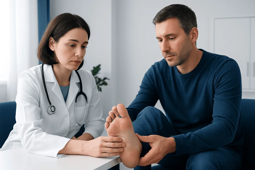
Most people look for images because they have already noticed symptoms. Athlete’s foot commonly causes itching, stinging, or burning, especially after shoes are removed. The skin may feel tight, tender, or unusually dry. Cracks can become uncomfortable when walking, particularly around the toes or heel.
Some patients mainly notice flaking and do not feel much itching. Others experience soreness, especially if the skin is split or if the area stays damp. In more irritated cases, there may be redness, swelling, and sensitivity. If a toenail also becomes thickened, yellow, brittle, or distorted, a fungal nail infection may be present as well.
Because symptoms overlap with other conditions, visual clues should be interpreted in context. A rash that affects only one foot, repeatedly returns after treatment, or spreads to the nails, groin, or hands deserves professional evaluation. Similar fungal infections can occur elsewhere on the body, such as ringworm, even though the appearance may differ by location.
Why athlete's foot develops and who is at higher risk
Athlete’s foot is caused by dermatophyte fungi that thrive in warm, moist environments. Locker rooms, communal showers, pool areas, and tight footwear create ideal conditions for these organisms. The fungi can spread through direct skin contact or through contaminated floors, towels, socks, and shoes.
Certain habits and situations make infection more likely. Sweaty feet, non-breathable shoes, staying in damp socks, and walking barefoot in shared wet areas all increase risk. Small breaks in the skin, such as from friction or dryness, can also make infection easier to establish.
Recurrence is common when the environment that allowed the fungus to grow has not changed. People who play sports, work long hours in closed shoes, or have conditions that affect healing may be more vulnerable. Patients with diabetes, poor circulation, or immune system problems should take foot rashes seriously because complications can be more significant for them.
How doctors diagnose athlete's foot when pictures are not enough
In many cases, a clinician can suspect athlete’s foot by examining the skin and reviewing symptoms. However, because several rashes look similar, diagnosis may need confirmation, especially if the rash is severe, recurring, or not responding to treatment. The goal is to make sure the patient is treating the correct condition.
A doctor may gently scrape some scale from the skin for microscopic examination or laboratory testing. These tests can help distinguish fungal infection from eczema, psoriasis, bacterial infection, or inflammatory skin disease. This is particularly useful when the appearance is atypical or when the patient has already tried multiple treatments.
If the skin is very inflamed, the doctor may also check for signs of secondary infection or involvement of the toenails. In persistent cases, dermatology input can be helpful. Patients who need specialist assessment may be evaluated through services related to dermatology care when the diagnosis is unclear or symptoms keep returning.
Treatment options and practical self-care
Treatment depends on the extent of the rash and whether the skin is dry, moist, cracked, or secondarily infected. Many uncomplicated cases improve with antifungal creams, sprays, powders, or solutions recommended by a clinician or pharmacist. Treatment usually needs to continue for the full advised period, even if the skin looks better sooner, because fungus can persist after symptoms start to fade.
Daily foot care supports treatment. Feet should be washed gently and dried carefully, especially between the toes. Socks should be changed when damp, and breathable shoes are generally preferable. Alternating shoes and allowing them to dry fully can reduce moisture build-up. Antifungal powders or shoe hygiene measures may help lower the chance of reinfection.
Patients should avoid sharing towels, nail tools, or footwear. Scratching can worsen irritation and increase the risk of broken skin. If the rash is widespread, recurrent, or associated with nail changes, stronger prescription treatment may be considered. In some situations, a specialist may also assess whether another skin condition such as psoriasis treatment or eczema management is more appropriate than antifungal therapy.
Prevention and when to seek medical care
Prevention focuses on reducing moisture and limiting exposure to fungi. Wearing shower shoes in communal wet areas, changing socks after exercise, choosing breathable footwear, and drying the feet well after bathing all help. People who have recurrent infection may benefit from reviewing shoe hygiene and treating any fungal nail infection at the same time, because nails can act as a reservoir for reinfection.
Medical care should be sought if a rash is very painful, rapidly spreading, oozing, bleeding, or accompanied by marked swelling. A patient should also speak with a doctor if over-the-counter treatment does not help, if the diagnosis is uncertain, or if the problem keeps coming back. Prompt assessment is especially important for people with diabetes, nerve damage, circulation problems, or reduced immunity.
If blisters are extensive, if there is fever, or if the skin becomes increasingly red and warm, a bacterial infection may need to be excluded. Near the end of the care pathway, some patients may benefit from multidisciplinary input; Acibadem International’s specialists in JCI-accredited hospitals diagnose and treat skin conditions for international patients when further evaluation is needed.
Frequently asked questions
What do images of athlete's foot usually look like?
They commonly show peeling, scaling, redness, or cracked skin, especially between the toes. Some cases look dry and flaky on the sole, while others appear white and soggy from trapped moisture.
Can athlete's foot look like dry skin?
Yes. The moccasin-type pattern can resemble simple dryness with fine scale on the sole and heel. If moisturizers do not help or itching and peeling continue, a fungal cause should be considered.
Is it possible to diagnose athlete's foot from a photo alone?
No photo can confirm the diagnosis with certainty. Athlete's foot can look similar to eczema, psoriasis, or contact dermatitis, so a clinician may need to examine the skin or perform a simple test.
When should a patient see a doctor for athlete's foot?
Medical advice is sensible if the rash is painful, spreading, recurrent, or not improving with standard treatment. Patients with diabetes, poor circulation, or weakened immunity should seek care early.
Can athlete's foot spread to other parts of the body?
Yes. Fungal infections can spread to the nails, hands, or groin, especially if the skin is scratched and then touched elsewhere. Good hygiene and prompt treatment help reduce this risk.
How can recurrence be prevented?
Feet should be kept clean and dry, socks changed when damp, and shoes allowed to dry fully between uses. Wearing footwear in communal wet areas and avoiding shared towels or shoes can also help prevent repeat infection.
References
- American Academy of Dermatology
- Centers for Disease Control and Prevention
- National Health Service
- Mayo Clinic
- Merck Manual Consumer Version
This article is for general information only and is not a substitute for professional medical advice. Please consult a qualified doctor about your individual situation.
Explore treatments in Turkey — costs, top hospitals & a free quote
JCI-accredited · board-certified surgeons · reply within 24h
Add us as a Preferred Source to see more of our trusted health content across Google Search, AI Overviews and Discover.

More from the Health Library

Excitatory: An Evidence-Based Guide for Patients

Forward Head Posture: A Complete Medical Overview

Prostate Fluid — Explained by Medical Evidence, Not Myths

Hemicrania Continua: A Complete Medical Overview

Staghorn Kidney Stone: An Evidence-Based Guide for Patients