Lytic Lesion — Explained by Medical Evidence, Not Myths

A lytic lesion means an area of reduced bone density, not a single disease. Some lytic lesions are harmless, but others need prompt evaluation.
Key Takeaways
- A lytic lesion means an area of reduced bone density, not a single disease.
- Some lytic lesions are harmless, but others need prompt evaluation.
- Symptoms may include pain, swelling, or fractures, though some lesions cause no symptoms.
- Diagnosis usually combines imaging, blood tests, and sometimes biopsy.
- Treatment depends on the underlying cause and may range from monitoring to surgery or cancer care.
A lytic lesion is a spot in bone where normal bone has been broken down or replaced, making the area look less dense on an X-ray, CT, or MRI. It is a radiology finding rather than a diagnosis itself, so the next step is to identify the cause, which may be benign, infectious, metabolic, or cancer-related.
What a lytic lesion means
A lytic lesion is an area in bone where bone tissue has been lost, destroyed, or replaced by another process. On imaging, this area looks darker or less dense than the surrounding bone. Doctors use the term to describe a pattern seen on a scan, not to name a final diagnosis.
This distinction is important because many different conditions can produce a lytic lesion. Some are benign, such as simple bone cysts or noncancerous tumors. Others may relate to infection, metabolic bone disease, or cancer that starts in bone or spreads there from another part of the body.
The meaning of a lytic lesion depends on several factors, including the person’s age, symptoms, medical history, and the lesion’s size, location, and appearance on imaging. A small lesion with smooth borders in a child may suggest a very different cause than a destructive lesion in an older adult with unexplained weight loss or persistent pain.
For patients, the most helpful way to think about a lytic lesion is as a finding that needs context. It should be interpreted by a qualified doctor, often with support from radiology, orthopedics, oncology, or internal medicine specialists, so the underlying cause can be identified and treated appropriately.
Symptoms and why some lesions are found by chance
Some lytic lesions cause no symptoms at all and are discovered incidentally during imaging for another reason, such as after a sports injury or back pain. Others produce symptoms because the affected bone becomes weaker, inflamed, or structurally unstable.
Common symptoms can include localized bone pain, tenderness, swelling, limited movement in a nearby joint, or pain that worsens with activity. If the lesion weakens the bone significantly, a fracture can occur with minor trauma or even normal daily movement. This is called a pathologic fracture.
Symptoms may also reflect the underlying disease rather than the lesion itself. For example, infection may cause fever and fatigue, while some cancers may be associated with weight loss, night sweats, or general weakness. In other cases, blood test abnormalities such as high calcium may cause thirst, constipation, confusion, or nausea.
- Bone pain that does not improve
- Swelling or a palpable lump
- Reduced ability to bear weight or use a limb
- A fracture after minimal injury
- General symptoms such as fever, fatigue, or unexplained weight loss
Common causes and risk factors

Lytic lesions can develop for many reasons. Benign causes include bone cysts, fibrous dysplasia, enchondroma, giant cell tumor, and other noncancerous bone growths. In children and younger adults, benign explanations are often more likely, although every lesion still needs proper review.
Infection of the bone, called osteomyelitis, can also create a lytic appearance. This may happen after an injury, surgery, bloodstream infection, or spread from nearby tissue. People with diabetes, poor circulation, weakened immunity, or recent orthopedic procedures may have a higher risk of bone infection.
Another important group of causes includes cancers. These may be primary bone cancers or cancers that have spread to bone, known as cancer. Multiple myeloma, a blood-related cancer involving plasma cells, is another classic cause of lytic bone lesions in adults. In older adults especially, a newly found lytic lesion often prompts doctors to consider metastatic disease or myeloma among the possibilities.
Metabolic and endocrine disorders can contribute as well. Severe osteoporosis, hyperparathyroidism, and some rare bone disorders can alter bone structure and mimic or cause lytic changes. Risk factors that make careful evaluation especially important include older age, a known history of cancer, persistent unexplained bone pain, immune suppression, smoking, or prior radiation exposure.
How doctors evaluate a lytic lesion
Evaluation begins with a detailed history and physical examination. Doctors ask when symptoms started, whether pain is constant or activity-related, whether there has been fever or weight loss, and whether the person has a history of cancer, fracture, infection, or bone disease. Age is a major clue because some lesions are more common in children and others in older adults.
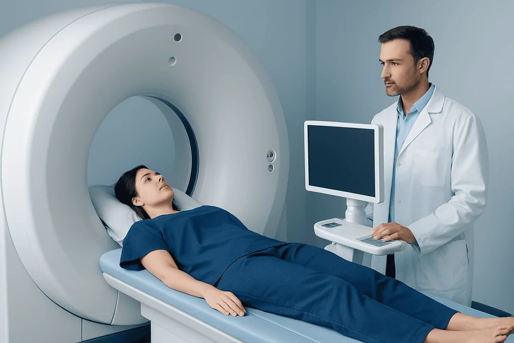
Imaging is central to the workup. A plain X-ray is often the first test, but CT or MRI may be needed to define the lesion more clearly, assess the bone cortex, and see whether nearby soft tissues are involved. If doctors suspect spread from another cancer, they may recommend broader imaging and PET-CT imaging or other scans to look for additional lesions.
Blood tests can help identify infection, inflammation, abnormal calcium levels, kidney problems, or markers that suggest multiple myeloma or another systemic illness. Depending on the clinical picture, testing may include complete blood count, inflammatory markers, calcium, kidney function, protein studies, and hormone-related measurements.
Sometimes imaging and laboratory findings strongly suggest a benign process, allowing careful monitoring. In other cases, a tissue sample is needed. A biopsy may be performed to confirm whether the lesion is benign, infectious, or malignant, and that information guides the treatment plan.
How imaging features help separate benign from serious causes
Radiologists do not look only at whether a lesion is lytic. They also assess the borders, shape, size, exact bone involved, and whether the cortex has been thinned or broken. A well-defined lesion with a narrow transition zone may suggest a slower, less aggressive process, while a poorly defined lesion with cortical destruction can raise concern for a more aggressive disease.
The location also matters. Some lesions tend to occur near joints, some in the shaft of long bones, and others in the spine, pelvis, or skull. Certain patterns, such as multiple punched-out lesions, may suggest multiple myeloma, while a single lesion in a growing child may point to a different set of diagnoses. MRI is especially useful for seeing bone marrow and surrounding soft tissue involvement.
Still, imaging alone does not provide all the answers. A lesion that appears nonaggressive may still need follow-up, and a suspicious lesion is not automatically cancer. The final interpretation comes from combining the scan appearance with symptoms, examination findings, and laboratory data.
This balanced approach helps avoid both overreaction and false reassurance. It is one reason patients are often referred to specialists in musculoskeletal radiology, orthopedics, or oncology when a lytic lesion is newly identified.
Treatment options depend on the cause
There is no single treatment for a lytic lesion because treatment is directed at the underlying condition. Some benign lesions only need observation with repeat imaging. This approach may be used when the lesion has a classic benign appearance, causes no symptoms, and does not put the bone at high risk for fracture.
If the lesion is painful, growing, weakening the bone, or uncertain in nature, treatment may involve surgery or a procedure to stabilize the bone. Orthopedic care can include curettage, bone grafting, or fixation if there is a fracture risk. When lesions affect major bones or joints, referral to orthopedic oncology may help guide safe and appropriate management.
Infectious causes are treated with antibiotics and sometimes surgical drainage or debridement. If the cause is cancer, treatment depends on the type and stage and may include systemic therapy, radiation therapy, surgery, or a combination of these. Some patients may also need bone marrow transplantation when the underlying diagnosis involves blood or marrow disease and specialists judge it appropriate.
Supportive care is also important. This can include pain control, mobility support, physical therapy, and steps to reduce fracture risk. At the end of the diagnostic pathway, the goal is not only to identify the lesion but also to protect bone strength and treat the person as a whole.
Self-care, prevention, and when to seek medical care
Not all lytic lesions can be prevented because many arise from conditions that are not under direct personal control. Still, bone health habits matter. Adequate calcium and vitamin D intake, regular weight-bearing exercise, smoking cessation, moderate alcohol use, and prompt treatment of infections all support stronger bones and may reduce some risks.
People with osteoporosis, prior cancer, chronic kidney disease, endocrine disorders, or long-term steroid use should keep regular medical follow-up. Reporting new bone pain early can help doctors investigate problems before a fracture occurs. If a doctor recommends repeat imaging, keeping those follow-up appointments is important even when symptoms are mild.
Medical care should be sought promptly for persistent bone pain, swelling, a new lump, inability to bear weight, or a fracture after a minor injury. Urgent assessment is also wise if bone symptoms occur together with fever, unexplained weight loss, night sweats, weakness, or a known history of cancer. A short delay is rarely dangerous, but ignoring symptoms for weeks or months can make evaluation more difficult.
For international patients who need coordinated assessment, Acibadem International’s multidisciplinary specialists and JCI-accredited hospitals evaluate bone lesions with advanced imaging, pathology, and specialty care. If symptoms overlap with broader bone cancer concerns or osteoporosis, specialist review can help clarify the diagnosis and next steps.
Frequently asked questions
Is a lytic lesion always cancer?
No. A lytic lesion can be caused by benign bone cysts, noncancerous tumors, infection, metabolic conditions, or cancer. Because the range of causes is wide, proper evaluation is needed before any conclusion is made.
Can a lytic lesion heal on its own?
Some benign lesions may remain stable or resolve over time, especially in younger people. Others need treatment or monitoring because they can grow, weaken bone, or reflect an underlying disease that should not be left untreated.
What tests are usually needed for a lytic lesion?
Doctors often start with an X-ray and may add CT or MRI for more detail. Blood tests are common, and some patients need a biopsy to confirm the exact cause.
What does it mean if a lytic lesion is found in the spine?
A spinal lytic lesion means there is an area of bone loss in a vertebra. Causes can include benign lesions, osteoporosis-related change, infection, or cancer, so the finding usually needs careful imaging review and clinical follow-up.
Are lytic lesions painful?
They can be, but not always. Some are found by chance and cause no symptoms, while others cause ongoing pain, tenderness, or pain with movement because they weaken or irritate the bone.
When is a biopsy necessary?
A biopsy may be recommended when imaging cannot confidently identify the lesion, when the lesion looks aggressive, or when cancer or infection is suspected. It helps doctors choose the safest and most effective treatment.
This article is for general information only and is not a substitute for professional medical advice. Please consult a qualified doctor about your individual situation.
Explore treatments in Turkey — costs, top hospitals & a free quote
JCI-accredited · board-certified surgeons · reply within 24h
Add us as a Preferred Source to see more of our trusted health content across Google Search, AI Overviews and Discover.

More from the Health Library

Images of Second Degree Burns: An Evidence-Based Guide for Patients

Virilization — Explained by Medical Evidence, Not Myths

Dysphasia: An Evidence-Based Guide for Patients

Transfeminine — Explained by Medical Evidence, Not Myths

Stingray Sting: A Complete Medical Overview