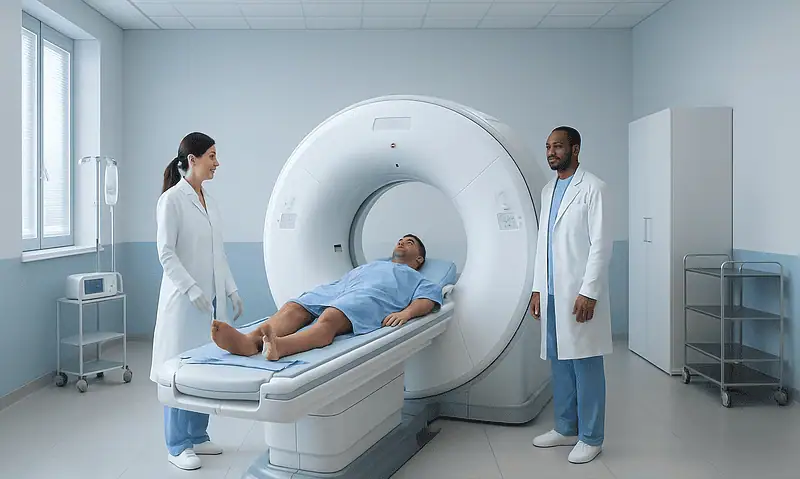
Mammography: Breast Screening, Dense Breasts, and Follow-Up Tests

Mammography can detect breast changes before they can be felt, making it an important tool for breast cancer screening. Dense breast tissue is common and not a diagnosis, but it can make cancers harder to see on mammograms and may slightly increase breast cancer risk.
Key Takeaways
- Mammography can detect breast changes before they can be felt, making it an important tool for breast cancer screening.
- Dense breast tissue is common and not a diagnosis, but it can make cancers harder to see on mammograms and may slightly increase breast cancer risk.
- A call-back after a mammogram is common and does not necessarily mean cancer; many follow-up findings are benign.
- Additional tests may include diagnostic mammography, breast ultrasound, breast MRI, or image-guided biopsy depending on the finding and personal risk.
- Screening recommendations vary by age, risk factors, and national guidelines, so patients should discuss the right plan with a qualified clinician.
Mammography is an X-ray imaging test used to screen for breast cancer and investigate breast symptoms. Understanding dense breasts, screening schedules, and follow-up tests can help patients feel prepared and make informed decisions with their doctor.
Overview
Mammography is a specialized X-ray examination of the breast. It is most often used as a screening test to look for early signs of breast cancer in people who have no symptoms. It can also be used as a diagnostic test when a person or clinician notices a lump, nipple discharge, skin change, breast pain in a specific area, or an abnormal result on another imaging test.
A screening mammogram usually takes images of each breast from two standard angles. Modern mammography may be performed as 2D digital mammography or as 3D mammography, also called digital breast tomosynthesis. In 3D mammography, multiple low-dose images are taken from different angles and reconstructed into thin slices, which may help radiologists assess overlapping breast tissue more clearly.
The goal of mammography is not to diagnose every breast condition on its own. Instead, it helps identify findings that appear normal, benign, probably benign, or suspicious enough to need more evaluation. When follow-up is recommended, it is a careful step to clarify the finding and guide the next decision.
Who Should Have Breast Screening?
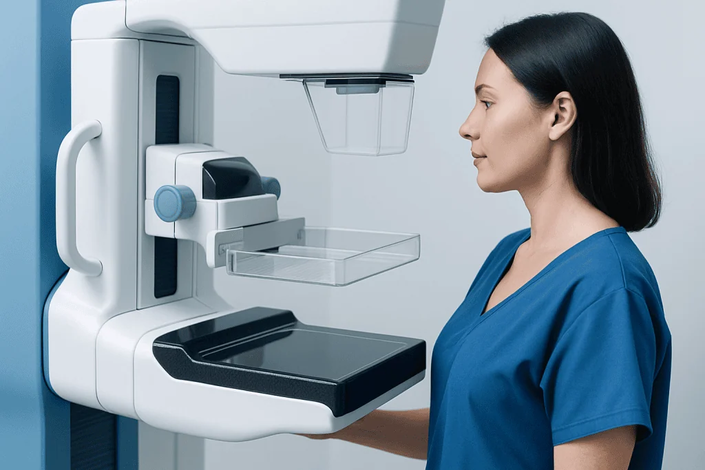
Breast screening recommendations vary between countries and medical organizations. In general, average-risk women are advised to begin regular screening mammography sometime in adulthood, often around the 40s or 50s, and to continue at intervals recommended by their health system or doctor. The exact starting age and frequency depend on personal risk, local guidelines, and shared decision-making.
Some people need an individualized screening plan. This may include those with a strong family history of breast cancer, known genetic mutations such as BRCA1 or BRCA2, previous chest radiation at a young age, a personal history of breast cancer or high-risk breast lesions, or certain inherited cancer syndromes. For higher-risk patients, screening may begin earlier and may include breast MRI in addition to mammography.
It is important to tell the doctor or imaging center about previous breast surgery, implants, hormone therapy, pregnancy or breastfeeding status, and any new breast symptoms. Patients should also bring or arrange transfer of prior mammogram images when possible. Comparing current and previous images is one of the most useful ways to identify whether a finding is stable or new.
Dense Breasts: What the Term Means
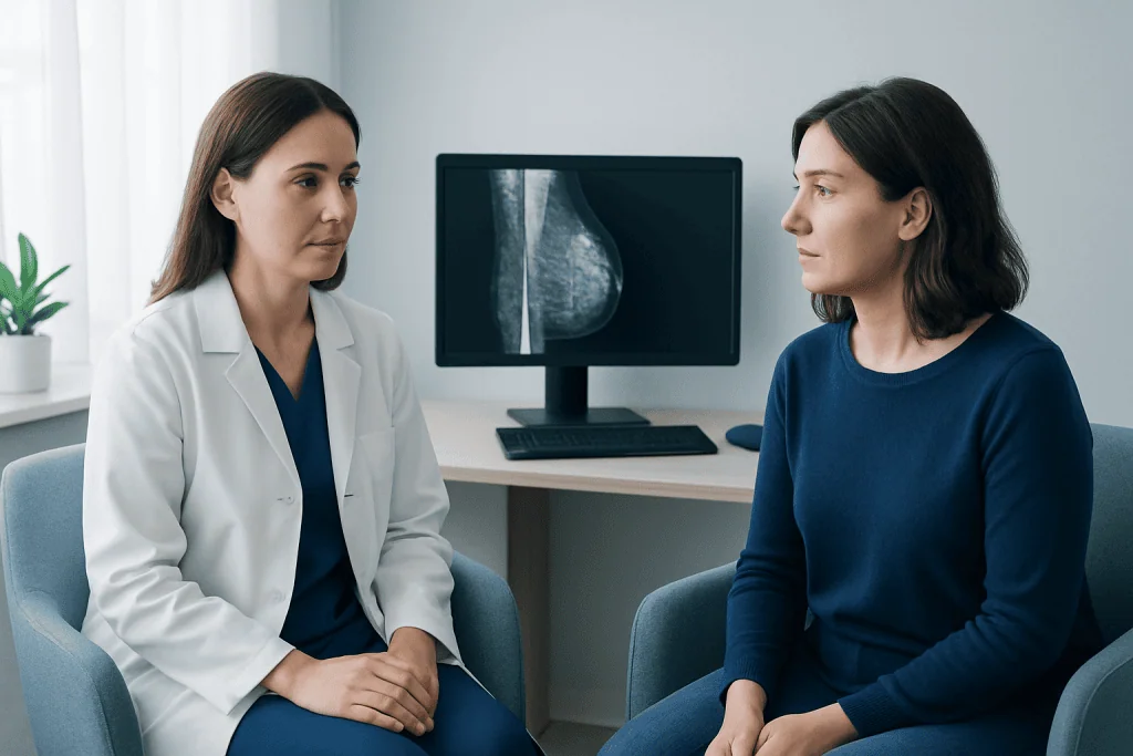
Breast density describes how breast tissue looks on a mammogram. Breasts contain fatty tissue as well as fibroglandular tissue, which includes milk glands, ducts, and supportive tissue. Dense breasts have more fibroglandular tissue and less fatty tissue. This is a normal variation and cannot be determined reliably by touch; it is assessed on mammography.
Radiology reports commonly classify density into four broad categories: almost entirely fatty, scattered areas of fibroglandular density, heterogeneously dense, and extremely dense. The last two categories are often described as dense breasts. Dense tissue appears white on a mammogram, and many breast cancers also appear white, which can make small abnormalities harder to see.
Having dense breasts does not mean a person has cancer. However, dense tissue can reduce the sensitivity of mammography and is also considered a modest risk factor for breast cancer. Because of this, some patients with dense breasts may be advised to consider supplemental screening, depending on their overall risk profile and the availability of tests.
What Happens During a Mammogram?
During a mammogram, a trained radiographer positions the breast on the imaging plate and gently compresses it for a few seconds while each image is taken. Compression is necessary because it spreads the tissue, reduces motion, improves image quality, and helps keep the radiation dose as low as reasonably achievable. The pressure can feel uncomfortable, but it is usually brief.
Patients are generally asked to undress from the waist up and wear a gown. It is best not to use deodorant, powder, lotion, or perfume on the chest or underarms on the day of the exam because some products can create spots on the images. If breasts are tender before menstruation, scheduling the exam at a less sensitive time of the cycle may make the experience easier.
People with breast implants can still have mammography, but they should inform the imaging center before the appointment. Additional implant-displacement views may be needed to see as much breast tissue as possible. Anyone who is pregnant, may be pregnant, or is breastfeeding should tell the clinician and imaging team so that the most appropriate imaging choice can be made.
Understanding Mammography Results and Follow-Up Tests
Mammogram results are reviewed by a radiologist, a doctor trained in medical imaging. Reports often use standardized categories, such as the Breast Imaging Reporting and Data System, to describe whether the result is negative, benign, probably benign, suspicious, or highly suggestive of cancer. The report also usually includes breast density and a recommendation for routine screening or follow-up.
A request to return for additional images is sometimes called a call-back. This can happen when an area is unclear, when there is overlap of normal tissue, when a new asymmetry is seen, or when tiny calcium deposits called calcifications need closer evaluation. A call-back can naturally cause worry, but it is a common part of screening and many findings turn out to be benign.
Follow-up tests may include a diagnostic mammogram with extra views, magnification images, or tomosynthesis. Breast ultrasound is often used to evaluate a lump, cyst, or dense tissue area. Breast MRI may be recommended for some high-risk patients or for specific diagnostic questions. If imaging shows a suspicious area, an image-guided biopsy may be performed to remove a small tissue sample for laboratory analysis.
Benefits, Limitations, and Safety
The main benefit of mammography is that it can detect breast cancer at an earlier stage, sometimes before a lump is felt. Earlier detection may allow a wider range of treatment options and can improve outcomes for many patients. Mammography also helps identify certain early breast changes, such as suspicious calcifications, that may not be visible on ultrasound or found by physical examination.
Like all screening tests, mammography has limitations. It may miss some cancers, especially in dense breasts, and it may identify findings that require additional testing but are ultimately benign. Occasionally, screening may detect cancers that would not have caused harm during a person’s lifetime, a concept called overdiagnosis. These limitations are part of the reason screening decisions should consider age, overall health, values, and risk factors.
Mammography uses a low dose of ionizing radiation. For most eligible patients, the potential benefit of screening is considered to outweigh the small radiation risk. Imaging centers follow safety standards to use the lowest dose needed for clear images, and patients should inform the team if they are pregnant or could be pregnant.
Preparing for the Appointment and When to See a Doctor
Patients can prepare by choosing an accredited imaging center, bringing prior breast imaging records, and sharing any symptoms or personal risk factors before the exam. It is also helpful to ask when and how results will be communicated. If a report mentions dense breasts, patients may wish to discuss whether their overall risk suggests routine mammography alone or supplemental screening.
A doctor should be contacted promptly for a new breast lump, nipple discharge that is bloody or occurs without squeezing, nipple inversion that is new, skin dimpling, persistent localized breast pain, redness or swelling, or a change in breast size or shape. These symptoms do not always mean cancer, but they deserve proper evaluation rather than waiting for the next routine screening exam.
For international patients seeking coordinated evaluation, Acibadem International’s multidisciplinary specialists and JCI-accredited hospitals provide diagnostic breast imaging, follow-up assessment, and treatment planning when needed. Patients should always review their options with a qualified doctor who can interpret imaging findings in the context of their personal history and risk.
Frequently asked questions
Is mammography painful?
Mammography can feel uncomfortable because the breast is compressed for a few seconds for each image. The pressure is brief and helps create clearer images with a lower radiation dose. Patients who have breast tenderness may prefer to schedule screening when their breasts are usually less sensitive.
Does a call-back after a mammogram mean cancer?
No. A call-back means the radiologist needs more information before giving a final interpretation. Many call-backs are due to overlapping tissue, benign cysts, or calcifications that need a closer look.
What should a patient do if the report says dense breasts?
Dense breasts are common and are not an abnormal result by themselves. The patient should discuss breast density together with age, family history, prior biopsies, and other risk factors. In some cases, the doctor may recommend supplemental ultrasound or MRI in addition to mammography.
Is 3D mammography better than 2D mammography?
3D mammography can help reduce tissue overlap and may improve evaluation in some patients, including those with dense breasts. However, the best choice depends on availability, the patient’s risk profile, and local clinical guidelines. A radiologist or referring doctor can advise which option is appropriate.
Can breast ultrasound replace mammography?
Ultrasound is very useful for evaluating certain findings, such as cysts or a palpable lump, and may be used as supplemental screening in selected patients. It usually does not replace mammography for routine screening because mammography can show findings, such as some calcifications, that ultrasound may not detect well.
How soon are mammography results available?
Timing varies by imaging center and whether the exam is screening or diagnostic. Diagnostic mammography results are often discussed sooner because the radiologist may review images while the patient is still at the center. Patients should ask when to expect results and how they will be notified.
This article is for general information only and is not a substitute for professional medical advice. Please consult a qualified doctor about your individual situation.
Oncology care in Turkey — second opinion and treatment plan
JCI-accredited · board-certified surgeons · reply within 24h
Add us as a Preferred Source to see more of our trusted health content across Google Search, AI Overviews and Discover.

More from the Health Library

Pediatric EEG: How Brain Wave Testing Is Done and How to Prepare

Neurophysiology Testing: When Doctors Order EEG, EMG, or Nerve Studies

Liver Ultrasound: Uses, Preparation and Results

What Happens During a Diagnostic Imaging Exam: Step-by-Step Guide

Abdominal Ultrasound: What It Shows and When Doctors Use It