Neurotrophic Keratitis Treatment: How It Works, Results and What to Expect

Neurotrophic keratitis can progress with little pain because the cornea has reduced sensation. Treatment is individualized according to corneal damage, healing response, and the underlying cause of nerve impairment.
Key Takeaways
- Neurotrophic keratitis can progress with little pain because the cornea has reduced sensation.
- Treatment is individualized according to corneal damage, healing response, and the underlying cause of nerve impairment.
- Cenegermin, also known by the brand name Oxervate, is a nerve growth factor eye drop used for persistent neurotrophic corneal defects.
- Corneal neurotization is a specialized operation that may restore nerve supply in selected people with severe or longstanding disease.
- Regular ophthalmology follow-up is essential, even when the eye feels comfortable or symptoms seem mild.
Neurotrophic keratitis treatment aims to heal the cornea and prevent vision-threatening damage caused by reduced corneal sensation. Care may range from preservative-free lubricating drops and protective contact lenses to cenegermin eye drops, amniotic membrane treatment, or corneal neurotization surgery, depending on disease stage and cause.
Overview: How Neurotrophic Keratitis Treatment Works
Neurotrophic keratitis treatment works by protecting the surface of the eye, encouraging the cornea to heal, and—when possible—addressing reduced nerve supply. The cornea is the transparent front layer of the eye. It needs healthy sensory nerves to maintain tears, blink responses, surface repair, and normal healing after minor injury.
In neurotrophic keratitis, corneal sensation is reduced or absent. As a result, a scratch, dry area, or ulcer may not be painful despite being clinically important. Treatment therefore focuses not only on how the eye feels, but also on careful examination of the corneal surface and frequent monitoring of healing.
The most suitable plan depends on the stage of disease. Early disease may respond to surface protection and removal of irritants. Persistent epithelial defects or ulcers may require biologic treatments such as cenegermin, protective procedures, or surgery. An ophthalmologist, preferably a corneal specialist, can determine which approach is appropriate.
Why Neurotrophic Keratitis Needs Timely Care
Neurotrophic keratitis is a potentially serious corneal condition because an untreated epithelial defect can deepen into a corneal ulcer, thinning, scarring, or, rarely, perforation. These complications can reduce vision and may require urgent treatment. However, early recognition and structured care can often support healing and protect the eye.
Common causes include herpes simplex or herpes zoster eye disease, prior eye surgery, long-term contact lens-related surface damage, diabetes, trigeminal nerve injury, tumors or surgery affecting the brain or face, and certain long-term topical eye medicines. Some people develop the condition after several factors affect corneal nerves at the same time.
A person with reduced sensation after shingles, facial nerve or trigeminal nerve problems, or previous corneal disease should have new redness, blurred vision, discharge, light sensitivity, or a visible spot on the eye checked promptly. The absence of pain does not rule out a significant corneal problem.
Who May Be a Candidate for Treatment

Nearly everyone diagnosed with neurotrophic keratitis needs some form of treatment and monitoring, but the type of care differs substantially. Mild surface changes may be managed with intensive lubrication and close review. A non-healing corneal epithelial defect, ulcer, or progressive thinning usually requires more active treatment.
Before choosing a treatment, the ophthalmologist evaluates visual acuity, tear film, eyelid closure, corneal staining, infection risk, corneal sensation, and the depth of any defect. Testing may also help identify contributing conditions, such as dry eye disease, eyelid abnormalities, diabetes, previous herpes infection, or a neurological cause.
Cenegermin may be considered for moderate to severe neurotrophic keratitis with a persistent epithelial defect or corneal ulcer. Corneal neurotization may be discussed for selected people with marked loss of corneal sensation, particularly when conventional surface-based treatments have not achieved durable healing or when the nerve injury is longstanding. Surgical suitability depends on overall health, eye anatomy, the cause of nerve damage, and specialist assessment.
Treatment Options and the Step-by-Step Care Pathway
Treatment commonly begins with measures that reduce friction and support the corneal surface. These may include frequent preservative-free artificial tears, lubricating ointment, moisture protection, and stopping or changing eye drops that may be toxic to the surface when clinically safe. Contact lens use is often paused unless a specialist recommends a therapeutic lens.
For a persistent defect, an ophthalmologist may use a bandage contact lens, temporary eyelid closure with tape or a procedure called tarsorrhaphy, or an amniotic membrane device. These approaches create a more protected environment for epithelial repair. If infection is suspected, the eye may need cultures and antimicrobial treatment before or alongside other measures.
Cenegermin is a recombinant human nerve growth factor eye drop, commonly known as Oxervate. It is intended to promote corneal healing in neurotrophic keratitis. The prescribed course requires careful storage, hygiene, and use exactly as instructed by the treating team. Follow-up visits assess whether the epithelial defect has closed and whether the cornea remains stable after treatment.
Corneal neurotization is a microsurgical procedure designed to bring functioning sensory nerve fibers toward the numb cornea. During the procedure, a surgeon uses a donor sensory nerve from the same side or opposite side of the face and connects it to a nerve graft or transfer pathway that reaches the affected eye. New nerve fibers grow gradually over time; it is not an immediate restoration of sensation. In appropriate cases, it may improve corneal sensation and support longer-term surface health.
Benefits, Risks and Recovery Timeline
The main benefit of treatment is closure of the corneal defect and prevention of deeper damage. Successful surface healing may reduce the chance of scarring, infection, thinning, and loss of vision. Vision may improve if corneal clarity is preserved, although visual results also depend on pre-existing scarring, other eye diseases, and the underlying neurological condition.
With lubrication, bandage contact lenses, or amniotic membrane treatment, surface changes may begin improving over days to weeks, but the follow-up schedule is individualized. Cenegermin treatment is typically delivered as a time-limited course, while corneal healing and long-term stability are assessed over subsequent weeks and months. Some people need ongoing lubrication even after the epithelium closes.
Corneal neurotization has a longer recovery timeline because nerve regrowth is gradual. The surface may heal sooner with protective treatment, while meaningful return of corneal sensation can take months and varies from person to person. Follow-up may include corneal sensation testing, surface examination, and adjustment of eye-surface treatment during recovery.
Risks depend on the chosen treatment. Eye drops may cause temporary discomfort, redness, tearing, or light sensitivity. A therapeutic lens can increase infection risk if it is not closely monitored. Surgical risks include bruising, swelling, scarring, numbness or altered sensation near the nerve donor site, infection, graft-related complications, and the possibility that sensation or corneal stability does not improve as hoped. The treating surgeon should explain individual risks before surgery.
What to Avoid While Taking Oxervate?
While using Oxervate, people should avoid touching the dropper tip to the eye, eyelid, fingers, or other surfaces, as this can contaminate the medicine. They should follow the storage, preparation, and single-use handling instructions supplied with the medication and use it at the times prescribed by their ophthalmologist.
Contact lenses should generally not be worn during treatment unless the eye specialist specifically advises otherwise. If other eye drops are needed, the patient should ask the prescribing clinician or pharmacist about the correct order and spacing between products. It is also important not to stop prescribed treatment early simply because the eye feels better, since corneal healing must be confirmed by examination.
People should avoid self-treating new redness, discharge, worsening blurred vision, or light sensitivity with leftover drops. These symptoms can indicate a complication requiring prompt assessment. Because neurotrophic keratitis may cause little discomfort, scheduled appointments remain important throughout treatment.
How Successful Is Oxervate?
Oxervate can help many people with moderate to severe neurotrophic keratitis achieve corneal epithelial healing. In clinical studies, more participants receiving cenegermin had complete corneal healing after the treatment course than those receiving the comparison treatment. However, individual outcomes vary based on the size and depth of the defect, the cause of nerve damage, eyelid and tear-film health, infection, and adherence to the treatment plan.
Healing of the corneal surface does not necessarily mean that corneal sensation has fully returned. Some people continue to need lubricants, protection from dryness, and regular ophthalmic reviews after treatment. Recurrence is possible, especially if the underlying nerve problem remains active or if other surface conditions are not controlled.
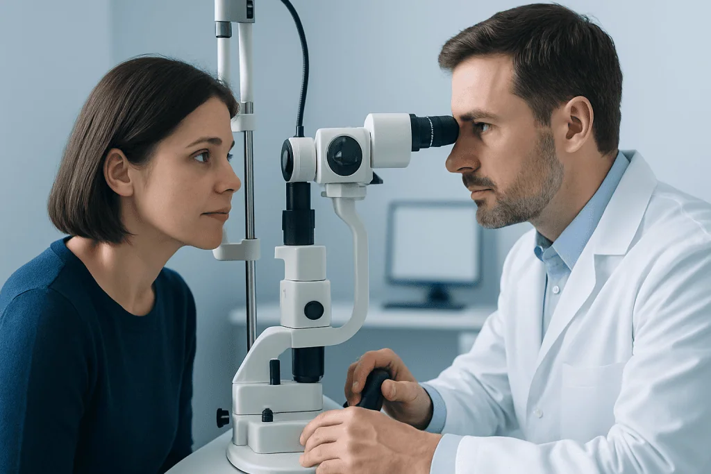
An ophthalmologist can judge response through slit-lamp examination, corneal staining, defect measurements, and sometimes corneal sensitivity testing. If healing is incomplete, the care plan may be adjusted to include additional protective measures, treatment of contributing problems, or discussion of other interventions.
What Is the Success Rate of Corneal Neurotization?
Corneal neurotization is a specialized procedure with encouraging results in published case series and small clinical studies, particularly for improving corneal sensation and supporting epithelial healing in severe neurotrophic keratitis. A single success rate is not reliable for every patient because studies use different surgical techniques, follow-up periods, definitions of success, and patient groups.
In general, many appropriately selected patients experience improved corneal sensitivity and a healthier, more stable ocular surface over time. Results can be influenced by how long the cornea has lacked sensation, the underlying cause of nerve injury, the presence of scarring or active inflammation, tear production, eyelid function, and whether there are other eye conditions affecting vision.
The procedure is usually considered part of a broader eye-surface management plan rather than a stand-alone cure. Even after surgery, lubricants, infection prevention when indicated, and regular corneal monitoring may remain necessary. A corneal surgeon can discuss the expected benefits and limitations for the individual eye.
When to Seek Medical Care
Urgent ophthalmology assessment is appropriate for sudden blurred or reduced vision, increasing redness, discharge, light sensitivity, a white or gray spot on the cornea, eye injury, or a visible open area on the eye. These signs may reflect a corneal ulcer, infection, or thinning and should not be watched at home.
People already diagnosed with neurotrophic keratitis should contact their eye team promptly if they notice changes in vision or the eye’s appearance, even without pain. They should also attend planned follow-up appointments, as a clinician may identify worsening corneal damage before it causes noticeable symptoms.
Acibadem International’s multidisciplinary specialists and JCI-accredited hospitals support international patients with evaluation and treatment for complex corneal conditions. Care may involve corneal ophthalmology, oculoplastic surgery, neurology, and other specialists when the underlying cause affects cranial nerves or overall health.
Frequently asked questions
How serious is neurotrophic keratitis?
Neurotrophic keratitis can be serious because reduced corneal sensation may allow damage to progress without significant pain. Without appropriate treatment, persistent defects can lead to ulcers, scarring, thinning, perforation, and vision loss. Early specialist care and regular follow-up can greatly reduce these risks.
Can neurotrophic keratitis be cured?
The corneal surface can often heal, but the underlying nerve problem may persist. Some people need long-term lubrication, protective measures, or periodic treatment to maintain corneal health. The outlook depends on the cause, stage of disease, and response to treatment.
How long does neurotrophic keratitis treatment take?
Treatment duration depends on the severity of corneal damage and the method used. Surface healing may take days to weeks in some cases, while recovery after corneal neurotization may involve months as nerve fibers grow and sensation gradually improves. Follow-up continues after apparent healing to check for recurrence.
Does neurotrophic keratitis cause pain?
It may cause surprisingly little pain because the corneal nerves that detect discomfort are impaired. Some people notice blurred vision, redness, dryness, light sensitivity, or a foreign-body sensation, while others have few symptoms. Lack of pain should never be used as a sign that the condition is mild.
Can corneal sensation return after neurotrophic keratitis?
Corneal sensation may improve when the underlying cause is reversible or after treatments designed to support nerve recovery. Corneal neurotization may restore some sensory input in selected patients, but the degree and timing of recovery vary. An ophthalmologist can monitor sensation along with surface healing.
What happens if a corneal epithelial defect does not heal?
A persistent epithelial defect increases the risk of infection, corneal ulceration, thinning, and scarring. The ophthalmologist may intensify lubrication, use a protective contact lens or amniotic membrane, prescribe cenegermin when suitable, or consider procedures that protect the eye. Prompt reassessment is important, even if the eye is not painful.
References
- American Academy of Ophthalmology
- European Medicines Agency
- United States Food and Drug Administration
- National Eye Institute
- American Society of Cataract and Refractive Surgery
This article is for general information only and is not a substitute for professional medical advice. Please consult a qualified doctor about your individual situation.
Explore treatments in Turkey — costs, top hospitals & a free quote
JCI-accredited · board-certified surgeons · reply within 24h
Add us as a Preferred Source to see more of our trusted health content across Google Search, AI Overviews and Discover.