Optic Nerve Atrophy: Causes, Diagnosis, and What Vision Changes Mean

Optic nerve atrophy means the optic nerve has been damaged and may no longer transmit visual signals normally. Common symptoms include blurred vision, reduced color vision, blind spots, and a pale-looking optic disc on examination.
Key Takeaways
- Optic nerve atrophy means the optic nerve has been damaged and may no longer transmit visual signals normally.
- Common symptoms include blurred vision, reduced color vision, blind spots, and a pale-looking optic disc on examination.
- Many different conditions can lead to optic nerve atrophy, including glaucoma, inflammation, poor blood flow, trauma, compression, and inherited disorders.
- Diagnosis usually involves a detailed eye exam, visual field testing, imaging, and sometimes blood tests or neurological evaluation.
- Treatment focuses on identifying and managing the underlying cause to protect remaining vision whenever possible.
- New or worsening vision loss always deserves medical attention, especially if it is sudden, painful, or affects one eye quickly.
Optic nerve atrophy refers to damage and loss of optic nerve fibers, which can reduce how clearly the eye sends visual information to the brain. It is not a disease by itself, but a sign of an underlying problem that should be assessed promptly by an eye specialist or neurologist.
Overview
Optic nerve atrophy is a term used when the optic nerve shows signs of damage and fiber loss. The optic nerve carries visual information from the retina to the brain, so when it is injured, vision may become less sharp or less complete. In many cases, the optic disc appears pale during an eye examination, reflecting changes in the nerve tissue.
This condition is not a single illness. Instead, it is a clinical finding that can result from many different problems affecting the eye, optic nerve, brain, or blood supply. These may include inflammation, pressure-related damage, reduced circulation, inherited disorders, trauma, tumors, or long-standing eye disease.
The amount of vision change varies widely. Some people notice only subtle difficulty with color vision or contrast, while others experience major vision loss or blind spots. Because some causes can continue to worsen if left untreated, timely evaluation is important to understand what the vision changes mean and whether the underlying problem can be treated.
Symptoms and vision changes
The symptoms of optic nerve atrophy depend on how much of the nerve is affected and what caused the damage. Vision loss may involve one eye or both eyes. It can develop suddenly in some conditions, but in many others it appears gradually over weeks, months, or even years.
People may describe blurry or dim vision, trouble seeing fine details, poor contrast, reduced side vision, or a dark or missing area in the visual field. Colors may seem less vivid, especially red tones. Some individuals notice increased sensitivity to glare or difficulty adjusting between bright and dim environments.
Not everyone has pain. Pain is more likely when optic nerve damage is related to active inflammation, but many causes of optic nerve atrophy are painless. Possible symptoms include:
- Blurred or decreased central vision
- Loss of peripheral vision
- Reduced color vision
- Blind spots or patchy missing areas
- Dimmer vision in one eye compared with the other
- Difficulty reading or recognizing faces
If vision changes are sudden, severe, or associated with headache, weakness, double vision, or eye pain, urgent assessment is especially important. These features can point to conditions that need prompt treatment.
Causes and risk factors

Optic nerve atrophy can develop when the nerve is injured directly or when it is affected by another disease process. One of the most common causes is long-term damage from glaucoma, in which pressure-related or pressure-sensitive injury affects optic nerve fibers over time. Other important causes include optic neuritis, poor blood flow to the nerve, severe retinal disease, head or eye trauma, and compression from masses such as tumors or enlarged blood vessels.
Inflammatory, infectious, toxic, nutritional, and inherited conditions can also damage the optic nerve. Examples include multiple sclerosis-related optic neuritis, vitamin deficiencies, exposure to certain toxins, and hereditary optic neuropathies. In some people, disorders affecting the brain and nervous system contribute to optic nerve injury, so a broader neurological work-up may be needed.
Risk factors depend on the underlying cause but may include older age, diabetes, high blood pressure, smoking, heavy alcohol use, poor nutrition, autoimmune disease, previous eye disease, family history of optic neuropathy, or a history of head injury. A person who already has visual problems or nerve-related symptoms may also be more likely to need detailed assessment for associated conditions such as brain tumors when compression is suspected.
Sometimes optic nerve atrophy is discovered after the original damaging event has already passed. In these cases, the focus shifts to understanding whether the problem is stable or ongoing, and whether remaining vision can be protected.
How doctors diagnose optic nerve atrophy
Diagnosis begins with a careful history. The doctor will ask when the vision changes started, whether one or both eyes are affected, whether symptoms are painful, and whether there have been headaches, neurological symptoms, trauma, infections, medication exposures, or a family history of eye disease. This context helps narrow the list of possible causes.
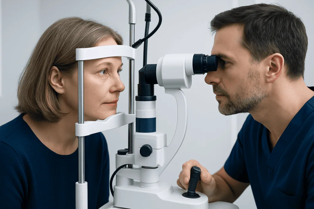
A complete eye examination usually includes visual acuity testing, pupil assessment, color vision testing, slit-lamp examination, and a detailed look at the optic nerve and retina after dilation. Visual field testing helps map any blind spots or peripheral loss. Optical coherence tomography may be used to measure the thickness of the retinal nerve fiber layer and support the diagnosis of optic nerve damage.
If the cause is not clear, additional testing may be recommended. This can include blood tests, orbital or brain imaging such as MRI, and sometimes neurological evaluation. Imaging is particularly useful when doctors need to look for inflammation, demyelination, structural abnormalities, or compression of the optic nerve. Depending on the findings, a patient may also need MRI imaging or other targeted investigations.
Because optic nerve atrophy can be linked to both eye and brain conditions, diagnosis is often multidisciplinary. Neuro-ophthalmologists, ophthalmologists, neurologists, and radiologists may all contribute to identifying the cause and planning care.
Treatment options and what they can achieve
There is no single treatment that reverses optic nerve atrophy itself once nerve fibers have been lost. Treatment is directed at the underlying cause and at protecting the remaining optic nerve function. The outlook depends on what caused the damage, how long it has been present, and how quickly treatment begins.
For example, if the damage is related to glaucoma, lowering eye pressure can help reduce further injury. If inflammation is suspected, treatment may focus on the inflammatory condition. Compression of the nerve may require a different approach, including specialist review and, in selected cases, neurosurgical treatment. If an optic nerve problem is linked to a broader eye condition, management may overlap with glaucoma treatment or retinal care.
Supportive care is also important. This may include low-vision aids, improved lighting, contrast-enhancing strategies, and rehabilitation to help with reading, mobility, and daily activities. Even when lost vision cannot be fully restored, practical support can make a meaningful difference in quality of life.
Near the end of the diagnostic process, some patients benefit from coordinated care in a tertiary center. Acibadem International’s multidisciplinary specialists and JCI-accredited hospitals evaluate and treat optic nerve disorders for international patients, particularly when neuro-ophthalmology, imaging, and neurological care are needed together.
Prevention and self-care
Not all cases of optic nerve atrophy can be prevented, but some causes can be addressed early. Regular eye examinations are especially important for people with glaucoma risk, diabetes, high blood pressure, autoimmune disease, or a family history of serious eye conditions. Early detection gives the best chance of limiting further damage.
General health habits also matter. Stopping smoking, limiting alcohol misuse, maintaining balanced nutrition, and managing chronic medical conditions may reduce risks related to circulation, toxic exposure, and nutritional deficiency. Medicines should be taken only as prescribed, and any concerns about side effects affecting vision should be discussed with a doctor.
People who already have optic nerve damage can protect daily function by using good lighting, magnification devices, larger print settings, and home safety adjustments. Keeping follow-up appointments is important, because the doctor may need to monitor for progression or changes in the underlying condition.
When to see a doctor
Any unexplained change in vision should be assessed by a qualified doctor, optometrist, or ophthalmologist. Prompt evaluation is especially important when symptoms are new, getting worse, or affecting normal activities such as reading, walking, or driving.
Urgent medical attention is needed if vision loss is sudden, painful, associated with severe headache, or accompanied by weakness, numbness, double vision, or trouble speaking. These symptoms may signal a condition affecting the optic nerve, eye, or brain that needs immediate care.
Even when vision changes seem mild, it is wise not to ignore them. Early assessment may reveal treatable causes such as inflammation, pressure-related damage, or compression. In some cases, referral for a comprehensive eye examination and specialist testing helps clarify whether optic nerve atrophy is present and what steps should come next.
Frequently asked questions
Is optic nerve atrophy the same as blindness?
No. Optic nerve atrophy means the optic nerve has been damaged, but the degree of vision loss can vary widely. Some people have mild changes, while others have severe and permanent visual impairment.
Can optic nerve atrophy be reversed?
Lost optic nerve fibers usually cannot be restored with current standard treatments. However, treating the underlying cause may help prevent further damage and preserve the vision that remains.
What does optic nerve atrophy look like on an eye exam?
Doctors often describe the optic disc as pale when optic nerve atrophy is present. This appearance is interpreted along with vision testing, visual field results, and imaging to understand the cause and extent of damage.
Does optic nerve atrophy always affect both eyes?
No. It can affect one eye or both eyes, depending on the underlying condition. Some causes are localized to one optic nerve, while others are inherited, inflammatory, or systemic and may involve both eyes.
Is optic nerve atrophy painful?
Often it is painless, especially when related to chronic eye disease or past injury. Pain is more likely when there is active inflammation, such as optic neuritis, but this varies from person to person.
What tests are usually needed?
Testing often includes a full eye exam, color vision testing, visual field assessment, and imaging of the optic nerve and retina. Depending on the suspected cause, blood tests and brain or orbit MRI may also be recommended.
When should someone seek urgent care for vision changes?
Urgent care is important for sudden vision loss, eye pain, severe headache, double vision, or any neurological symptoms such as weakness or difficulty speaking. These signs can point to conditions that need immediate medical evaluation.
References
- American Academy of Ophthalmology
- National Eye Institute
- National Institute of Neurological Disorders and Stroke
- Mayo Clinic
- Merck Manual Professional Edition
This article is for general information only and is not a substitute for professional medical advice. Please consult a qualified doctor about your individual situation.
Explore treatments in Turkey — costs, top hospitals & a free quote
JCI-accredited · board-certified surgeons · reply within 24h
Add us as a Preferred Source to see more of our trusted health content across Google Search, AI Overviews and Discover.
Check your numbers in seconds
BMI, calories, due date, blood pressure and 30+ more clinical calculators — free, instant, doctor-reviewed ranges.