Thyroid Cancer Ultrasound Image: Preparation, Procedure and Results

Thyroid ultrasound uses sound waves, not radiation, to create images of the thyroid gland and nearby lymph nodes. Most thyroid nodules are benign, and a suspicious-looking ultrasound finding does not by itself mean cancer is present.
Key Takeaways
- Thyroid ultrasound uses sound waves, not radiation, to create images of the thyroid gland and nearby lymph nodes.
- Most thyroid nodules are benign, and a suspicious-looking ultrasound finding does not by itself mean cancer is present.
- Features such as irregular margins, microcalcifications, a taller-than-wide shape, and suspicious lymph nodes may prompt further assessment.
- Most people do not need special preparation and can return to normal activities immediately after the scan.
- Fine-needle aspiration biopsy is commonly used when a nodule meets size and ultrasound-risk criteria.
A thyroid cancer ultrasound image can show features that may raise or lower concern for cancer in a thyroid nodule, but ultrasound alone cannot provide a definite diagnosis. This painless imaging test helps clinicians decide whether monitoring, blood tests, or a fine-needle aspiration biopsy is appropriate.
Overview: What a Thyroid Cancer Ultrasound Image Can Show
A thyroid cancer ultrasound image is a detailed picture of the thyroid gland made with high-frequency sound waves. It can show whether a thyroid nodule is solid, fluid-filled, mixed, smooth, irregular, small or large. It also allows the clinician to examine lymph nodes in the neck, which can be relevant when thyroid cancer is suspected.
Ultrasound is one of the main tests used to assess a thyroid lump, enlarged thyroid, unexplained neck swelling, or a nodule found during another scan. It does not expose the person to ionizing radiation and is generally quick and comfortable. Importantly, an ultrasound can estimate the likelihood that a nodule may need further testing, but it cannot confirm cancer on its own.
Many nodules are common and noncancerous, particularly with increasing age. A radiologist may use a standardized reporting system, such as TI-RADS, to describe the nodule’s appearance and guide recommendations for follow-up or biopsy. When results suggest a need for additional evaluation, the next step is often a needle biopsy rather than immediate surgery.
How Thyroid Ultrasound Works and Who May Need It
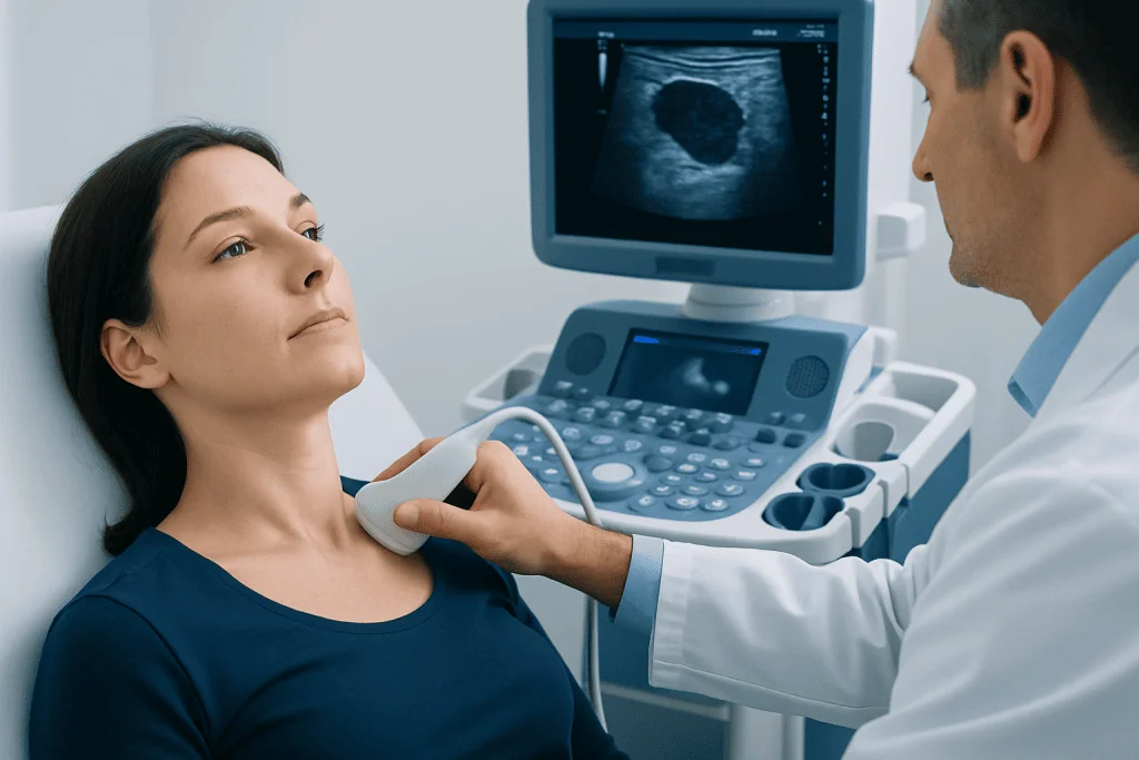
During a thyroid ultrasound, a small handheld device called a transducer sends sound waves through the skin of the neck. The waves reflect differently from normal thyroid tissue, nodules, cysts, blood vessels, and lymph nodes. A computer converts these reflections into moving images that the sonographer and radiologist can assess.
A clinician may recommend this examination when a person or doctor notices a neck lump, when thyroid enlargement is present, or when a nodule is detected on CT, MRI, PET, or another imaging test. It may also be used to monitor an already known nodule, evaluate neck lymph nodes, or guide a fine-needle aspiration biopsy.
Ultrasound is suitable for most people, including pregnant people and those who cannot receive contrast dye. It evaluates anatomy rather than thyroid hormone function, so blood tests such as thyroid-stimulating hormone may be ordered alongside imaging when clinically appropriate.
- Palpable lump, fullness, or swelling in the front of the neck
- Thyroid nodule found incidentally on another scan
- Known thyroid nodules needing surveillance
- Enlarged or abnormal neck lymph nodes
- Planning or guidance for needle biopsy
Do I Need to Do Anything to Prepare for a Thyroid Ultrasound?

Most people need no special preparation for a thyroid ultrasound. They can usually eat, drink, and take regular prescribed medicines as usual unless their healthcare team has given different instructions for another test taking place on the same day.
It may help to wear a loose, open-necked top and avoid necklaces or scarves that could need to be removed. People should bring previous ultrasound reports, biopsy results, thyroid blood test results, or imaging discs if they are available, as comparisons over time can help the radiologist assess whether a nodule has changed.
If lying with the neck extended is difficult because of pain, dizziness, breathing difficulties, or reduced mobility, it is helpful to tell the imaging team beforehand. They can often adjust positioning and provide support to make the examination more comfortable.
Step by Step: What Happens During the Procedure
The examination is usually performed in an imaging department or outpatient clinic. The person lies on their back, often with a small support under the shoulders so the head tilts gently backward. This position makes the thyroid easier to scan.
A clear gel is placed on the front of the neck. The sonographer moves the transducer over the skin to capture images from different angles. The examination includes both thyroid lobes, the central neck area, and often lymph nodes on each side of the neck. Mild pressure may be applied to obtain clear images.
The scan commonly takes around 15 to 30 minutes, although the time can vary depending on the number of nodules and whether lymph nodes require closer assessment. If a biopsy is needed, it may be scheduled for another visit or performed with ultrasound guidance, depending on the clinical plan.
For nodules that meet criteria for sampling, ultrasound-guided thyroid biopsy can collect cells for laboratory examination. This helps provide a more reliable diagnosis and supports an individualized treatment plan.
What Are the Red Flags on a Thyroid Ultrasound?
Radiologists assess several ultrasound features together rather than relying on one finding. A nodule may be described as higher suspicion when it is solid and very dark compared with surrounding thyroid tissue, has irregular or lobulated edges, contains tiny bright spots called punctate echogenic foci or microcalcifications, or is taller than it is wide on a cross-sectional view.
Other findings that may need closer assessment include evidence that a nodule may extend beyond the thyroid boundary, suspicious-looking lymph nodes, or significant growth on follow-up imaging. Abnormal lymph nodes may have features such as a rounded shape, cystic changes, calcifications, unusual blood flow, or loss of their typical internal appearance.
These findings are not a diagnosis of cancer. Some benign nodules can have concerning features, while some cancers can appear less striking on ultrasound. The radiology report combines appearance and size to recommend observation, repeat imaging, or needle biopsy when appropriate.
- Irregular, spiculated, or poorly defined margins
- Taller-than-wide shape
- Very low echogenicity in a solid nodule
- Punctate echogenic foci or certain calcification patterns
- Suspicious lymph nodes in the neck
What Are the Signs That Thyroid Cancer Is Present on an Ultrasound?
There is no single ultrasound sign that proves thyroid cancer is present. Instead, a combination of suspicious nodule features and abnormal lymph nodes can increase concern enough to recommend a fine-needle aspiration biopsy. The final diagnosis is usually based on cells or tissue examined by a pathologist, and sometimes on surgical pathology if surgery is required.
Ultrasound can also help identify patterns linked with particular thyroid conditions, but interpretation must account for the person’s age, clinical history, prior radiation exposure, family history, thyroid function, and changes in the nodule over time. A small nodule with highly suspicious features may be managed differently from a larger nodule with reassuring features.
If cancer is diagnosed, further imaging and specialist assessment help determine the appropriate next steps. Management may include active surveillance in selected low-risk cases, surgery, or other treatments depending on the cancer type and extent. Learn more about thyroid cancer and the evaluation pathway with an endocrinology and oncology team.
How Uncomfortable Is a Thyroid Ultrasound?
A thyroid ultrasound is usually painless. The gel may feel cool, and the transducer can create mild pressure on the neck, especially when images are taken near a tender lump or when the head is tilted back. Most people tolerate the procedure well.
Some people find the neck position mildly uncomfortable, particularly if they have neck arthritis, recent surgery, swallowing difficulties, or anxiety about lying flat. Informing the sonographer can help, as they may use cushions, modify the angle of the examination, or allow short breaks.
There are no injections, needles, or recovery restrictions for a routine diagnostic ultrasound. If a needle biopsy is performed separately, there may be brief pressure, a small pinch, or mild local soreness afterward, and the care team will explain what to expect.
Results, Recovery and When to Seek Medical Care
There is no recovery period after a standard thyroid ultrasound. The person can normally return to work, driving, meals, exercise, and other daily activities immediately. A radiologist reviews the images and sends a report to the referring clinician, who explains the findings in the context of the individual’s health history.
Results may describe normal thyroid tissue, one or more nodules, thyroid enlargement, inflammation-related changes, cysts, or lymph node findings. A recommendation may be for no further action, repeat ultrasound after a defined interval, thyroid blood tests, or fine-needle aspiration biopsy. A recommendation for biopsy does not mean cancer has been confirmed; it is a way to clarify the nature of a nodule.
Medical review should be arranged promptly for a new or enlarging neck lump, persistent hoarseness, increasing difficulty swallowing, unexplained breathing discomfort, or enlarged lymph nodes in the neck. Urgent assessment is appropriate for sudden breathing difficulty or rapidly developing neck swelling. Acibadem International’s multidisciplinary specialists in JCI-accredited hospitals can assess thyroid nodules and coordinate imaging, biopsy, endocrine care, and cancer treatment for international patients.
Frequently asked questions
Can a thyroid ultrasound diagnose cancer?
No. Ultrasound can identify features that make a thyroid nodule more or less suspicious, but it cannot confirm cancer by itself. When indicated, fine-needle aspiration biopsy is used to examine cells and clarify the diagnosis.
How long does a thyroid ultrasound take?
A standard thyroid ultrasound often takes about 15 to 30 minutes. It may take longer when there are multiple nodules, detailed lymph node imaging is needed, or comparison with previous findings is required.
Can I eat before a thyroid ultrasound?
Yes, most people can eat and drink normally before a thyroid ultrasound. Individual instructions may differ if another procedure or test is planned on the same day, so it is sensible to follow the appointment guidance.
What happens if a thyroid nodule looks suspicious on ultrasound?
The clinician considers the nodule's size, ultrasound pattern, symptoms, and medical history. A follow-up scan or fine-needle aspiration biopsy may be recommended, depending on established clinical criteria. Many suspicious nodules are ultimately found to be benign.
Does a thyroid ultrasound use radiation?
No. Thyroid ultrasound uses sound waves to create images and does not use ionizing radiation. This makes it suitable for repeated monitoring when follow-up is needed.
When should a thyroid nodule be checked?
A person should arrange medical evaluation for a new neck lump, noticeable thyroid enlargement, persistent voice changes, swallowing difficulty, or enlarged neck lymph nodes. Known nodules should be monitored according to the interval recommended by the treating clinician.
References
- American Thyroid Association
- American College of Radiology
- National Cancer Institute
- Radiological Society of North America
- World Health Organization
This article is for general information only and is not a substitute for professional medical advice. Please consult a qualified doctor about your individual situation.
Thyroid Cancer in Turkey — costs, top hospitals & a free quote
JCI-accredited · board-certified surgeons · reply within 24h
Add us as a Preferred Source to see more of our trusted health content across Google Search, AI Overviews and Discover.

Check-up & Preventive Medicine
Comprehensive health screenings designed for early detection and prevention.
2 specialists in this unit