Understanding Seborrhoeic Keratosis or Melanoma: A Complete Patient Guide

Seborrhoeic keratoses are benign growths that often appear waxy, raised, rough, or as if they are stuck onto the skin. Melanoma may be flat or raised and can change in size, shape, color, or sensation over time.
Key Takeaways
- Seborrhoeic keratoses are benign growths that often appear waxy, raised, rough, or as if they are stuck onto the skin.
- Melanoma may be flat or raised and can change in size, shape, color, or sensation over time.
- The ABCDE and “ugly duckling” checks can help identify concerning spots, but they cannot diagnose melanoma.
- A dermatologist may use dermoscopy and, when needed, a biopsy to distinguish a benign lesion from skin cancer.
- Do not try to remove, burn, or freeze an uncertain lesion at home before it has been medically assessed.
- Regular skin awareness and sun protection support early detection and help reduce ultraviolet-related skin damage.
Seborrhoeic keratosis is a common harmless skin growth, whereas melanoma is a potentially serious skin cancer that needs early diagnosis. Because appearance alone can sometimes be misleading, any new, changing, bleeding, or unusual-looking spot should be assessed by a qualified clinician.
Seborrhoeic Keratosis or Melanoma: The Essential Difference
Seborrhoeic keratosis and melanoma are very different conditions, although some spots can appear similar at first glance. A seborrhoeic keratosis is a benign, non-cancerous skin growth that is especially common with increasing age. Melanoma is a type of skin cancer that develops from pigment-producing cells called melanocytes and should be assessed and treated without unnecessary delay.
In many cases, a clinician can recognize a typical seborrhoeic keratosis during a skin examination. However, an irritated, unusually dark, unevenly colored, or changing lesion may need closer evaluation. A person should not rely on online images or self-diagnosis to decide whether a spot is harmless.
The most useful question is not whether a mark perfectly matches a checklist, but whether it is new, changing, different from a person’s other marks, or causing symptoms such as persistent bleeding or non-healing. Early professional assessment provides clarity and is particularly important for people with a personal or family history of melanoma.
What Does Seborrhoeic Keratosis Usually Look and Feel Like?
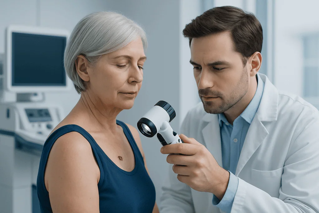
Seborrhoeic keratoses can develop almost anywhere except the palms of the hands and soles of the feet. They commonly occur on the face, scalp, chest, back, abdomen, or shoulders. They may be one lesion or many, and their number often increases over the years.
These growths are often round or oval, slightly raised, and well defined. Their surface may be waxy, scaly, crumbly, rough, or wart-like. They can range from skin-colored or pale tan to brown, dark brown, or nearly black. A classic description is that they look as though they have been “stuck on” the skin.
Most seborrhoeic keratoses do not hurt and do not turn into melanoma. They may itch, become irritated by clothing or jewelry, or bleed if scratched or rubbed. Itching or irritation alone does not prove that a lesion is cancerous, but a spot that repeatedly bleeds, changes, or cannot be confidently identified should be examined.
Several benign skin conditions can resemble one another, including warts, moles, lentigines, and seborrhoeic keratoses. This is one reason a clinician may examine the entire skin surface rather than assessing only one noticeable spot.
How Melanoma May Differ From a Benign Growth
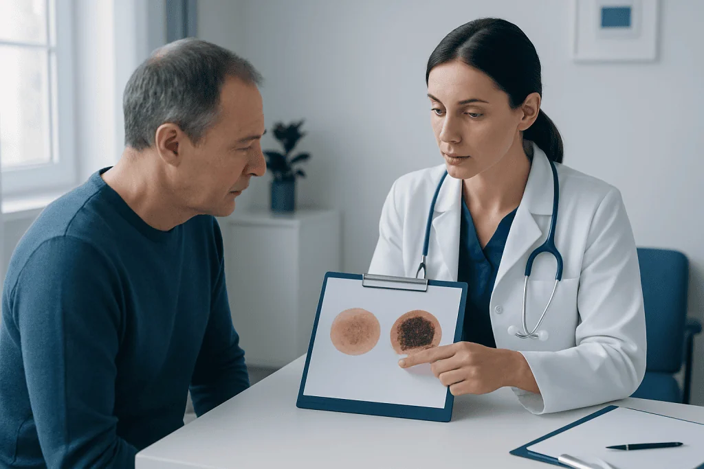
Melanoma may develop in an existing mole or arise as a new mark on previously normal skin. It can appear anywhere, including areas that receive little sun exposure, such as the scalp, nails, soles, genital area, or between the toes. Melanoma does not always look dark; some lesions are pink, red, skin-colored, or only lightly pigmented.
Clinicians often teach the ABCDE guide as a practical way to notice changes: asymmetry, irregular borders, uneven color, a diameter that is increasing or larger than many ordinary moles, and evolution over time. “Evolution” is particularly important and may include a change in shape, color, thickness, surface, or symptoms.
The “ugly duckling” sign is another helpful concept. A mole or spot that looks noticeably different from a person’s other lesions deserves attention, even if it does not meet every ABCDE feature. For example, a single very dark or rapidly changing lesion among many similar-looking moles may warrant prompt review.
These signs are screening clues rather than a diagnosis. Some melanomas do not follow the usual pattern, and some harmless lesions can have irregular features. A dermatologist can assess uncertainty using clinical examination, magnified skin imaging, and, if appropriate, tissue testing.
Why These Lesions Develop and Who May Be at Risk
Seborrhoeic keratoses are associated mainly with age and inherited tendency. They are common in adults and may run in families. They are not contagious, are not caused by poor hygiene, and are not generally considered a warning that skin cancer will develop.
Melanoma risk is influenced by a combination of genetics, skin type, mole pattern, and ultraviolet exposure. Risk may be higher in people with fair skin that burns easily, many moles, atypical moles, a weakened immune system, previous skin cancer, or a close relative with melanoma. However, melanoma can occur in people of every skin tone and at any age.
Intense intermittent sun exposure and sunburns can increase melanoma risk, especially when they occur repeatedly. Tanning beds also expose the skin to ultraviolet radiation and are not a safe alternative to natural sunlight. Protecting skin from excessive ultraviolet exposure is useful regardless of a person’s complexion.
A sudden appearance of multiple itchy seborrhoeic keratosis-like growths is uncommon and has several possible explanations. It should be discussed with a doctor, particularly if it occurs alongside other concerning symptoms, rather than being interpreted independently.
How Doctors Tell the Difference
A medical assessment usually begins with questions about when the lesion appeared, whether it has changed, and whether it itches, hurts, crusts, or bleeds. The clinician will also ask about sun exposure, past skin cancers, family history, medications, and immune health. Examining the surrounding skin and other moles helps provide useful context.
Dermatoscopy, also called dermoscopy, is a painless examination using a handheld device that magnifies the skin and reduces surface glare. It lets trained clinicians see pigment patterns and structures that are not visible to the unaided eye. This can improve diagnostic accuracy, but it does not replace biopsy when a lesion remains suspicious.
If melanoma or another skin cancer cannot be ruled out, the clinician may remove all or part of the lesion for laboratory examination. This is called a biopsy. A pathologist examines the cells under a microscope and provides the definitive diagnosis. The type of biopsy depends on the lesion’s size, location, and clinical appearance.
If melanoma is diagnosed, further evaluation is individualized according to features such as the depth of the tumor and whether it has spread. Early melanoma is often treated with surgery to remove the lesion and a margin of surrounding skin. The care team will explain any recommended tests, treatment steps, and follow-up plan clearly.
Treatment, Skin Care, and Prevention
Typical seborrhoeic keratoses do not need treatment. Removal may be considered when a growth is irritated, catches on clothing, interferes with shaving, repeatedly bleeds after friction, or causes cosmetic concern. Depending on the lesion, a dermatologist may use methods such as cryotherapy, curettage, electrosurgery, or minor surgical removal.
Removal methods should be selected after a diagnosis has been made. Over-the-counter wart treatments, home freezing products, acids, or attempts to cut off a lesion can cause burns, infection, scarring, or delayed cancer diagnosis. It is safest to have an unfamiliar or changing mark checked before any removal is attempted.
There is no guaranteed way to prevent seborrhoeic keratoses. For melanoma prevention, sensible sun protection is important: seek shade when ultraviolet levels are high, wear protective clothing and a wide-brimmed hat, use broad-spectrum sunscreen as directed, and avoid tanning beds. Sunscreen is one part of protection and should not be used to extend time in strong sun.
Monthly skin awareness can help a person recognize change. Looking at the scalp, back, nails, soles, and between the toes with a mirror or a trusted person’s help may be useful. People with numerous moles, a past melanoma, or a strong family history may benefit from a personalized skin-check schedule set by a dermatologist.
When to Seek Medical Care
A person should arrange a medical appointment for any new or changing mole, patch, or raised growth that does not have a clear explanation. Prompt assessment is appropriate if a lesion changes in color, size, shape, or texture; has an irregular border; looks unlike the person’s other spots; or persists despite healing time.
Medical advice is also important for a spot that bleeds without obvious injury, forms a persistent crust, becomes painful, or develops a sore that does not heal. These features do not automatically mean melanoma, but they should not be ignored. A clinician can decide whether observation, dermoscopy, biopsy, or treatment is needed.
People who have previously had melanoma or other skin cancer should follow their specialist’s surveillance plan and report new changes between appointments. Those with a first-degree relative with melanoma, many atypical moles, or immune suppression may also need more regular professional skin examinations.
Acibadem International’s multidisciplinary specialists and JCI-accredited hospitals support international patients who need assessment and treatment for suspicious skin lesions. A dermatologist or appropriate skin cancer specialist can help establish an accurate diagnosis and an individualized plan.
Frequently asked questions
Can seborrhoeic keratosis turn into melanoma?
Seborrhoeic keratosis is benign and does not usually transform into melanoma. However, a melanoma can occasionally resemble a seborrhoeic keratosis, or a person may have both types of lesions. A changing or unusual growth should therefore be assessed rather than assumed to be harmless.
How can a person tell whether a spot is seborrhoeic keratosis or melanoma?
A typical seborrhoeic keratosis may look waxy, rough, raised, and stuck onto the skin, but appearance alone is not always reliable. Changes in color, shape, size, surface, or symptoms should prompt medical review. Dermoscopy and, when needed, biopsy are the most dependable ways to distinguish them.
Is a dark seborrhoeic keratosis always dangerous?
No. Seborrhoeic keratoses can naturally be dark brown or nearly black, particularly in some skin types. Still, a newly dark lesion or one that is changing, unevenly colored, bleeding, or unlike other spots should be examined by a clinician.
Should seborrhoeic keratoses be removed?
Most do not require removal because they are harmless. A dermatologist may remove one if it is irritated, repeatedly traumatized, bothersome, or diagnostically uncertain. Removal should follow a clinical assessment, especially if the lesion has changed.
Can melanoma be pink or skin-colored?
Yes. Some melanomas have little or no dark pigment and may appear pink, red, or skin-colored. A persistent new bump, patch, or sore that changes or does not heal should be medically assessed, regardless of its color.
How often should skin be checked for melanoma?
Regular personal skin awareness is useful, and many people choose to look over their skin about once a month. The best schedule for professional examinations depends on individual risk factors, including previous melanoma, family history, mole pattern, and immune status. A dermatologist can recommend an appropriate follow-up plan.
References
- American Academy of Dermatology
- American Cancer Society
- National Cancer Institute
- NHS
- World Health Organization
This article is for general information only and is not a substitute for professional medical advice. Please consult a qualified doctor about your individual situation.
Melanoma in Turkey — costs, top hospitals & a free quote
JCI-accredited · board-certified surgeons · reply within 24h
Add us as a Preferred Source to see more of our trusted health content across Google Search, AI Overviews and Discover.

Medical Oncology Department
Medical treatment of cancer with chemotherapy, immunotherapy and targeted therapies under a multidisciplinary tumor board.
60 specialists in this unitMore from the Health Library

Related Specialists
Prof. Dr. Mehmet Serdar Binnet
Orthopedic Surgery & Traumatology
Prof. Dr. Gökhan Demir
Medical Oncology
Dr. Gökçe Gökkaya
Gynecology & Obstetrics
Dr. Halime Bayram
Oral & Dental Health