Visual Field Loss: Causes, Diagnosis, and When Brain Imaging Is Needed

Visual field loss may affect side vision, central vision, or create patchy blind spots. Common causes include glaucoma, retinal disease, optic nerve disorders, stroke, and brain lesions.
Key Takeaways
- Visual field loss may affect side vision, central vision, or create patchy blind spots.
- Common causes include glaucoma, retinal disease, optic nerve disorders, stroke, and brain lesions.
- A visual field test helps map missing areas of vision and guides further evaluation.
- Brain imaging is often needed when the pattern suggests a neurological cause or symptoms began suddenly.
- Prompt medical assessment is important, especially with sudden vision changes, headache, weakness, or double vision.
Visual field loss means part of the normal area of sight is missing, blurred, or less sensitive. It can come from problems in the eye, optic nerve, or brain, so careful testing is important to find the cause and decide whether brain imaging is needed.
Overview of Visual Field Loss
Visual field loss refers to a reduction in the normal area a person can see while looking straight ahead. This may involve side vision, central vision, the upper or lower part of sight, or small blind spots scattered across the visual field. Some people notice bumping into objects, difficulty reading, trouble driving, or missing part of words or faces. Others may not notice any change until formal testing is done.
The visual field depends on healthy structures all along the visual pathway. Light must be received by the retina, carried through the optic nerve, cross partially at the optic chiasm, and then travel through brain pathways to the visual cortex. A problem at any point along this pathway can create a characteristic pattern of vision loss. For this reason, visual field loss is an important clue in both eye care and neurology.
Visual field changes are not a diagnosis by themselves. Instead, they are a sign that helps doctors narrow down the cause. In some cases, the cause is within the eye, such as glaucoma or retinal disease. In others, the pattern points to an optic nerve condition or a brain-related problem that may require urgent assessment.
Symptoms and Types of Visual Field Loss
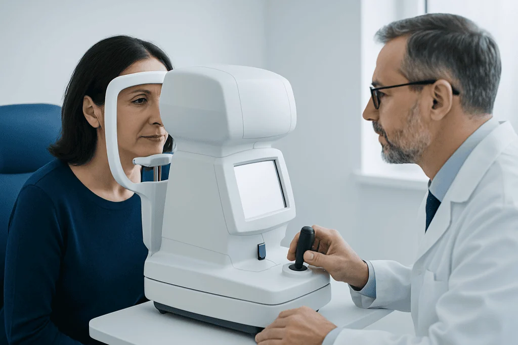
The way visual field loss feels can vary widely. Some people describe tunnel vision, while others notice a shadow, curtain, dark patch, or missing area off to one side. Central field loss can make reading, recognizing faces, or seeing fine detail more difficult. Peripheral field loss may interfere with walking, driving, or noticing objects approaching from the side.
Doctors often describe visual field loss by its pattern. Common patterns include central scotoma, which affects the center of vision; peripheral constriction, often called tunnel vision; altitudinal defects, which affect the upper or lower half of sight; and hemianopia, where the same half of the visual field is missing in both eyes. These patterns can suggest whether the issue is in the retina, optic nerve, or brain.
Other symptoms may occur at the same time and can help guide diagnosis. These may include blurred vision, reduced color vision, eye pain, flashes, floaters, headache, double vision, weakness, numbness, or difficulty speaking. Sudden symptoms, especially when combined with neurological changes, need prompt medical attention.
- Central vision loss: trouble reading or focusing on faces
- Peripheral vision loss: reduced side vision or tunnel vision
- Patchy blind spots: missing small areas within the field of sight
- Half-field loss: missing the right or left side of vision in both eyes
Causes and Risk Factors

There are many possible causes of visual field loss. Eye conditions are common causes, especially glaucoma, which can slowly damage the optic nerve and reduce peripheral vision. Retinal disorders such as retinal detachment, vascular blockage, diabetic eye disease, or inherited retinal conditions may also affect the field of vision. In some people, the first clue may appear during routine eye testing before symptoms are obvious.
Optic nerve disorders are another important group of causes. Optic neuritis, ischemic optic neuropathy, compression from tumors, and other inflammatory or autoimmune conditions can all produce visual field defects. Disorders affecting the optic chiasm, such as pituitary tumors, may classically reduce the outer halves of vision in both eyes. The broader category of optic neuritis is especially relevant when pain with eye movement and color vision changes are present.
Brain-related causes include stroke, traumatic brain injury, multiple sclerosis, and tumors involving the visual pathways. A stroke affecting the occipital lobe, for example, may cause a sudden homonymous hemianopia, where the same side of the visual field is missing in both eyes. Migraine aura can also produce temporary field symptoms, though this diagnosis should be made carefully after more serious causes are considered.
Risk factors depend on the underlying condition. They may include older age, diabetes, high blood pressure, smoking, vascular disease, autoimmune disease, prior eye disease, head injury, or a family history of glaucoma or neurological disorders. Because the range of causes is broad, persistent or unexplained visual field loss should be assessed by a qualified eye specialist or neuroophthalmologist.
How Visual Field Loss Is Diagnosed
Diagnosis begins with a detailed history and eye examination. The doctor will ask when the symptoms started, whether they came on suddenly or gradually, and whether one or both eyes are affected. Questions about headache, eye pain, weakness, speech changes, recent infection, vascular risk factors, or previous eye problems can be especially helpful.
The physical examination usually includes checking visual acuity, pupils, eye movements, color vision, and the front and back of the eye. The appearance of the optic nerve and retina may provide important clues. For example, optic disc swelling, optic nerve pallor, retinal bleeding, or signs of glaucoma can help guide the next steps.
A visual field test is central to the evaluation. Automated perimetry is commonly used to map which parts of vision are reduced or missing. The exact pattern may suggest whether the cause is likely to be retinal, optic nerve, chiasmal, or brain-related. Additional tests such as optical coherence tomography may measure the retina and optic nerve in more detail, while blood tests may be used when inflammation, infection, or nutritional causes are suspected.
In some situations, doctors may recommend further eye imaging or specialized assessment in a comprehensive eye examination or neuro-ophthalmology evaluation. The goal is to identify the site of the problem accurately, because treatment depends on the underlying cause rather than the visual field defect alone.
When Brain Imaging Is Needed
Brain imaging is not necessary for every person with visual field loss, but it becomes important when the findings suggest a neurological cause. Imaging is commonly considered when the pattern of field loss matches damage behind the eye, such as homonymous hemianopia, bitemporal field loss, or other defects that localize to the optic chiasm or visual pathways in the brain. It is also more likely to be needed when the eye examination does not fully explain the symptoms.
Urgent imaging may be required if visual field loss begins suddenly, especially with headache, weakness, numbness, confusion, speech difficulty, imbalance, or double vision. These features can point to stroke, bleeding, or another acute neurological problem. Progressive symptoms, optic nerve swelling, unexplained optic atrophy, or concern for a compressive lesion are other common reasons for imaging.
MRI is often preferred because it provides detailed views of the optic nerves, optic chiasm, brain, and surrounding structures. In emergency settings, CT may be used first, especially if stroke or bleeding is suspected. In some cases, imaging of the blood vessels may also be recommended. If a structural cause is found, management may involve neurology, neurosurgery, endocrinology, or ophthalmology depending on the diagnosis.
Patients may hear about imaging in the setting of MRI scanning or neurological work-up for conditions such as stroke. When visual field loss appears to come from the brain rather than the eye, timely imaging helps doctors confirm the diagnosis and begin appropriate treatment without delay.
Treatment Options
Treatment for visual field loss depends entirely on the cause. For glaucoma, lowering eye pressure can help slow further damage, although vision already lost is usually difficult to restore. Retinal conditions may need medical treatment, laser procedures, or surgery. If the cause is inflammatory, autoimmune, or infectious, medicines directed at that condition may improve outcomes.
When the optic nerve or brain is involved, treatment may include corticosteroids for some inflammatory disorders, vascular risk management after stroke, or surgery and other targeted treatment for compressive lesions such as pituitary tumors. In selected cases, the underlying issue can be stabilized or improved, but some field loss may remain. Early diagnosis often offers the best chance to protect remaining vision.
Rehabilitation is also an important part of care. People with lasting field loss may benefit from visual aids, occupational therapy, scanning techniques, and practical strategies for reading, mobility, and home safety. Driving advice depends on the degree and pattern of vision loss as well as local legal requirements, so patients should ask their doctor before continuing to drive.
Near the end of the care pathway, some people may be referred for multidisciplinary support that can include ophthalmology, neurology, radiology, and rehabilitation services. Acibadem International’s multidisciplinary specialists and JCI-accredited hospitals diagnose and treat visual pathway disorders for international patients when specialist assessment is needed.
Self-care, Monitoring, and Prevention
Not all causes of visual field loss can be prevented, but regular eye care and attention to general health can reduce risk in some cases. Routine eye examinations are especially important for people with diabetes, high blood pressure, a family history of glaucoma, or increasing age. Some conditions progress quietly, so screening can identify problems before a person notices symptoms.
Good control of blood pressure, blood sugar, and cholesterol supports both eye and brain health. Avoiding smoking and following treatment plans for known medical conditions can also help lower the risk of optic nerve and vascular damage. If a person has migraine, autoimmune disease, or previous neurological illness, ongoing follow-up may help detect changes early.
For those already diagnosed with visual field loss, self-care includes keeping follow-up appointments, taking medicines as prescribed, and reporting new symptoms promptly. Practical steps such as improving lighting, reducing fall hazards, and using visual scanning techniques may make daily activities easier and safer. Family support can also be helpful, particularly when reading, navigation, or confidence with mobility has been affected.
When to See a Doctor
A doctor should assess any persistent or unexplained visual field loss. Even gradual changes deserve attention because some causes, such as glaucoma or compressive lesions, can worsen over time if left untreated. Prompt evaluation helps protect remaining vision and identify whether the problem comes from the eye or the brain.
Urgent medical care is needed when visual field loss starts suddenly or is accompanied by severe headache, weakness, numbness, difficulty speaking, confusion, or double vision. These symptoms can indicate a stroke or another emergency. Sudden flashes, floaters, or a curtain-like shadow over vision also need urgent assessment because they may suggest retinal problems.
If vision changes occur together with eye pain, color vision changes, or pain when moving the eye, specialist review is also important because optic nerve inflammation may be involved. A person should not ignore even mild symptoms if they are new, recurrent, or affecting daily life. Early evaluation is the safest approach.
Frequently asked questions
What is visual field loss?
Visual field loss means that part of the normal area a person can see is missing or less clear. It may affect side vision, central vision, or create isolated blind spots. The problem can come from the eye, optic nerve, or brain.
Can visual field loss happen without obvious symptoms?
Yes. Some people do not notice gradual visual field loss at first, especially if it affects only one eye or the outer parts of vision. That is one reason regular eye examinations are important.
Does visual field loss always mean a brain problem?
No. Many cases are caused by eye conditions such as glaucoma or retinal disease. However, certain patterns of field loss suggest a problem in the optic nerve or brain and may require neurological assessment and imaging.
Why might a doctor order an MRI for visual field loss?
An MRI may be ordered when the pattern of vision loss suggests damage in the visual pathways behind the eye, or when the eye exam does not fully explain the symptoms. It is also useful if doctors suspect stroke, inflammation, or a compressive lesion such as a tumor.
Can visual field loss be treated?
Treatment depends on the cause. Some conditions can be stabilized or improved, especially when diagnosed early, while others may cause permanent changes. Even when vision cannot be fully restored, treatment may help prevent further loss and rehabilitation can improve daily function.
Is visual field loss an emergency?
It can be. Sudden visual field loss, especially with headache, weakness, speech difficulty, numbness, or double vision, needs urgent medical care. Gradual symptoms are not always an emergency, but they still should be assessed promptly.
References
- American Academy of Ophthalmology
- National Eye Institute
- National Institute of Neurological Disorders and Stroke
- Royal College of Ophthalmologists
- Mayo Clinic
This article is for general information only and is not a substitute for professional medical advice. Please consult a qualified doctor about your individual situation.
Explore treatments in Turkey — costs, top hospitals & a free quote
JCI-accredited · board-certified surgeons · reply within 24h
Add us as a Preferred Source to see more of our trusted health content across Google Search, AI Overviews and Discover.
Check your numbers in seconds
BMI, calories, due date, blood pressure and 30+ more clinical calculators — free, instant, doctor-reviewed ranges.

More from the Health Library



Related Specialists

Dr. Burcu Duran Tak
Acibadem Life Clinical Service
Dt. Akif Aydın Şen
Oral & Dental Health
Prof. Dr. Şahin Şenay
Cardiovascular Surgery
Dr. Babak Hekmatjou
Emergency Service