What Is a Cardiology Echo? Echocardiography Types and When They Are Used

A cardiology echo uses sound waves, not radiation, to examine the heart. Different echo types are used for different questions, including transthoracic, transesophageal, stress, and Doppler echo.
Key Takeaways
- A cardiology echo uses sound waves, not radiation, to examine the heart.
- Different echo types are used for different questions, including transthoracic, transesophageal, stress, and Doppler echo.
- An echocardiogram can help diagnose valve disease, heart failure, congenital heart problems, and fluid around the heart.
- Most echo tests are painless and done without special recovery time.
- The best type of echocardiography depends on symptoms, medical history, and what the doctor needs to see.
A cardiology echo, also called echocardiography, is a safe ultrasound test that creates moving images of the heart. It helps doctors assess heart structure, blood flow, pumping strength, and valve function in many common and complex heart conditions.
Overview: What Is a Cardiology Echo?
A cardiology echo is another name for echocardiography, a test that uses sound waves to create pictures of the heart. Because it is based on ultrasound technology, it does not use radiation. The images allow doctors to see the heart beating in real time and to evaluate how well it is working.
This test can show the size and shape of the heart, how strongly the heart muscle pumps, and how the valves open and close. It can also help assess blood flow through the chambers and major vessels. For many people, a cardiology echo is one of the first and most useful tests in heart evaluation.
Echocardiography is used in both routine and urgent situations. A doctor may request it for symptoms such as shortness of breath, chest discomfort, swelling in the legs, fainting, or a heart murmur. It is also often used to monitor known conditions such as heart failure, aortic regurgitation, or cardiomyopathy.
How Echocardiography Works
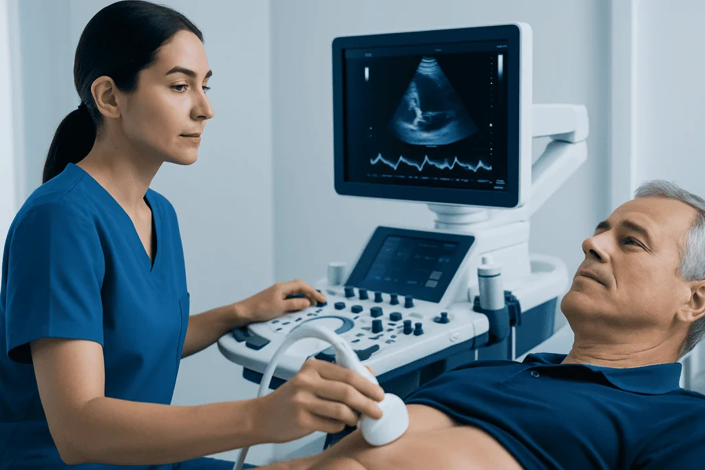
During an echo, a small handheld device called a transducer sends high-frequency sound waves into the body. These sound waves bounce off the heart structures and return to the machine, which turns them into moving images. This lets the care team view the heart muscle, valves, chambers, and surrounding tissues.
Standard ultrasound images show anatomy and movement, while Doppler techniques show how blood flows through the heart. Doppler can help identify narrowing, leakage, abnormal pressure patterns, or unusual flow between chambers. In many cases, this added information is essential for understanding whether a valve or pumping problem is mild, moderate, or severe.
Some echocardiograms are done through the chest wall, while others use a small probe in the esophagus for closer images. The test chosen depends on the patient’s symptoms, body structure, medical history, and what details the cardiologist needs to clarify.
Types of Echocardiography and When They Are Used

The most common type is transthoracic echocardiography, often called TTE. In this test, the transducer is placed on the chest with gel to improve contact. TTE is usually the first choice because it is noninvasive, widely available, and provides valuable information about chamber size, heart pumping, valve function, and fluid around the heart.
Transesophageal echocardiography, or TEE, uses a flexible probe passed gently into the esophagus after the throat is numbed and sedation is usually given. Because the esophagus sits close to the heart, TEE can produce especially detailed images of valves, the atria, and nearby structures. It is often used when chest-wall images are not clear enough, when infection such as endocarditis is suspected, or before procedures related to rhythm disorders such as ablation procedures.
Stress echocardiography shows how the heart performs during exercise or with medication that makes the heart work harder. It is commonly used when doctors are evaluating symptoms that may be related to reduced blood supply to the heart muscle, including possible coronary artery disease. Stress echo can also help assess exercise-related valve problems or changes in pumping function.
Other specialized forms include Doppler echo, color Doppler, contrast echo, 3D echocardiography, fetal echo, and pediatric echo. These may be used to evaluate leaks or narrowing in heart valves, structural differences present at birth, or blood flow patterns in more detail. In children and unborn babies, a heart specialist may use pediatric cardiology or fetal cardiology assessment when a congenital condition is suspected.
Why Doctors Order a Cardiology Echo
An echocardiogram may be ordered to investigate symptoms or to monitor a known diagnosis. It is especially useful when a doctor hears a heart murmur, suspects a valve problem, or needs to understand the cause of shortness of breath, unexplained fatigue, palpitations, swelling, or dizziness. In emergency settings, it may help identify fluid around the heart, severe valve dysfunction, or major changes in heart pumping.
Doctors also use echo to look for changes after a heart attack, to evaluate the effects of long-standing high blood pressure, and to monitor heart muscle weakness or enlargement. It is an important test in assessing heart failure, cardiomyopathies, congenital heart disease, infections involving the heart lining or valves, and diseases affecting the sac around the heart.
In some people, echocardiography helps guide treatment planning. For example, it can help determine whether a valve problem needs close follow-up, medication, or a procedure. It may also be used before and after surgery or catheter-based treatment, including some forms of congenital repair and cardiothoracic surgery.
What to Expect Before, During, and After the Test
For a standard transthoracic echo, very little preparation is usually needed. The person typically lies on an exam table while a sonographer places gel on the chest and moves the transducer to obtain images from different angles. The test is painless, though some pressure from the probe may be felt briefly.
A transesophageal echo requires more preparation. Patients are commonly asked not to eat or drink for a period before the test. Sedation is often used, and the throat is numbed to make the procedure more comfortable. Because of sedation, someone may need assistance getting home afterward, depending on local protocols and the patient’s overall condition.
Stress echo preparation depends on whether the test involves exercise or medicine. The care team may advise avoiding heavy meals, caffeine, or certain medicines beforehand. After most echocardiograms, people can return to normal activities quickly, although instructions may vary after TEE or stress testing.
- Transthoracic echo: usually no special recovery time
- Transesophageal echo: temporary throat discomfort may occur
- Stress echo: heart rate and blood pressure are monitored closely during the test
- Results may be available the same day or after specialist review
What the Results Can Show
An echocardiogram can reveal whether the heart chambers are normal in size, whether the heart muscle is thickened, and how effectively the heart pumps blood. One commonly reported measurement is the ejection fraction, which helps estimate pumping strength. Although one number never tells the whole story, it can be helpful in understanding overall heart function.
Echo results can also show valve narrowing or leakage, abnormal blood flow, blood clots in some situations, signs of infection, and fluid around the heart. In people with congenital heart disease, the test can define structural differences and help guide follow-up. If the doctor suspects disease of the heart sac, valves, or major vessels, echo may provide important clues that shape the next step in care.
Sometimes the images are completely normal, which can be reassuring and still very useful. In other cases, the findings suggest that additional tests are needed, such as electrocardiography, cardiac MRI, CT, angiography, or rhythm studies. Echo is often one part of a broader cardiac evaluation rather than a stand-alone answer.
Limitations, Safety, and Follow-Up
Echocardiography is considered very safe, and standard transthoracic echo has no known radiation exposure. Even so, every test has limits. Image quality can sometimes be reduced by body shape, lung disease, prior chest surgery, or the position of the heart. In such cases, a transesophageal echo or another imaging method may provide clearer information.
Stress echocardiography and TEE involve additional considerations. Stress testing can occasionally trigger symptoms such as chest discomfort, shortness of breath, or changes in heart rhythm, which is why it is done under medical supervision. TEE has small procedure-related risks related to sedation and passage of the probe, so doctors use it when the expected benefit is meaningful.
Follow-up depends on the reason for the test and the findings. Some people need only reassurance, while others require monitoring over time to watch a valve problem, changes in heart muscle function, or the effect of treatment. Near the end of the care pathway, patients who need coordinated evaluation can be assessed by multidisciplinary specialists; Acibadem International’s JCI-accredited hospitals diagnose and treat heart conditions for international patients.
When to See a Doctor
A person should speak with a doctor if they have ongoing shortness of breath, chest discomfort, swelling in the ankles or abdomen, new palpitations, fainting, or unusual fatigue. These symptoms do not always mean a serious heart problem, but they deserve medical assessment, especially if they are new, worsening, or interfering with daily life.
An echocardiogram may also be appropriate when a clinician detects a new murmur, when blood pressure has affected the heart over time, or when there is a family history of certain inherited heart muscle conditions. People who already have a diagnosed heart condition should attend follow-up visits as advised, even if they feel well, because echo can sometimes detect changes before symptoms become obvious.
Urgent medical care is important for severe chest pain, sudden shortness of breath, fainting, or symptoms of stroke. In these situations, doctors may use echocardiography as part of a rapid evaluation to understand how the heart is functioning and what treatment may be needed.
Frequently asked questions
Is a cardiology echo the same as an echocardiogram?
Yes. A cardiology echo is a common way of referring to an echocardiogram or echocardiography. All of these terms describe an ultrasound test that creates images of the heart.
Does an echocardiogram hurt?
A standard transthoracic echocardiogram is usually painless. A person may feel cool gel on the skin and light pressure from the probe, but the test is generally well tolerated. Transesophageal echo can cause temporary throat discomfort, though sedation is commonly used for comfort.
How long does an echo test take?
A routine transthoracic echo often takes about 30 to 60 minutes, depending on how many images are needed. A transesophageal or stress echo may take longer because of preparation, monitoring, and recovery time.
Why would a doctor order a transesophageal echo instead of a standard echo?
A transesophageal echo is often chosen when the standard chest-wall images are not detailed enough. It can provide clearer views of certain valves, the atria, and nearby structures, and it is useful in situations such as suspected infection, blood clots, or planning some procedures.
Can an echocardiogram detect heart failure?
An echocardiogram is one of the key tests used to evaluate heart failure. It can show how well the heart pumps, whether the chambers are enlarged, and whether valve disease or another structural problem may be contributing to symptoms.
Is there any radiation in a cardiology echo?
No. Echocardiography uses sound waves rather than X-rays, so there is no radiation exposure in a standard echo. This makes it a useful test for repeated follow-up when necessary.
This article is for general information only and is not a substitute for professional medical advice. Please consult a qualified doctor about your individual situation.
Echocardiography in Turkey — costs, top hospitals & a free quote
JCI-accredited · board-certified surgeons · reply within 24h
Add us as a Preferred Source to see more of our trusted health content across Google Search, AI Overviews and Discover.

Cardiology Department
Diagnosis and treatment of heart and vascular conditions, from prevention to advanced interventional procedures.
93 specialists in this unitRelated Treatments
Related Conditions
More from the Health Library

Anemia Before Heart Bypass Surgery: Testing, Transfusion Risk, and Optimization

CABG After a Heart Attack: Timing, Benefits, and Recovery Planning

Cardiac Ablation: Who It Helps and What to Expect During Recovery
Nuclear Stress Test vs Stress Echocardiogram: Which Heart Test May Be Better?

Premature Ventricular Contractions: When PVCs Are Benign and When to Treat Them

Heart Failure: Shortness of Breath, Swelling, and Modern Treatment
Cardiology Specialists at Acibadem

Prof. Dr. Fuat Bilgen
Cardiovascular Surgery
Asst. Prof. Dr. Funda Helvacıoğlu
Cardiology
Asst. Prof. Dr. Gökhan Arslanhan
Cardiovascular Surgery
Prof. Dr. Gültekin Karakuş
Cardiology