When Imaging Finds an Incidentaloma: What Unexpected Scan Results May Mean

An incidentaloma is a finding discovered by chance on imaging such as CT, MRI, ultrasound, or PET scans. Most incidentalomas are not cancer and do not cause symptoms, but some require additional tests or monitoring.
Key Takeaways
- An incidentaloma is a finding discovered by chance on imaging such as CT, MRI, ultrasound, or PET scans.
- Most incidentalomas are not cancer and do not cause symptoms, but some require additional tests or monitoring.
- The next steps depend on the finding’s size, location, imaging features, and whether it may affect hormone function.
- Follow-up can include repeat imaging, blood or urine tests, or occasionally a biopsy or surgery.
- Patients should ask what was found, how concerning it appears, and whether specialist review is recommended.
An incidentaloma is an unexpected abnormality found on an imaging test done for another reason. Many incidental findings are benign, but some need careful follow-up to determine whether they are harmless, hormone-producing, or related to a more significant condition.
Overview: What an Incidentaloma Is
An incidentaloma is an unexpected finding seen on an imaging test that was ordered for a different reason. For example, a person may have a CT scan after an injury, an MRI for back pain, or an ultrasound for abdominal discomfort, and the scan may reveal a small mass, nodule, cyst, or area of change unrelated to the original problem.
The term does not describe one single disease. Instead, it is a general label for a finding discovered by chance. Incidentalomas can appear in many organs, including the adrenal glands, thyroid, kidneys, liver, lungs, pituitary gland, ovaries, and brain. Some are tiny and clinically unimportant, while others need more attention.
Modern imaging is very detailed, so incidental findings have become more common. This can be unsettling, but it is important to remember that “unexpected” does not mean “dangerous.” Many incidentalomas are benign growths, cysts, or age-related changes that never cause harm.
The main goal after an incidental finding is not to assume the worst, but to identify whether it is clearly harmless, whether it should be watched over time, or whether it needs more testing. A careful, step-by-step review by a doctor helps avoid both unnecessary worry and unnecessary procedures.
Why Incidental Findings Happen So Often
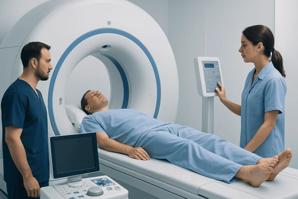
Imaging tests can show structures in great detail, including very small abnormalities that older technologies might not have detected. CT, MRI, and high-resolution ultrasound can reveal tiny nodules, cysts, and tissue changes that may have been present for years without causing symptoms.
Incidentalomas are especially common when imaging covers a large area of the body. A scan performed to evaluate one issue may also capture nearby organs. For instance, a chest CT may show part of the thyroid or adrenal glands, and an abdominal MRI may reveal a kidney cyst or liver lesion.
Age also plays a role. As people get older, benign changes such as cysts, nodules, and noncancerous growths become more frequent. This does not necessarily indicate disease. It often reflects normal variation, prior inflammation, or wear-and-tear changes over time.
Radiologists are trained to identify and describe these findings carefully. Their report often includes details such as size, shape, density, borders, and whether follow-up imaging is suggested. These details help the referring doctor decide if the incidentaloma can be ignored safely or should be evaluated further.
What Unexpected Scan Results May Mean

An incidentaloma may represent several different possibilities. In many cases, it is a benign cyst, adenoma, scar, or harmless nodule. These findings may never cause symptoms and may not need treatment. Common examples include simple kidney cysts, benign liver hemangiomas, or small adrenal adenomas.
Some incidental findings are not harmful but still deserve follow-up because their appearance is not completely specific. A doctor may recommend repeat imaging after a period of time to see whether the lesion remains stable. Stability over time is often reassuring.
Less commonly, an incidentaloma may be functionally important. For example, an adrenal or pituitary lesion may produce excess hormones even if it causes no obvious symptoms. In these situations, blood or urine tests may be needed to assess hormone levels and decide whether endocrine treatment is necessary.
A smaller proportion of incidentalomas may represent a precancerous change or a cancerous tumor. This possibility is one reason doctors assess imaging features carefully rather than relying on the word incidentaloma alone. Risk depends on the organ involved, the appearance on the scan, personal medical history, and whether the lesion changes over time. When needed, a doctor may evaluate for related conditions such as adrenal gland cancer or thyroid cancer.
Symptoms, Hormones, and Other Clues Doctors Consider
Most incidentalomas do not cause symptoms and are found in people who feel well apart from the issue that led to the scan. Even so, doctors will ask about symptoms because some findings can be clinically important despite being discovered by chance.
Symptoms depend on the location of the lesion. A thyroid nodule may be associated with hoarseness or difficulty swallowing, though many cause no symptoms. An adrenal incidentaloma may raise concern if a person has high blood pressure, unexplained weight changes, headaches, easy bruising, muscle weakness, or episodes of sweating and palpitations that suggest hormone excess.
Other important clues include a history of cancer, smoking, inherited syndromes, hormone disorders, or previous abnormal imaging. Family history can matter as well, especially if there are endocrine tumors or multiple relatives with certain cancers.
Doctors also look at the wider clinical picture, including age, medications, and the reason the scan was done. An incidentaloma seen during imaging for trauma is often approached differently from a lesion found during cancer staging. The same imaging appearance can have different significance depending on the patient’s background.
How Incidentalomas Are Evaluated
Evaluation begins with the radiology report and a review of the actual images. Important features include size, shape, edges, internal structure, calcification, fat content, density, and contrast enhancement. In some organs, these features can strongly suggest a benign diagnosis without invasive testing.
Doctors may compare the finding with any previous scans. A lesion that has remained unchanged for years is often less concerning than a new or enlarging one. This is why patients should mention prior imaging done at other hospitals or clinics whenever possible.
Depending on the organ involved, the next step may be a more targeted imaging test. Examples include a dedicated adrenal CT protocol, thyroid ultrasound, breast imaging, or MRI scan for better tissue characterization. Sometimes PET-CT scan is used when there is concern about metabolic activity, especially in selected oncology settings.
Laboratory testing is important for lesions that may produce hormones. Adrenal or pituitary incidentalomas, for instance, may lead to blood and urine testing even if the person feels well. In some cases, doctors may recommend biopsy to analyze cells directly, but biopsy is not appropriate for every incidentaloma and is usually chosen only after imaging and clinical assessment suggest it will be useful and safe.
Treatment Options and Follow-Up
Treatment depends on what the incidentaloma turns out to be. Many incidental findings require no treatment at all. If a lesion has classic benign features, a doctor may simply reassure the patient and document that no further action is needed.
Some incidentalomas are monitored with follow-up imaging. This approach is common when a finding is probably benign but not completely definitive, or when size influences risk. Repeat scans help show whether the lesion stays stable, shrinks, or grows. The timing of follow-up varies by organ and by established clinical guidance.
If the lesion affects hormone production, treatment focuses on the underlying endocrine issue. This may involve specialist care, medication, or surgery. If there are concerning imaging features, progressive growth, or a strong suspicion of cancer, a doctor may recommend referral to a surgeon, endocrinologist, oncologist, or another relevant specialist. In selected cases, treatment may include robotic surgery or another procedure suited to the lesion’s location.
Patients often find the waiting period stressful. It can help to ask for a clear plan: what the lesion most likely is, what tests are needed, when the next scan should be done, and what changes would alter management. Near the end of the evaluation process, some people may also benefit from multidisciplinary review; Acibadem International’s multidisciplinary specialists in JCI-accredited hospitals diagnose and treat incidental findings for international patients when specialist assessment is needed.
Prevention, Self-Care, and Questions to Ask
There is no specific way to prevent an incidentaloma, because it is a descriptive term for a finding rather than a single disease. However, patients can help reduce confusion and unnecessary testing by keeping records of prior imaging and sharing them with their doctor. This allows comparison over time and may prevent duplicate scans.
Self-care after an incidental finding mainly involves staying informed without becoming overwhelmed. Reading the radiology report alone can be confusing, because the language is technical and often lists possibilities rather than conclusions. It is best to review the report with the doctor who knows the full medical history.
Helpful questions include:
- What exactly was found, and in which organ?
- Does it look clearly benign, probably benign, or uncertain?
- Do I need blood tests, urine tests, or another imaging study?
- Should I see a specialist such as an endocrinologist, surgeon, or oncologist?
- What follow-up interval is recommended, and what changes would be concerning?
General healthy habits still matter. Not smoking, attending routine checkups, managing blood pressure, and discussing any new symptoms promptly can support overall health. However, most incidentalomas are managed based on imaging characteristics and specialist guidance rather than lifestyle changes alone.
When to See a Doctor Promptly
Any incidentaloma should be discussed with the doctor who ordered the scan, even if the report sounds reassuring. Prompt review is especially important if the report recommends additional imaging, specialist referral, or laboratory testing.
Medical attention should also be sought sooner if there are symptoms that could relate to the finding. Examples include unexplained weight loss, persistent pain, new lumps, ongoing hoarseness, blood in the urine, severe headaches, visual changes, unusual fatigue, or symptoms of hormone excess such as episodes of palpitations, sweating, or difficult-to-control blood pressure.
People with a personal history of cancer, a strong family history of endocrine or hereditary tumor syndromes, or abnormal blood test results may need a more urgent and structured evaluation. In these situations, timing matters because the finding must be interpreted in the context of the broader medical history.
In many cases, the best next step is not emergency care but a timely outpatient appointment with a primary doctor or specialist. A calm, organized evaluation usually provides the clearest answers and helps ensure that significant findings are addressed while harmless ones are not overtreated.
Frequently asked questions
Is an incidentaloma usually cancer?
No. Many incidentalomas are benign cysts, nodules, or adenomas that never cause harm. The purpose of follow-up is to identify the smaller number of findings that need closer attention.
Why did my doctor order more tests if the finding was incidental?
An incidental finding may still need clarification. Additional tests help determine whether it is clearly harmless, whether it affects hormone production, or whether it should be monitored over time.
Can an incidentaloma go away on its own?
Some incidental findings remain unchanged for years, and some cysts or inflammatory changes may resolve or become less noticeable. Others persist without causing problems. Follow-up imaging can show whether a lesion is stable, shrinking, or growing.
What kinds of scans can find an incidentaloma?
Incidentalomas can be found on CT, MRI, ultrasound, PET-CT, and sometimes X-rays. They are often discovered when imaging is done for another reason, such as pain, injury, or routine evaluation.
Will I always need a biopsy?
No. Many incidentalomas can be assessed accurately with imaging features, comparison with prior scans, and blood or urine tests. Biopsy is usually reserved for findings where tissue sampling would meaningfully guide diagnosis or treatment.
Should I be worried if the radiology report sounds uncertain?
Not necessarily. Radiology reports often list possibilities because imaging findings can overlap. A report that sounds cautious does not automatically mean serious disease; it usually means the doctor should interpret it together with symptoms, history, and sometimes follow-up tests.
This article is for general information only and is not a substitute for professional medical advice. Please consult a qualified doctor about your individual situation.
Explore treatments in Turkey — costs, top hospitals & a free quote
JCI-accredited · board-certified surgeons · reply within 24h
Add us as a Preferred Source to see more of our trusted health content across Google Search, AI Overviews and Discover.
Check your numbers in seconds
BMI, calories, due date, blood pressure and 30+ more clinical calculators — free, instant, doctor-reviewed ranges.

More from the Health Library

Diagnostic Mammogram: Why It Is Done and What to Expect

How Long Do Lab and Imaging Results Take to Come Back?

Blood Test or Imaging First? How Doctors Decide

What Happens During a Diagnostic Imaging Exam: Step-by-Step Guide

Understanding Abnormal Lab Results: Common Causes and Next Steps