

Quick answer
Digital subtraction angiography (DSA) is an X-ray imaging technique that uses contrast material and digital image processing to display blood vessels clearly by removing background structures. At Acibadem in Turkey, DSA is used to assess vascular conditions and to guide selected minimally invasive diagnostic and interventional procedures.
Digital subtraction angiography (DSA) does something almost theatrical to an X-ray image: it deletes the body. A mask image is taken before contrast; then, as contrast flows through the vessels, the computer subtracts the mask, bone, soft tissue and everything static vanish, leaving the vascular tree alone in luminous detail. Half a century on, DSA remains the reference standard for vascular imaging, and, in modern hybrid suites, the live map on which aneurysms are coiled, clots retrieved and bleeding vessels sealed in the same session that finds them.
What is digital subtraction angiography?
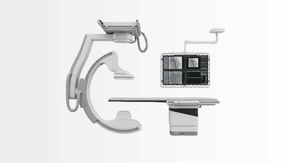
DSA is catheter angiography with digital subtraction: through a small puncture, wrist or groin, a thin catheter is guided along the vessels to the territory in question; contrast is injected while rapid X-ray sequences run; and subtraction removes everything but the flowing contrast. The result outresolves CT and MR angiography where fine detail decides, tiny aneurysms, vessel-wall irregularities, the real-time direction and speed of flow through a malformation, which is why non-invasive angiography screens, and DSA confirms and treats.
What DSA is used for
Diagnostically: definitive mapping of brain aneurysms and arteriovenous malformations; carotid and peripheral artery disease where intervention is being planned; unexplained bleeding hunts; and pre-surgical vascular road-mapping. Therapeutically, the larger half of modern practice, DSA is the imaging backbone of interventional procedures: aneurysm coiling and flow-diversion, stroke thrombectomy, embolisation of bleeding vessels, fibroids and tumours, and the stents and balloons of cardiovascular and peripheral work. Diagnosis and treatment share one table, one catheter and often one hour.
The procedure: what to expect
DSA is usually done awake under local anaesthesia with sedation as needed. After the puncture site is numbed, catheter navigation itself is painless, vessels have no touch sensors, though contrast injections can bring seconds of warmth or flashing sensations. A diagnostic study typically takes 30–60 minutes; interventions run longer. Afterwards the puncture site is sealed and observed, a few hours of flat rest for groin access, less for wrist, with many diagnostic patients home the same day. Kidney function and allergies are screened beforehand because iodinated contrast is used; blood thinners are coordinated, not simply stopped.
DSA vs CT and MR angiography
CT angiography and MR angiography answer most vascular questions non-invasively, and they rightly come first. DSA’s territory is what remains: findings that need finer resolution or true flow dynamics, disagreements between scans, and, decisively, every case where treatment is likely, since the catheter that confirms can treat in the same session. The honest sequence in a modern centre is screen non-invasively, catheterise with intent.
Honest risks
DSA is invasive and honest pages say so: puncture-site bruising is common and minor; serious complications, vessel injury, stroke in cerebral work, contrast reactions, kidney strain, are uncommon in experienced hands and quantified for your case beforehand. Experience concentrates safety: high-volume interventional teams, anaesthesia backup and post-procedure observation are what turn a powerful tool into a routinely safe one.
DSA at Acibadem
DSA suites operate across the group’s interventional radiology and cardiovascular practice, corporate-verified angiography installations run from Istanbul’s hybrid floors to Bursa’s coronary and DSA units and City Clinic Tokuda’s imaging floors, working beside neurosurgery and neurology for cerebrovascular cases. Records-first applies: your CT/MR angiography travels ahead, so a catheter study happens only with a defined question or a treatment plan attached.
The thrombectomy era: DSA as emergency medicine
Nothing recast angiography’s role like stroke thrombectomy. For large-vessel ischaemic stroke, a major brain artery blocked by clot, catheter retrieval of the clot on the DSA table is among modern medicine’s most effective interventions, with benefit measured in whole grades of independence and treatment windows that imaging, not the clock alone, now defines. The chain is unforgiving: recognition, imaging that confirms salvageable brain, and a team that moves from door to reopened artery in minutes that matter. This is why angiography suites live inside stroke pathways rather than beside them, and why neurology, neurointervention and anaesthesia rehearse the sequence as one organism. The same emergency logic serves bleeding: embolisation stops haemorrhage, gastrointestinal, obstetric, traumatic, through a puncture, replacing open operations in shocked patients for whom surgery itself was the second injury.
Preparing for an angiogram: the details that matter
The checklist that smooths the day: recent kidney values, since contrast dosing is planned around them and hydration protocols protect borderline kidneys; a complete medication list, metformin, anticoagulants and antiplatelets each carry specific timing instructions rather than blanket stopping; allergy history, because prior contrast reactions are pre-treated, not merely noted; fasting per instructions when sedation is planned; and, practical but real, arranging the quiet day afterwards, since puncture sites dislike stairs, lifting and long drives on day one. Bring prior vascular imaging or have it sent: the diagnostic question shapes catheter choice and projections before you arrive. Afterwards, the honest self-monitoring is simple: a growing, hard or painful lump at the puncture site, a cold or pale limb, or any new neurological symptom earns an immediate call: your discharge sheet says exactly whom.
Frequently Asked Questions
What is stroke thrombectomy?
Catheter removal of the clot blocking a major brain artery, performed on the DSA table, one of modern medicine’s most effective emergency interventions when the chain from door to artery moves fast.
Do I stop metformin or blood thinners before a DSA?
Neither is simply stopped, each carries specific timing instructions coordinated with the team; bring your full medication list early.
What symptoms after the procedure need an immediate call?
A growing or painful puncture-site lump, a cold or pale limb, or any new neurological symptom: the discharge sheet lists them and the number to call.
Is DSA painful?
The puncture site is numbed and catheter navigation itself is painless; contrast injections can bring brief warmth or light flashes, and sedation keeps you comfortable.
Am I awake during the procedure?
Usually yes, with sedation as needed, being responsive helps in some cerebral studies; deeper anaesthesia is used where the plan calls for it.
How long does a DSA take?
Diagnostic studies typically 30–60 minutes; interventional procedures longer, by their nature.
Wrist or groin, where does the catheter go in?
Both routes are standard; the team chooses by target territory and your anatomy. Wrist access typically means shorter rest afterwards.
How long until I can go home?
After a diagnostic study, commonly the same day following observation and puncture-site rest; interventions may add an overnight stay.
Why do a DSA if I already had a CT or MR angiogram?
Because DSA resolves what they leave open, finer detail, true flow dynamics, and because the confirming catheter can treat in the same session.
What are the risks?
Minor bruising commonly; serious complications, vessel injury, stroke in cerebral work, contrast or kidney effects, uncommonly, and quantified for your case before consent.
Is the contrast dye dangerous?
Iodinated contrast is screened against allergies and kidney function beforehand; hydration and dose protocols manage the risk, and alternatives are discussed where needed.
Can aneurysms really be treated during the same procedure?
Often yes, coiling and flow-diversion are performed through the same catheter system on the DSA table, plan permitting and consented in advance.
Do I stop my blood thinners?
They are coordinated, not reflexively stopped, tell the team early; the plan is adjusted around them.
What is a hybrid angiography suite?
An operating room with a built-in DSA system, surgery and catheter imaging at one table, used for combined procedures.
How should I prepare?
Fasting per your instructions, kidney-function bloods, an allergy and medication list, and your prior vascular imaging sent ahead, which shapes the entire study.
Related Medical Units
Available at these Hospitals


















Related Treatments
Speak with our medical team
Find out how this technology could support your diagnosis or treatment.