

Quick answer
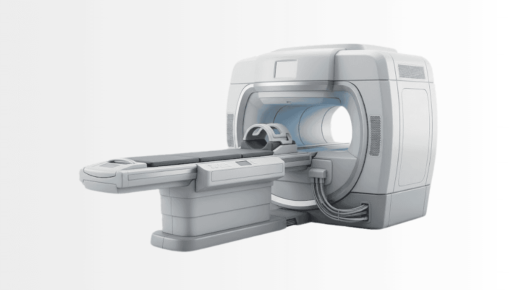
Whole Body MR is an MRI examination that captures images of multiple body regions in one session without ionizing radiation. At Acibadem in Turkey, imaging specialists tailor the scan protocol to the clinical question and use the results to support assessment or follow-up when appropriate.
A whole-body MRI is the full body scan without the radiation: one session that images from head to toe using magnetic fields alone, looking for what silent disease leaves behind, masses, marrow changes, early structural trouble, before symptoms announce them. It has become the thinking person’s screening question: what can a full body MRI actually find, who genuinely benefits, and where does honest medicine draw the line? This page answers all three, the way Acibadem’s radiologists answer them in the reporting room.
What is a whole-body MRI?
Magnetic resonance imaging builds pictures from the behaviour of hydrogen atoms in a strong magnetic field, no X-rays, no radioactive tracer. A whole-body protocol chains regional acquisitions, head and neck, chest, abdomen, pelvis, spine, long bones, into one continuous examination, typically 45–75 minutes on the couch depending on protocol depth. Diffusion-weighted sequences, the workhorse of modern whole-body imaging, highlight tissue where cells are packed abnormally tight, one of the ways early tumours betray themselves.
What a full body MRI scan can find
The examination’s genuine strengths: solid organ masses in the liver, kidneys and pancreas; brain and spine abnormalities; bone-marrow disease, which MRI sees earlier than almost anything else; lymph node enlargement; and the incidental aneurysm or structural variant worth knowing about. In people with hereditary cancer predisposition, Li-Fraumeni syndrome is the textbook case, whole-body MRI is an established, guideline-supported surveillance tool precisely because it screens widely without accumulating radiation year after year.
What it cannot do: the honest half
A whole-body MRI is not a guarantee, and a centre that says otherwise is selling rather than screening. The method’s real limits: lung detail belongs to CT, small airway and lung nodules are not MRI’s strength; the bowel hides polyps from any scanner, which is why colonoscopy keeps its job; microcalcifications in the breast belong to mammography; and slow-growing findings of no consequence can trigger cascades of follow-up: the “incidentaloma” problem every honest radiologist names. Screening MRI complements organ-specific screening; it does not replace it.
Who genuinely benefits?
Strongest case: people with hereditary tumour syndromes or strong family cancer histories, where wide, radiation-free surveillance is the point. Reasonable case: adults who want a thorough structural survey as part of an executive-level check-up, understood as one instrument in a panel rather than an oracle. Weak case: replacing targeted diagnostics when a specific symptom deserves a specific work-up: a headache work-up, a breast question, a bowel question each have better-fitting tools. The screening conversation before the scan is part of the service, and the physician’s honest job is occasionally to advise against it.
The scan: what to expect
No radiation, usually no injection, most screening protocols run without contrast, and where contrast would add value the radiologist says so beforehand. You lie in the scanner while sequences run; the machine is loud (hearing protection provided) and the tunnel is a real consideration for claustrophobic patients, sedation-free coping strategies, and where appropriate wider-bore systems, are discussed in advance. Metal implants need screening before the appointment: most modern implants are MRI-compatible, but the checklist exists for the exceptions.
What determines the cost of a whole-body MRI?
Pricing varies with protocol depth (a targeted screening protocol versus a long multi-sequence study), whether contrast is used, and whether the scan sits inside a broader check-up programme with consultations and laboratory work. At Acibadem, planned imaging follows the group’s written-estimate practice, the price is stated before the appointment, and international patients typically take the scan within a records-first plan so the reporting consultation lands in the same visit.
Whole-body MRI at Acibadem
Whole-body protocols run on the group’s high-field MRI fleet within radiology departments, with corporate-verified whole-body MRI capability at campuses including City Clinic Tokuda in Sofia and the Varna medical centre, and screening use woven into the group’s check-up programmes. Every study is reported by radiologists into your group file, comparable year to year, readable at every campus, and framed by the honest screening conversation above.
How a whole-body protocol is actually built
“Whole-body MRI” is not one product; it is a protocol decision, and knowing its anatomy helps you ask better questions. A screening study typically chains T1 and fluid-sensitive sequences region by region with diffusion-weighted imaging as the tumour-sensitive thread: the sequence that makes densely cellular tissue glow against the background. Depth varies deliberately: a hereditary-risk surveillance protocol runs longer and finer than an executive-screening survey; a myeloma protocol weights marrow; an oncological staging study may add contrast and organ-specific blocks. The honest question to any provider is therefore not “do you do whole-body MRI?” but “which protocol, and read by whom?”, because a fast gradient sweep and a surveillance-grade study fit the same marketing phrase and are not the same examination.
When the scan finds something: the incidentaloma pathway
Screening’s statistical truth: image enough healthy people head to toe and findings appear in most of them, cysts, haemangiomas, nodules, variants. The value of a mature centre is the pathway that receives them. Findings are graded against established criteria: most earn the honest label “benign, no action”; a minority earn a defined follow-up interval; a small few earn immediate work-up: the targeted scan, the specialist referral, occasionally the biopsy. What patients deserve to be spared is the fourth category: vague alarm with no plan. At Acibadem the report closes with a disposition for every finding, the group file routes anything actionable to the right unit with images attached, and follow-up intervals land in the calendar rather than in goodwill. Screening without that machinery is anxiety with pictures; with it, it is medicine.
Frequently Asked Questions
What is an incidentaloma?
An unexpected finding on a scan done for another reason, mostly benign, always deserving a clear disposition: dismissed, dated for follow-up, or worked up.
Are all whole-body MRI protocols the same?
No, depth, sequences and reading vary with purpose; surveillance, screening and staging protocols are different examinations sharing one name, and it is fair to ask which you are getting.
Can whole-body MRI be combined with other check-up tests?
Yes: it commonly anchors broader programmes alongside laboratory panels and organ-specific screening, with one physician synthesising the whole picture.
Does a whole-body MRI use radiation?
No: MRI uses magnetic fields and radio waves only, which is exactly why it suits repeated surveillance.
How long does a full body MRI scan take?
Typically 45–75 minutes in the scanner depending on protocol depth; plan for a little longer at the department.
Can a whole-body MRI detect cancer?
It can find many structural signs of tumours, especially in solid organs, marrow and nodes, but it is not a guarantee and misses what CT, mammography or colonoscopy see better; it complements organ-specific screening.
Do I need contrast dye?
Most screening protocols run without it; where contrast would genuinely add value, the radiologist says so before the scan, not after.
Is the scan suitable for claustrophobic patients?
Often yes, with preparation, coping strategies, breaks between sequences and, where appropriate, wider-bore scanners are discussed in advance.
Can I have the scan with a metal implant?
Most modern implants are MRI-compatible, but every implant is checked against the safety list before the appointment, bring your implant card.
What is diffusion-weighted imaging?
A sequence highlighting tissue where cells are abnormally densely packed, one of the main ways early tumours stand out on whole-body studies.
Who should consider regular whole-body MRI?
People with hereditary tumour syndromes or strong family cancer histories have the clearest, guideline-supported case; for others it is one instrument in a thorough check-up, agreed with a physician.
What happens if something is found?
The report grades findings honestly, most incidental findings are innocent, and anything needing action is routed to the right specialist within the group, with your images already in the file.
Is whole-body MRI better than a PET-CT?
They answer different questions: MRI surveys structure without radiation; PET-CT maps metabolic activity with a tracer. Your physician chooses by the question, not by ranking.
Does it replace mammography or colonoscopy?
No, microcalcifications belong to mammography and bowel polyps to colonoscopy; the honest scan report says so rather than implying total coverage.
How often should a screening whole-body MRI be repeated?
In hereditary-risk surveillance, on the schedule your specialist sets; in general check-up use, intervals are agreed individually, repetition without a reason is not screening, it is habit.
Will my previous scans be compared?
Yes, comparison is where surveillance earns its value, and prior imaging is integrated into every report wherever available.
Related Medical Units
Available at these Hospitals


















Related Treatments
Speak with our medical team
Find out how this technology could support your diagnosis or treatment.