Dental Anatomy
Dental anatomy evaluation examines tooth structure, alignment, bite, and oral tissues to guide preventive, restorative, orthodontic, or aesthetic dental care planning.
Quick answer
Adults normally have 32 permanent teeth, 28 if the wisdom teeth are removed or never erupt, and children have 20 primary teeth. Dental anatomy describes the structure of each tooth, from enamel to root, and how the teeth meet in the bite. A dental anatomy evaluation examines these structures to diagnose problems and plan restorative, orthodontic or implant treatment.
Dental Anatomy: Understanding Your Teeth Before a Treatment Decision
Dental anatomy is the structure of your teeth and everything that supports them: the enamel, dentine and pulp inside each tooth, the roots anchored in the jaw, the gums and bone that hold each tooth in place, and the way the upper and lower teeth meet when you bite. A dental anatomy evaluation applies that knowledge to your own mouth: it maps every tooth, records its condition, and studies the bite and supporting tissues before any treatment is planned. It is the diagnostic groundwork for veneers, crowns, implants, orthodontics and full-mouth rehabilitation.
Many people arrive at this subject through a simple search, how many teeth do humans have, and then discover that the answer matters more than expected. Whether you have 32, 28 or 24 teeth changes how your bite distributes force, which teeth are being overworked, and which treatment options are realistic. A chipped front tooth may be related to grinding. Aesthetic concerns may be shaped by tooth proportions, gum levels, bite forces or previous restorations. Sensitivity may involve enamel loss, gum recession, fracture lines, cavities or inflammation of the pulp inside the tooth. None of these things can be judged from a single symptom or a photograph of a smile.
You may be considering veneers, crowns, implants, orthodontic treatment, whitening or a complete smile rehabilitation. You may have discomfort when chewing, visible wear, spacing, crowding, gum recession, or a bite that no longer feels balanced. There is often an added layer of concern as well: whether the diagnosis is complete and whether every proposed treatment is truly necessary. A detailed evaluation of your dental anatomy is how those concerns are answered, not with reassurance, but with findings. Instead of focusing on one visible problem, it treats the mouth as a functional system and asks what should be treated, in what sequence, and with what long-term consequences.
How Many Teeth Do Humans Have?
Adult humans have 32 teeth when all four wisdom teeth are present, and 28 when the wisdom teeth have been removed or never developed. Children have 20 primary teeth, often called baby teeth or milk teeth. That is the short answer to how many teeth do humans have; the fuller answer depends on age, development and dental history, because the count changes across a lifetime and no two mouths follow exactly the same path. Some people are born without certain teeth, some develop extras, and many adults have lost one or more teeth to decay, gum disease or injury.
Do we have 32 or 36 teeth?
Thirty-two is the full complement for a typical adult mouth; 36 is not standard human anatomy. When people ask how many teeth do humans have at the absolute maximum, the answer for normal development is 32, eight incisors, four canines, eight premolars and twelve molars, including the wisdom teeth. A small number of people develop extra teeth beyond this, a condition called hyperdontia or supernumerary teeth. A common example is a small extra tooth between the upper front teeth. Supernumerary teeth are usually found on X-rays, and they matter clinically because they can crowd the arch, block the eruption of normal teeth or complicate orthodontic planning.
How many teeth do adults have?
The direct answer to how many teeth do adults have is 32, or 28 once the wisdom teeth are removed or if they never erupt. The teeth are arranged as 16 in the upper jaw and 16 in the lower, mirrored left and right. In practice, the number of teeth actually working in an adult mouth is often lower than 28, because extractions, failed teeth and unreplaced gaps are common. Part of any thorough dental examination is simply establishing which teeth are present, which are functional, and which spaces are affecting the rest of the bite.
Is having 24 teeth okay?
It can be: the honest answer depends on which teeth are missing and why. Some people function comfortably with a shortened set of teeth, particularly when the front teeth and premolars are present and the bite is stable. But missing back teeth shift chewing load onto teeth that were not designed to carry it, and over time neighbouring teeth can drift into gaps while opposing teeth over-erupt into the space. A mouth with 24 teeth is therefore not automatically a problem, and not automatically fine either. An anatomical evaluation looks at how the remaining teeth are coping: signs of overload, drifting, wear and gum stress tell the dentist whether the current count is sustainable or whether replacing missing teeth would protect what remains.
Do 70-year-olds still have all their teeth?
Some do, and the difference is rarely age itself. Teeth are not programmed to fall out in older age; they are lost to untreated decay, gum disease, fracture and the cumulative effect of years without maintenance. People who keep their gums healthy, manage decay early and protect their teeth from grinding can keep natural teeth throughout life. This is one of the strongest arguments for anatomical assessment at any age: the factors that end a tooth’s life are usually identifiable, and often manageable, long before the tooth is lost.
Which animal has the most teeth?
Not humans, by a wide margin. Snails and slugs carry a ribbon-like structure called a radula lined with thousands of microscopic tooth-like points, which is the origin of the enormous tooth counts often quoted online. Sharks continuously grow and replace teeth in rows throughout their lives. Humans get exactly two sets, 20 primary teeth and up to 32 permanent ones, and the second set has no replacement. That biological limit is why preserving natural tooth structure sits at the centre of modern treatment planning.
What Is Dental Anatomy?
Dental anatomy is the branch of dentistry that describes the form, structure and position of the teeth and their supporting tissues, and it is the vocabulary in which every diagnosis and treatment plan is written. When a dentist notes a fractured cusp, a deep fossa with staining, recession past the neck of the tooth or bone loss around a root, each of those terms refers to a specific anatomical structure. Understanding the basics helps you read your own treatment plan rather than take it on trust.
What are the parts of a tooth?
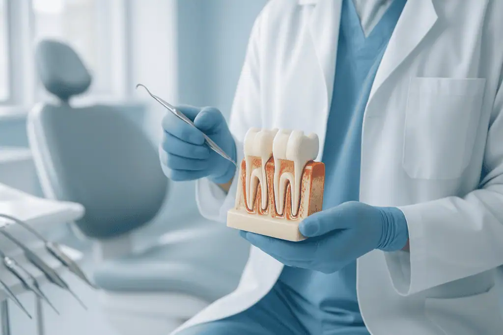
Every tooth has three broad zones: the crown, which is the visible part above the gum; the neck, where crown meets root at the gumline; and the root, anchored in the jawbone. Within these zones lie distinct tissues, each with a different job:
- Enamelthe outer shell of the crown and the hardest tissue in the human body. It has no nerve supply and cannot regrow once lost, which is why worn or eroded enamel is treated so seriously in planning.
- Dentinethe layer beneath the enamel, making up most of the tooth. It is softer, contains microscopic tubules connected to the nerve, and is the usual source of sensitivity when exposed by wear, decay or recession.
- Pulpthe soft core of the tooth, carrying nerves and blood vessels through the pulp chamber in the crown and the root canals below. Deep decay or cracks that reach the pulp are what make root canal treatment necessary.
- Cementuma thin, bone-like covering over the root surface.
- Periodontal ligamenta cushion of fibres connecting cementum to bone, allowing tiny movements and distributing chewing forces.
- Alveolar bonethe part of the jaw that holds the tooth socket. Its height and thickness determine tooth stability and, later, whether implants can be placed without grafting.
The chewing surfaces of back teeth carry their own micro-landscape: raised points called cusps, connecting ridges, and depressions between them. These details are not academic: they determine where opposing teeth touch, where plaque collects and where decay begins.
What is a fossa in dental anatomy?
A fossa is a shallow, rounded depression on the surface of a tooth. Molars and premolars have fossae between their cusps, the central fossa of a molar is where the opposing cusp rests when you bite, and the tongue-facing surfaces of upper front teeth carry a lingual fossa. Fossae matter clinically for two reasons. First, they are part of how the bite fits together: cusps closing into fossae is what makes a bite stable. Second, the pits and fissures at the depths of fossae trap plaque and are among the most common places for decay to start, which is why dentists probe these areas carefully and sometimes recommend sealing them before cavities can form.
The Four Types of Teeth and What Each One Does
The 32 teeth of a complete adult set divide into four types, and each type has a distinct shape because it has a distinct job. Incisors, the eight flat-edged teeth at the front, four upper and four lower, cut food and shape speech and the smile. Premolars, sometimes called bicuspids, are the eight teeth behind the canines; with two cusps each, they bridge the transition from tearing to grinding. Molars are the twelve broad teeth at the back, including the four wisdom teeth; their wide, multi-cusped surfaces do the heavy grinding that breaks food down for digestion. Children have no premolars at all: their primary molars are later replaced by the adult premolars, which is one of several reasons a child’s tooth count differs from an adult’s.
What are canine teeth?
Canine teeth are the four pointed teeth at the corners of the dental arches, one in each quadrant, between the incisors and the premolars. They are built for gripping and tearing, and they typically have the longest roots of any teeth, which makes them exceptionally stable anchors. In many bites the canines also act as guides: when you slide your jaw sideways, the canines take the contact and separate the back teeth, protecting molars from grinding forces. This is why worn, chipped or missing canines are a red flag in bite analysis, and why orthodontists and restorative dentists treat the canine position as a cornerstone of the whole arch.
Tooth Chart: How Dentists Name and Number Your Teeth
A tooth chart is the standardised map dentists use to identify each tooth by number, so that findings and treatment plans can be recorded and shared without ambiguity. The most widely used system internationally is the FDI two-digit notation: the mouth is divided into four quadrants numbered 1 to 4, and the teeth in each quadrant are numbered 1 to 8 from the front, so tooth 11 is the upper right central incisor and tooth 36 is the lower left first molar. Dentists in the United States commonly use the Universal system instead, numbering all adult teeth 1 to 32 starting from the upper right wisdom tooth, and some clinics use the older Palmer notation. This is worth knowing when comparing treatment plans: two plans from different clinics or countries may describe the same tooth with different numbers, and a correct comparison starts with translating the chart.
| Tooth type | Number in a full adult set | Position in each quadrant (FDI) | Main function | Typical eruption of permanent tooth |
|---|---|---|---|---|
| Incisors (central and lateral) | 8 | 1–2 | Cutting food; speech and smile aesthetics | Around 6–9 years |
| Canines | 4 | 3 | Tearing; guiding the bite during jaw movement | Around 9–12 years |
| Premolars | 8 | 4–5 | Crushing; transition between front and back teeth | Around 10–12 years |
| First and second molars | 8 | 6–7 | Grinding; carrying most chewing force | Around 6 years and 12 years |
| Third molars (wisdom teeth) | 4 | 8 | Grinding; frequently absent, impacted or removed | Around 17–25 years, if at all |
From Baby Teeth to Wisdom Teeth: How the Count Changes
The first primary teeth usually erupt at around six months of age, and by roughly age three a child has the full set of 20. These teeth are not disposable placeholders: they hold space for the permanent teeth, guide their eruption, and support speech and nutrition through childhood. Between about ages 6 and 12 the mouth is in mixed dentition, primary teeth are lost as permanent teeth erupt, and the answer to how many teeth do humans have is genuinely a moving target during those years. By the early teens most people have 28 permanent teeth, and the four wisdom teeth, when they come at all, typically arrive between 17 and 25.
The primary set is organised differently from the adult one. Each quadrant of a child’s mouth holds five teeth, two incisors, one canine and two molars, for twenty in total, with no premolars anywhere in the arch. The lower central incisors usually arrive first, followed by the upper centrals, and the second primary molars complete the set last. When the permanent teeth take over, they do so in a broadly predictable order: the first permanent molars erupt behind the primary teeth at around age six without replacing anything, the incisors change over next, and the canines and premolars follow in the years before the second molars arrive at around twelve. Dentists watch this sequence closely, because teeth that erupt far out of order, or a primary tooth that refuses to loosen long after its partner on the other side has gone, can be the first sign of a missing, extra or impacted permanent tooth beneath the gum.
Wisdom teeth are where individual anatomy diverges most. Some erupt normally and function for life. Some never form. Many become trapped against the neighbouring molar or in the bone: an impacted toothwhere they can cause pressure, decay in the adjacent tooth, or cyst formation, and removal may be recommended even without symptoms. Separately, some people are congenitally missing one or more teeth other than wisdom teeth (hypodontia), most often lateral incisors or second premolars. This is why a competent evaluation never assumes a standard mouth: it counts, charts and images what is actually there.
What Is a Dental Anatomy Evaluation?
A dental anatomy evaluation is a comprehensive assessment of tooth form, tooth position, bite relationship, oral tissues and the supporting structures of the mouth. It is not a single test. It is a clinical and diagnostic process that brings together visual examination, functional analysis, imaging, measurements and treatment planning into one coherent picture.
The scope covers the visible crown of each tooth, the root structure below the gum, the enamel and dentine layers, the pulp chamber, the contours of the biting surfaces, and the contact points between neighbouring teeth. It also covers the relationship between teeth and gums, the jawbones that hold the teeth, and the temporomandibular joints and muscles involved in chewing. Dental problems tend to develop where structure, function and biology intersect, so a thorough evaluation refuses to look at any of these in isolation.
In practical terms, the dentist establishes whether your teeth are properly shaped, aligned, supported and functioning under normal bite forces. The assessment may examine tooth wear, cracks, cavities, previous fillings, crowns, bridges, implants, root-canal-treated teeth, missing teeth, gum levels, bone support, tooth mobility and bite interference. If aesthetic treatment is being considered, it extends to smile line, tooth proportions, colour, symmetry, facial balance and how the teeth show during speech and smiling.
This groundwork matters most before major work. Veneers require enough healthy enamel and a stable bite. Implants require adequate bone and healthy surrounding tissue. Orthodontics must account for tooth shape, root position, gum condition and jaw relationship. Full-mouth rehabilitation depends on an accurate understanding of bite height, wear, jaw movement and restorative needs. The evaluation is the map; treatment is the journey that follows it.
Who May Need a Dental Anatomy Evaluation?
The evaluation is useful for three broad groups: people with symptoms, people with visible concerns, and people planning significant dental treatment. It is also valuable when previous dental work has not lasted as expected, or when different dentists have given different opinions, because anatomy is the common ground on which any recommendation can be tested. The findings clarify whether a concern is primarily cosmetic, structural, functional, periodontal, or a combination of several factors.
Common reasons for assessment include tooth pain, sensitivity to hot or cold, discomfort when biting, chipped or cracked teeth, worn edges, uneven tooth length, crowding, spacing, gaps, jaw fatigue, headaches related to clenching, gum recession, bleeding gums, loose teeth, missing teeth, or dissatisfaction with the appearance of the smile. Some patients have no pain at all but notice that teeth are shifting, restorations keep breaking, food traps between teeth, or the bite feels different from how it used to.
Diagnosis begins with a detailed conversation. The dentist asks about symptoms, previous dental treatment, medical history, medications, habits such as grinding or clenching, any history of trauma, and aesthetic goals. Where earlier records exist: X-rays, photographs, orthodontic or implant documentation, written treatment plans from another clinic: they can be reviewed as part of the evaluation. This helps establish whether previously proposed treatment is consistent with current findings and whether anything further needs to be examined before decisions are made.
The clinical examination typically includes inspection of every tooth surface, gum assessment, bite analysis, evaluation of jaw movement, and screening of the oral tissues. Imaging is used for what cannot be seen directly: roots, bone levels, impacted teeth, hidden decay, and the relationship between teeth and nearby anatomical structures. Intraoral scanning or impressions may be used to study alignment and bite contacts in finer detail, and three-dimensional imaging may be considered for implant planning, impacted teeth, complex root anatomy or jawbone assessment.
Aesthetic procedures deserve particular mention. A smile design cannot be responsibly planned by appearance alone. The dentist must understand enamel thickness, tooth position, bite forces, gum symmetry, the health of supporting tissues, and whether orthodontic or periodontal treatment should come before veneers, bonding, crowns or whitening. A result worth having is biologically sound and functionally stable, not just photogenic on the day it is fitted.
Conditions and Indications Addressed by Dental Anatomy Assessment
Anatomical evaluation supports diagnosis and planning across most of dentistry: preventive care, restorative dentistry, prosthodontics, orthodontics, periodontics, endodontics, implant dentistry, oral surgery and aesthetic work. It can identify early disease, explain symptoms, set treatment sequence, and, just as importantly, prevent unnecessary or poorly timed procedures.
Tooth wear. Worn teeth may result from grinding, acid erosion, an imbalanced bite, abrasive brushing, or several of these together. Wear patterns are diagnostic: they show how teeth contact during chewing and clenching. If the cause is not recognised before cosmetic or restorative care, new restorations are simply exposed to the same forces that damaged the natural teeth, and they fail the same way.
Cracked or weakened teeth. Cracks may be visible, hidden under old fillings, or detectable only through symptoms and careful testing. The dentist assesses whether the tooth can be protected conservatively, whether a dental crown is needed to hold it together, or whether the damage has already reached the nerve or root. Early identification sometimes allows treatment before a crack progresses into a far more complex problem.
Decay and old restorations. Cavities develop between teeth, around crown margins, under existing fillings and near the gumline, often invisibly. Previous work is examined for leakage, fracture, poor fit, over-contouring and bite-related stress. The aim is a graded verdict: what needs a new filling or restoration now, what can be monitored, and what will influence future planning.
Orthodontic concerns. Crowding, spacing, rotated teeth, deep bite, open bite, crossbite and jaw relationship problems all require understanding of tooth shape, root position and periodontal support. In adults, orthodontic planning must also work around existing crowns, implants, recession, bone levels and missing teeth. The evaluation determines whether alignment alone will solve the problem or whether restorative and periodontal care must be integrated with it.
Missing teeth. The assessment allows an honest comparison of optionsdental implants, bridges, removable prostheses, or orthodontic space closure, based on bone volume, gum health, the condition of adjacent teeth, bite forces, aesthetic zone requirements and general health. In implant planning, anatomy is decisive: the position of nerves, sinuses, bone contours and neighbouring roots sets the boundaries of what is safe.
Gum and bone support. Periodontal disease changes tooth stability, gum levels and aesthetic proportions. Before veneers, crowns, implants or orthodontics, the foundation must be assessed, because treating the visible tooth without understanding what holds it up compromises the long-term result.
How the Evaluation Is Performed: From Preparation to Treatment Planning
Before your appointment
Preparation is mostly information. Your dental history, current symptoms, allergies, medications, medical conditions and previous records all feed into the assessment. Recent X-rays, photographs, orthodontic or implant records and written treatment plans are particularly useful when a second opinion is being formed, because they let the dentist compare what was proposed with what is actually found. No special physical preparation is usually needed. Acute pain, swelling, fever, recent trauma or difficulty opening the mouth changes the character of the visit: urgent assessment takes priority over elective planning. Pregnancy, heart conditions, blood-thinning medication, immune concerns and a history of bone-modifying medications are all relevant to imaging decisions and future planning, so the dental team needs to know about them.
Clinical examination
The dentist examines the mouth visually and with instruments. Each tooth is assessed for shape, colour, surface texture, decay, fractures, wear, existing restorations and sensitivity. The gums are inspected for inflammation, recession, pocketing, bleeding and changes in contour. The tongue, cheeks, palate, floor of the mouth and lips are screened for lesions or abnormalities.
Bite evaluation is central. You may be asked to bite together, slide your jaw side to side, or close on marking material that shows exactly where teeth touch. This identifies teeth carrying excessive force, restorations that sit too high, unstable contacts, and wear patterns that correspond to clenching or grinding. Jaw joint sounds, muscle tenderness, limited opening and deviation during movement may also be assessed. Measurements around the gums evaluate periodontal support, and tooth mobility is checked. Where aesthetic planning is involved, facial and smile photographs, shade evaluation, gumline analysis and tooth proportion assessment build the record that specialists and laboratory technicians will later work from.
Imaging and digital records
Imaging is selective, driven by the clinical question rather than routine. Digital radiographs reveal cavities between teeth, root shape, bone levels, infections, impacted teeth and the condition of previous root canal treatments. Panoramic imaging shows the jaws, missing teeth, wisdom teeth and broader anatomical relationships in one view. Three-dimensional imaging is reserved for questions that need it, implant planning, complex extractions, jawbone evaluation, impacted teeth, unusual root anatomy.
Intraoral scanning can create a digital model of your teeth and bite, supporting orthodontic planning, restorative design, occlusal analysis and communication between dentist, specialist and laboratory. Unlike traditional impressions, digital scans can be reviewed immediately and compared over time to track wear or movement. Photographs and digital smile analysis add the relationship between teeth, lips, gums and facial features. The point of the technology is not complexity for its own sake: it makes anatomy visible and measurable, and it gives you something concrete to look at while options are discussed, which matters most when a plan involves several specialists working from the same records.
Planning the next steps
After examination and imaging, the dentist explains the findings in plain terms: which teeth are healthy, which need monitoring, which need treatment, and which problems are connected. Sensitivity may trace back to recession and enamel loss. A repeatedly broken filling may be a bite-overload problem, not a filling problem. An aesthetic concern may require alignment before veneers. Missing teeth may be quietly destabilising the whole bite.
Plans range from simple to staged. A preventive plan might include professional cleaning, fluoride support, a bite guard, improved daily dental hygiene and scheduled monitoring. A restorative plan may involve fillings, inlays, crowns, root canal therapy or replacement of failing work. A complex plan may combine periodontal treatment, orthodontics, implants, gum contouring, prosthetic rehabilitation and aesthetic dentistry. Sequence is the discipline that holds it together: in most cases gum health and bite stability come before final cosmetic restorations, not after.
The evaluation itself may be a brief focused visit or a longer comprehensive appointment, depending on complexity and the records required; full-mouth planning sometimes spans more than one visit. There is no recovery period after the assessment, although periodontal probing can leave mild, short-lived gum tenderness, and keeping the mouth open for imaging can cause temporary jaw fatigue.
Why Acting Early Matters
Dental problems begin quietly. Early enamel changes, small cracks, mild recession, subtle bite instability and early bone loss rarely hurt. By the time discomfort is persistent, treatment is usually more complex. Anatomical evaluation identifies risk patterns while the conservative options are still on the table.
Delay lets decay progress from enamel into dentine and towards the nerve: what could have been a small filling becomes root canal treatment, a crown, or an extraction. A crack confined to the crown of a tooth can be driven towards the root by continued chewing forces. Undiagnosed gum disease leads to bone loss, mobility, recession and a weaker foundation for any future restoration or implant.
Postponement also narrows aesthetic and orthodontic options. Shifting teeth create more crowding, bite interference and gum stress. Excessive wear shortens teeth and changes facial proportions. Unreplaced missing teeth allow neighbours to drift and opposing teeth to over-erupt, degrading chewing function: the practical sequence and timing of replacement is covered in detail in our guide to dental implants after tooth extraction. Every one of these changes makes later treatment more involved and can rule out the most conservative approaches.
Early assessment also buys the one thing that cannot be added later: planning time. A careful evaluation before committing to treatment shows whether care fits into a single phase, whether staged appointments are more sensible, and whether preparatory work, gum treatment, decay control, a bite guard, should come first. That reduces last-minute changes and keeps the plan aligned with medical safety, personal circumstances and long-term maintenance.
Benefits of a Dental Anatomy Evaluation
A structured evaluation connects symptoms, anatomy, function and long-term planning, so that decisions rest on findings rather than impressions.
| Benefit | What it means for you |
|---|---|
| Clear diagnosis | Identifies whether concerns stem from decay, cracks, gum health, bite forces, tooth position, previous restorations, or several of these together. |
| More appropriate treatment planning | Establishes whether preventive care, restorative treatment, orthodontics, implants, periodontal care or aesthetic dentistry is the right next step. |
| Better sequencing of care | Clarifies what must come first, gum inflammation, active decay, bite instability or infection, before final cosmetic or prosthetic work. |
| Reduced risk of unnecessary procedures | Grounds decisions in clinical findings and imaging rather than appearance alone or a single isolated symptom. |
| Improved long-term maintenance | Identifies risk factors such as grinding, erosion, recession or plaque retention, so preventive strategies can protect future dental work. |
| A solid second-opinion foundation | Provides structured, documented information for anyone comparing recommendations from different clinicians. |
Recovery and Follow-Up Timeline
Because the evaluation is diagnostic, most patients return to normal activities immediately; what follows depends on the findings and any recommended treatment.
| Time period | What patients can expect |
|---|---|
| Day 1 | Examination, imaging and digital records may all be completed. You can usually eat, work and travel as normal unless an urgent dental condition is identified. |
| First week | Findings are reviewed, treatment priorities discussed, and specialist input coordinated where orthodontic, periodontal, implant, endodontic or aesthetic planning is needed. |
| First month | Preventive care or initial treatment may begin: cleaning, decay control, bite guard planning, replacement of failing restorations, or management of gum inflammation. |
| Longer term | Complex plans progress in stages, orthodontics, implants, prosthetic rehabilitation or aesthetic restorations, with periodic reassessment of bite, gum health and stability. |
What Influences the Quality of the Result?
The value of the evaluation depends on the completeness of the assessment, the accuracy of the diagnosis, and how well the plan that follows fits your anatomy and daily life. A good outcome is not measured by how teeth look on the day treatment finishes. It is measured in comfort, function, tissue health and maintainability over years.
The condition of the teeth at assessment. Teeth with extensive decay, deep cracks, short roots, repeated restorations or advanced wear have fewer conservative options than teeth evaluated earlier. The amount of healthy enamel and dentine determines whether bonding, veneers, crowns or other restorations are appropriate; root and bone anatomy shape implant, orthodontic and extraction planning.
Gum and bone health. Inflamed gums, untreated periodontal disease and reduced bone support compromise restorative and aesthetic work. Periodontal stabilisation frequently has to precede final restorations, and the position of the gumline itself governs smile aesthetics, particularly around front teeth.
Bite forces. Patients who clench or grind place high stress on natural teeth and restorations alike. Unrecognised, this leaves fillings, veneers, crowns and implants vulnerable to chipping, loosening and wear. Bite analysis, protective appliances, careful material selection and sometimes orthodontic adjustment are how long-term planning answers that risk.
Medical health and lifestyle. Diabetes, smoking, certain medications, dry mouth, reflux, eating patterns and oral hygiene habits all influence decay risk, gum health, healing and implant planning. A responsible plan treats the mouth as part of the body, not in isolation from it.
Realistic, clearly discussed goals. Some patients want the most conservative treatment possible; others are seeking a major aesthetic change. Some need fast stabilisation; others can commit to staged care over months. The best plan balances clinical need, anatomy, longevity, appearance, time and your willingness to maintain the result through regular care.
Communication among providers. When treatment crosses specialties, coordination decides the outcome. Orthodontic movement changes restorative space. Periodontal treatment changes gum levels. Implant placement must be planned around the final crown, not the other way round. Endodontic findings can alter whether a tooth is worth restoring at all. The evaluation earns its keep when it is integrated into one coordinated plan rather than ticked off as a checklist.
How Acibadem Approaches Dental Anatomy Assessment
At Acibadem, dental anatomy evaluation sits within a hospital-based dental and oral health service rather than a standalone cosmetic clinic. That setting shapes how assessment works: documentation, infection control, interdisciplinary communication and evidence-based decision-making are built into the routine, and when a patient’s needs extend beyond everyday dentistry, the relevant specialists can review the findings and plan together.
Multidisciplinary collaboration matters most in complex cases. Full-mouth rehabilitation may need input from restorative dentists, prosthodontic-focused clinicians, periodontists, endodontists, orthodontists, oral surgeons and radiology teams. Implant planning may require evaluation of bone anatomy, gum tissue, bite forces, medical risk factors and the final prosthetic design before any surgery is scheduled. Aesthetic requests benefit from honest assessment of tooth position, gum symmetry, enamel quality, smile dynamics and long-term bite stability. Coordinated case discussions exist to align those perspectives before treatment begins, not after problems appear.
The diagnostic pathway is digital where that genuinely helps: digital radiography for rapid, reviewable images; three-dimensional imaging when clinically indicated for bone volume and anatomical relationships; intraoral scanning for accurate models of teeth and bite; clinical photography and digital planning tools for aesthetic and restorative design. None of this replaces clinical judgement: it makes anatomy visible and measurable so that judgement has something solid to work on.
The hospital environment also matters for patients with medical complexity. Heart disease, diabetes, bleeding disorders, immune concerns, cancer history, bone-modifying medications or significant anxiety about procedures can all require additional precautions or medical consultation before dental surgery or extensive treatment. Coordinated access to medical specialties supports safer decision-making where dental care intersects with general health.
The evaluation’s purpose is to clarify priorities, not to maximise treatment. Not every patient who asks for veneers should receive veneers first. Not every missing tooth is best replaced the same way. Not every worn tooth needs immediate restoration. What is urgent, what is elective, what can be monitored, and what should be finished before final aesthetic or prosthetic work: those distinctions are the whole point of the exercise.
What a Good Evaluation Gives You
A dental anatomy evaluation is usually the first step toward well-planned care, whether the goal is relief from symptoms, a second opinion, implant planning, orthodontic guidance or an aesthetic smile plan. Careful assessment reveals the difference between a simple correction and a condition that needs staged, multidisciplinary treatment, and it does so before decisions are locked in. For most patients, the most valuable outcome of a first evaluation is clarity: a structured, documented explanation of what is healthy, what requires attention, in what order, and which options genuinely fit the anatomy they have.
Preparation
- Patients should bring previous dental records, X-rays, or treatment plans if available. Brush and floss before the appointment, and inform the dentist about medications, allergies, oral pain, or past dental procedures.
Aftercare
- The dentist may recommend preventive care, imaging, hygiene treatment, restorative procedures, or specialist referral depending on findings. Continue daily oral hygiene and follow the personalized dental care plan provided after the evaluation.
Frequently Asked Questions
What affects the cost of a dental anatomy evaluation?
The cost depends on the scope of the examination, imaging needs, specialist involvement, digital scans, bite analysis, and whether a multidisciplinary treatment plan is required. International patient services and follow-up coordination may also affect the final quote.
How can I get a personalised quote?
You can request a free consultation and share your dental concerns, previous images, treatment history, and photos if available. A dental team can then advise which assessments are needed and provide a personalised estimate without offering a diagnosis remotely.
Is dental imaging always required?
Not always. A dentist may recommend imaging when it is needed to evaluate roots, bone, hidden decay, impacted teeth, infections, or planning for orthodontic, implant, or restorative care. The decision is based on clinical need.
Can the evaluation be combined with treatment during the same trip?
In many cases, diagnostic assessment and treatment planning can be coordinated efficiently for international patients. Whether treatment can begin during the same visit depends on oral health status, imaging findings, laboratory needs, and the specialist’s recommendation.
Does accreditation matter when choosing where to have the evaluation?
Accreditation, such as JCI for hospitals, can indicate structured quality and safety processes. Patients may also consider dentist qualifications, infection control, digital dentistry capabilities, communication support, and continuity of care.
Is this information medical or financial advice?
No. This is general educational information. A dental specialist should assess your anatomy, oral health, and goals before recommending tests or treatment, and a personalised quote should be requested through consultation.
Medically reviewed by the Acıbadem International Medical Board September 13, 2026
See our medical review board →
Update history
- PublishedJune 8, 2026
- Medical review approvedSeptember 13, 2026
- Last content updateSeptember 12, 2026
References1
- Dental Health medlineplus.gov
Trusted care for international patients
From the Acibadem Blog
Bringing Old X-Rays and Records to a Dental Anatomy Assessment: Why History Helps Planning
Bringing old X-rays and records to a dental anatomy assessment gives the clinician a timeline rather than a single snapshot. Prior radiographs, periodontal charts,…
Why Enamel, Dentin and Pulp Matter: Dental Anatomy Explained Layer by Layer
A tooth has three main living and non-living layers. Enamel is the hard outer shell that resists chewing forces and acid but cannot regrow…