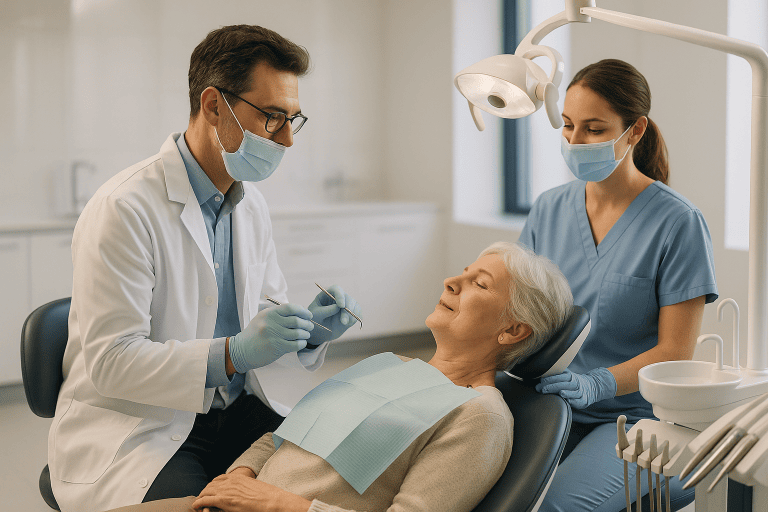
Digital Dentistry
Digital dentistry uses intraoral scanning, 3D imaging and CAD/CAM workflows to plan and produce restorations with high precision. It can support crowns, veneers, implants and smile design.
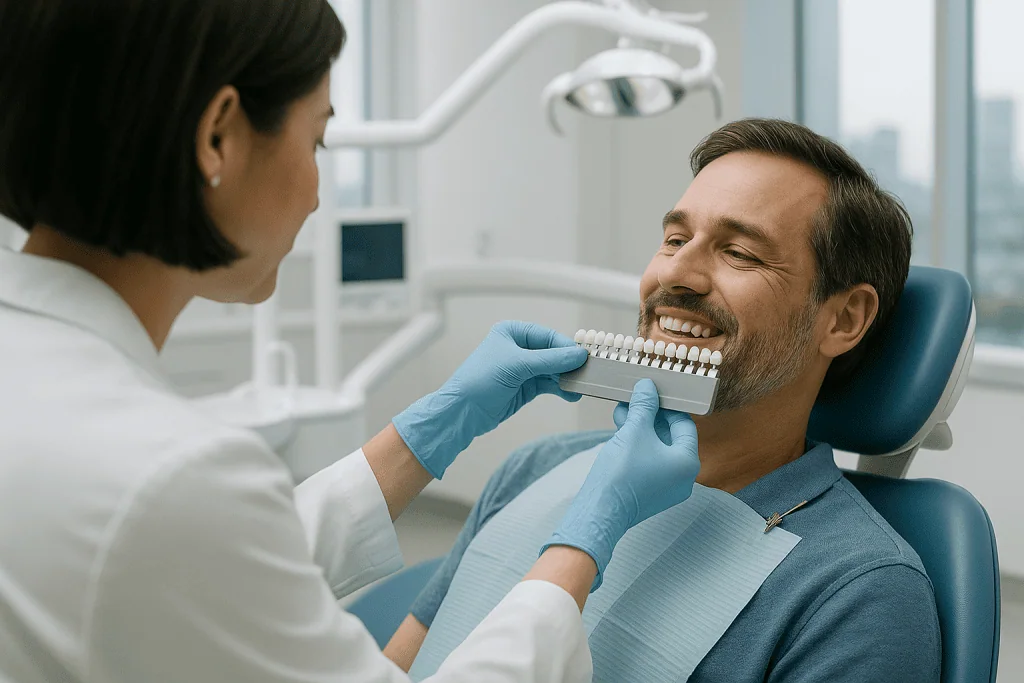
Quick answer
Digital dentistry uses intraoral scanners, three-dimensional imaging and computer-aided design and manufacturing (CAD/CAM) to diagnose dental problems, plan treatment and produce restorations such as crowns, veneers and implant prostheses. Instead of conventional impression trays and manual laboratory steps, the dentist works from precise digital records, which typically improves comfort during impressions and supports more detailed, better-communicated treatment planning.
Digital Dentistry: Precision, Comfort and Clearer Planning
Digital dentistry is the use of digital technologies, intraoral scanners, three-dimensional imaging, digital radiography and computer-aided design and manufacturing, to diagnose dental problems, plan treatment and produce restorations such as crowns, veneers, bridges and implant prostheses. It replaces or supplements conventional moulds, two-dimensional film X-rays and manual laboratory steps with scanned data, virtual planning and digitally guided production. It is relevant to anyone considering restorative, cosmetic or implant-related dental care, and particularly to patients who want their treatment planned from precise records rather than estimation.
Choosing dental treatment is surprisingly personal. Your teeth affect how you eat, speak, smile and present yourself every day, and the details that matter, whether a crown will look natural, whether implant planning will be accurate, whether veneers will suit your face, are hard to assess from a clinic’s promises alone. Many patients also worry about the practical side: how many visits the process will take, and whether they will need to sit through repeated conventional impressions. Digital dentistry addresses a good part of these concerns by making the planning stage visible, measurable and easier to discuss before anything irreversible is done to a tooth.
At Acibadem, digital dentistry sits within a wider dental and oral health service, where diagnostics, planning and treatment are coordinated around the individual patient rather than around a single procedure. Digital records support clearer treatment discussions, more predictable planning and efficient communication between dentists, specialists and dental laboratories. The aim is not simply to produce attractive teeth. It is to restore function, protect oral health and design results that look appropriate for the person wearing them.
What Is Digital Dentistry?
Digital dentistry means using digital tools across the whole treatment cycle: diagnosis, planning, design and delivery. In practice, a digital dental workflow can include intraoral scanners that create a digital impression of the teeth, three-dimensional imaging that shows bone and anatomical structures, software that designs restorations on screen, and CAD/CAM systems that help manufacture crowns, veneers, inlays, onlays, implant-supported restorations and other prostheses. Some clinics use every element of this chain; others use parts of it alongside conventional methods. Both approaches can be clinically sound, and the balance between them should follow the needs of the case.
In a traditional workflow, the dentist takes physical impressions with trays and impression material, then sends them to a laboratory where restorations are built by hand through several manual stages. This approach can still be effective, but it depends heavily on each manual step being executed well, and many patients find the trays uncomfortable. Digital dentistry replaces or supplements those stages with scanned data and digitally guided production, which reduces the number of points where small inaccuracies can creep in: a scanner captures the surface of the teeth and gums within minutes, and the dentist can check the model on screen immediately rather than discovering a flawed impression days later.
What digital dentistry does not mean is standardised treatment. The value of digital planning lies in customisation. The dentist assesses tooth colour, shape, alignment, gum contours, bite relationships, facial features and underlying oral health before recommending anything. The digital workflow supports clinical judgement; it does not replace it. A good result still depends on careful diagnosis, an experienced team and a realistic understanding of what can be achieved safely for a particular mouth.
What does a digital dentist do differently?
A digital dentist plans treatment from measurable records, scans, three-dimensional images, calibrated photographs, instead of relying only on physical moulds and visual estimation. In daily terms, this changes three things. First, data capture: impressions become scans that can be checked, repeated and stored without degradation. Second, communication: the dentist, the laboratory technician and the patient can all look at the same on-screen model, so decisions about shape, length and bite are discussed concretely rather than described in words. Third, production: restorations are designed in software and manufactured under digital control, with the design file kept on record. The clinical skills remain the same; the information the dentist works from becomes more precise and more shareable.
The Core Technologies, Explained Plainly
Most of what is marketed as digital dentistry comes down to a handful of specific tools. Understanding what each one actually does makes it easier to judge whether a proposed workflow is genuinely useful for your case or simply fashionable.
What is a digital impression used for in dentistry?
A digital impression is a three-dimensional model of your teeth and gums captured by an intraoral scanner, and it is used wherever a conventional mould would have been used: designing crowns, veneers, inlays, onlays, bridges, implant restorations, night guards, orthodontic appliances and study models. The scanner is a small handheld camera that takes many overlapping images and stitches them into a single model on screen. Because the dentist can see the result immediately, gaps or unclear areas can be rescanned on the spot. Digital impressions are also used to record how the upper and lower teeth meet, which matters for any restoration that will carry chewing force. For patients with a strong gag reflex, scanning is usually considerably easier to tolerate than trays filled with impression material.
What is digital radiography in dentistry?
Digital radiography in dentistry means taking X-ray images with electronic sensors instead of photographic film, producing an image that appears on screen within seconds. The images can be enlarged, adjusted for contrast and compared side by side with earlier ones, which helps the dentist spot decay between teeth, changes around roots, bone levels and problems under existing restorations. Digital radiographs are stored in the patient record and can be shared with a specialist or laboratory without physical copies. Like all radiography, it uses ionising radiation, so the dentist takes images when there is a clinical reason to do so, weighing diagnostic benefit against exposure.
What is digital imaging in dentistry?
Digital imaging in dentistry is the umbrella term for all electronically captured visual records: intraoral scans, digital radiographs, three-dimensional cone-beam imaging, intraoral camera photographs and calibrated facial photographs. Each layer answers a different question. Surface scans show tooth shape and gum contours; radiographs show what is happening inside teeth and immediately around them; cone-beam imaging shows bone volume, sinus position, nerve pathways and root orientation in three dimensions, which is essential for implant planning and complex surgery; photographs record colour, smile line and how the teeth relate to the lips and face. Combined, these records let the dentist study the mouth as a whole rather than one isolated tooth at a time.
Which dentist office technology will you actually encounter?
Dentist office technology is a broad label, and not every device a clinic advertises will be relevant to your treatment. In a digitally equipped practice you are most likely to meet:
- An intraoral scannerreplaces or reduces conventional impression trays.
- Digital radiography sensorsroutine X-ray images viewed on screen within seconds.
- Cone-beam three-dimensional imagingused selectively for implants, surgery and complex anatomy, not for every patient.
- Design softwareused by the dentist and technician to plan restorations and smile changes.
- Milling units and digitally guided laboratory productionused to manufacture the final restoration, either in-house or in a partner laboratory.
- Intraoral cameras and clinical photographyused to document and explain findings.
The presence of a device matters less than whether the team uses it routinely and knows its limits. A clinic that owns a scanner but rarely uses it will not deliver the benefits of a genuinely digital workflow.
Who May Benefit from a Digital Dental Workflow?
A digital dental workflow can suit most patients who need restorative, cosmetic or implant-related care. Some people seek treatment because a tooth is damaged, cracked, worn or carrying an old filling or crown that has reached the end of its useful life. Others want to improve the appearance of their smile through veneers, bonding or a broader smile design. Patients with missing teeth may need implants, bridges or implant-supported prostheses, all of which benefit from accurate digital planning because the position of the final tooth can be decided before surgery rather than after it.
Common situations that bring patients to this kind of care include tooth wear, broken restorations, gaps between teeth, discolouration that does not respond to whitening, uneven tooth shape, missing teeth, bite problems and dissatisfaction with older crowns or veneers. Some patients also have functional complaints, difficulty chewing, jaw fatigue, food trapping, sensitivity or discomfort from poorly fitting restorations. Digital records are particularly helpful here because they let the dentist study how the whole bite works before deciding which tooth to treat first.
Digital workflows also help specific groups of patients. Those with a strong gag reflex or anxiety about impressions usually tolerate scanning far better than trays; for patients whose anxiety extends to treatment itself, sedation dentistry can be discussed as a separate option. Patients with complex bite relationships, multiple restorations or implant treatment requiring careful anatomical assessment gain the most from three-dimensional planning. And patients who simply want to understand their own treatment tend to value seeing their scans and proposed designs on screen instead of taking descriptions on trust.
Diagnosis always begins with a clinical examination. The dentist evaluates the teeth, gums, bite, jaw function and existing restorations; photographs record the smile from different angles; scanning creates a model of the mouth; and three-dimensional imaging is added when bone, root position or implant placement must be evaluated. Conventional radiographs may still be used where they answer the question best. No digital tool removes the need for this examination: it makes the examination more informative.
Conditions and Treatments Supported by Digital Dentistry
Digital dentistry is not a single procedure but a set of methods that adapt to the condition being treated. In restorative dentistry, digital systems are routinely used to design and produce crowns, bridges, inlays, onlays and overlays, restorations needed when teeth are weakened by decay, fracture, root canal treatment, wear or large old fillings. Because the design is checked on screen against the neighbouring teeth and the opposing bite, margins, contact points and chewing surfaces can be refined before manufacture rather than adjusted extensively in the mouth.
In cosmetic dentistry, digital tools support veneers, smile design, tooth contour planning and aesthetic mock-ups. A digital smile design process evaluates tooth size, symmetry, gum line, incisal edges and the relationship between the smile and the face. This matters most when the patient wants a natural result: software makes it easier to design teeth with individual character than to default to an overly uniform template.
In implant dentistry, digital workflows plan the placement of implants in relation to available bone and the intended final restoration. Cone-beam imaging shows bone volume, sinus position, nerve pathways and neighbouring roots; where bone is insufficient, dental bone grafts may be planned as part of the sequence. When appropriate, the virtual implant plan can be transferred to the mouth with a printed surgical guide, so the surgery follows the restorative plan rather than the other way round.
Digital methods also support orthodontic assessment, bite analysis, night guard production, full-mouth rehabilitation and the replacement of ageing dental work. Patients with worn teeth, collapsed bite height or multiple missing teeth need a plan that considers the entire mouth; digital models let the dentist study how the arches meet, how space is distributed and how restorations can be sequenced in a stable way. That said, not every dental concern requires a fully digital workflow. Some straightforward treatments are managed perfectly well with conventional techniques, and the dentist’s job is to decide where digital tools add value and where they add only cost and complexity.
How Digital Dentistry Is Performed
The exact sequence varies with the treatment, but a typical digital case moves through the following stages:
- Step 1: Consultation and clinical examination: health history, oral examination, discussion of goals.
- Step 2: Records: intraoral scanning, radiographs or three-dimensional imaging as indicated, clinical photographs.
- Step 3: Planning and design: restorations or smile changes designed in software, often with a mock-up or trial stage.
- Step 4: Production: CAD/CAM manufacture of the restoration from a clinically appropriate material.
- Step 5: Placement and adjustment: fit, colour and bite checked, then bonding or cementation.
- Step 6: Follow-up and maintenance: review, hygiene guidance and long-term protection of the result.
Initial Consultation and Clinical Evaluation
The process begins with a consultation covering both health and personal goals. The dentist asks about symptoms, previous dental treatment, medical history, medications, allergies and expectations, then performs a detailed oral examination to identify gum disease, decay, bite issues, failing restorations or missing teeth. This stage is essential because digital dentistry cannot compensate for untreated infection, unstable gums or unrealistic planning. A well-made restoration still needs a healthy foundation, and sometimes the honest recommendation is to treat the gums or a diseased tooth first and design restorations afterwards.
Digital Scanning and Imaging
An intraoral scanner captures the teeth and gums without conventional trays, combining many images into a three-dimensional model on screen. The dentist reviews the scan immediately and confirms that the details are sufficient before moving on. Three-dimensional imaging is added when treatment involves implants, complex anatomy, root assessment or jawbone evaluation, because surface scanning alone cannot show what lies beneath the gum. Photographs record tooth colour, smile line, facial proportions and gum display, giving the design team the context a model alone cannot provide.
Digital Planning and Smile Design
With the records collected, the dentist and technical team design restorations or simulate changes in planning software. For crowns and veneers this involves tooth shape, thickness, contact points, bite relationships and margin position. For smile design, the team assesses how proposed tooth shapes align with the lips, face and natural expression. In many cases a mock-up or temporary trial restoration lets the patient preview proportions in their own mouth before final restorations are made: a valuable step for veneers and full smile rehabilitation, because shape and length can be discussed concretely rather than imagined. A preview is a design aid, not a certainty; small refinements are normal between mock-up and final result.
CAD/CAM Production of Restorations
CAD/CAM stands for computer-aided design and computer-aided manufacturing. Once a restoration is designed digitally, the file guides its manufacture from a suitable dental material, restorations may be milled from ceramic or zirconia blocks, or produced through other digitally guided laboratory methods, with composite and further material options used where clinically appropriate. Material choice depends on tooth location, bite forces, aesthetic goals, remaining tooth structure, gum health and the patient’s habits: front teeth demand careful attention to translucency and shape, back teeth must withstand chewing force, and implant restorations must harmonise with implant position and the surrounding gum.
What is a digital dental studio?
A digital dental studio is a laboratory or design workspace where restorations are planned and manufactured digitally, technicians work from the dentist’s scan files in design software, then produce the restoration with milling units, printers or digitally guided layering rather than purely manual modelling. Some clinics operate a studio in-house, allowing certain single restorations to be designed and milled during a compressed schedule; others collaborate with an external studio, exchanging files rather than shipped moulds. For the patient, the relevant point is that the dentist and technician are looking at identical data, which reduces the interpretation gaps that plague conventional handovers.
Placement and Adjustment
When the restoration is ready, the dentist checks its fit, colour, contact with neighbouring teeth and bite relationship, making minor adjustments before final bonding or cementation. For veneers and certain ceramic restorations, bonding technique matters greatly because it affects strength, longevity and appearance, so tooth surface and restoration are prepared according to established protocols. For implant-related treatment, the restoration is usually attached after the implant has integrated with the bone, unless immediate provisionalisation is appropriate for the case; gum shape, implant stability, bite forces and hygiene access are all assessed before the final restoration goes in.
How long does treatment take?
Duration depends on scope. A single crown or inlay is completed more quickly than a multi-tooth smile design or implant rehabilitation. Veneers typically require planning, tooth preparation, temporary restorations and final placement across more than one visit. Implant treatment may run in stages over months, especially when bone grafting, healing or integration time is needed. Digital workflows shorten certain steps, record-taking, laboratory communication, remakes, but responsible dentistry does not rush biological healing or quality checks, and a schedule that promises everything in a weekend deserves scrutiny.
Recovery and Aftercare
Recovery after scanning, crown placement, veneers or smile design work is usually mild. Some patients notice temporary sensitivity after tooth preparation or bonding, which often settles as the teeth adjust, and gum tenderness can occur when margins sit near the gum line or while temporary restorations are worn. Implant treatment, extractions or bone procedures involve a different recovery: mild swelling, bruising or discomfort may last several days, and the treating dentist gives individual guidance on diet, hygiene and any medication. Follow-up visits confirm healing and protect the final result.
Recovery Timeline After Digital Dentistry
Recovery depends on the procedure performed, but the following timeline describes what many patients can expect after common digital restorative and aesthetic treatments.
| Time Period | What Patients Can Expect |
|---|---|
| Day 1 | After scanning alone, no recovery time is needed. After tooth preparation, bonding or crown placement, mild sensitivity or gum tenderness may occur. |
| First week | Most patients return to normal daily activities quickly. Very hard or sticky foods are best avoided while temporary restorations are in place. |
| First month | Final restorations typically feel more natural as the bite settles. Persistent discomfort, a high bite sensation or gum irritation should be assessed by the treating dentist. |
| After implant procedures | Where implants, extractions or grafting are involved, healing takes longer. Swelling and tenderness usually improve gradually, and follow-up visits matter. |
| Longer term | Good oral hygiene, routine check-ups, night guard use when recommended and avoiding harmful habits protect the longevity of restorations. |
Benefits of Digital Dentistry
The benefits vary by case, but most patients value the combination of comfort, precision and planning they can actually see.
- More comfortable impressions. Intraoral scanning reduces or eliminates conventional impression trays: a genuine relief for people with a strong gag reflex or dental anxiety.
- Detailed treatment planning. Digital models and imaging let the dentist evaluate tooth shape, bite, gum contours and bone anatomy before treatment begins, rather than discovering problems mid-treatment.
- Personalised aesthetic design. Smile design is planned in relation to your face, lips and natural tooth proportions, supporting a result that looks individual rather than generic.
- Careful fit of restorations. Digitally designed crowns, veneers and other restorations are produced with close attention to margins, contact points and bite alignment, and the design can be checked before manufacture.
- Efficient communication. Scans, images and design files are shared between the dentist, specialists and the laboratory, keeping everyone working from the same information, particularly useful in complex, multi-stage cases.
- Better-informed implant planning. Three-dimensional imaging and digital planning position implants according to bone anatomy and the planned final tooth, and surgical guides can transfer that plan to the mouth.
- Durable records. Digital files do not distort or degrade the way physical moulds can, and they remain available for comparison, repairs or remakes years later.
What Are the Downsides of Digital Dentistry?
The main downsides of digital dentistry are its limits, not hidden dangers: the technology cannot fix poor diagnosis, it is not ideal for every clinical situation, and it adds equipment cost that clinics must recover. Being clear about these limits is more useful than pretending they do not exist.
- It does not replace clinical judgement. A precise scan of a badly planned restoration produces a precise, badly planned restoration. Diagnosis, material choice and bite management remain human decisions.
- Scanners have physical limits. Deep margins below the gum line, bleeding, saliva and limited mouth opening can make accurate scanning difficult; in some situations a conventional impression is still the better tool.
- Imaging still involves radiation. Digital radiography and cone-beam imaging use ionising radiation, so they should be taken when there is a clinical question to answer, not as a routine spectacle.
- Previews show intent, not certainty. A digital smile simulation is a design aid; biological factors such as gum response, tooth colour beneath restorations and bite adaptation influence the final outcome.
- Equipment does not equal expertise. A clinic can own every device and still lack the experience to use it well; conversely, an experienced team with a partly conventional workflow can achieve excellent results.
- Not every case needs it. Simple fillings and straightforward treatments gain little from an elaborate digital pipeline, and adding technology for its own sake adds expense without benefit.
How Much Does Digital Dentistry Cost?
There is no single price for digital dentistry, because it is a set of methods rather than one procedure: the cost of any treatment planned digitally depends on what is actually being done to your teeth. The main drivers are the number of teeth involved, the type of restoration (a single inlay sits at a different level from full-mouth rehabilitation), the material chosen, whether implants, extractions or bone grafting are part of the plan, the imaging required, and the laboratory work behind the restorations. Digital methods themselves cut some costs, such as remakes caused by distorted impressions, while adding others, such as the equipment and expertise a clinic invests in. A meaningful figure can only follow a clinical examination and a written treatment plan that itemises each stage, and any quotation offered before an examination should be treated as an estimate at best.
Why Acting Early Matters
Dental problems tend to progress quietly. A cracked tooth seems manageable until the fracture extends deeper. A worn restoration lets bacteria in beneath it. Missing teeth allow neighbouring teeth to shift, the bite to drift out of balance and bone to change over time. Gum disease erodes the foundation that crowns, veneers and implants all depend on. Early evaluation gives the dentist more options and can reduce the need for complex intervention later.
Digital dentistry rewards early planning in particular. If enough healthy tooth structure remains, a conservative restoration may be possible instead of a full crown. If bone volume is assessed soon after tooth loss, implant planning is often more favourable and grafting may be simpler or avoidable. If bite problems are recognised before multiple restorations fail, the plan can address the underlying forces rather than repeatedly patching individual teeth, exactly the kind of whole-mouth analysis that digital records make practical.
Delaying care raises the risk of infection, pain, tooth loss, bite collapse, gum recession and more extensive treatment. It also makes aesthetic planning harder once gum levels, bone support or tooth position have changed. A timely assessment, made while the situation is stable, allows a measured plan instead of a rescue.
Factors That Influence the Outcome
A good digital dentistry result depends on more than advanced equipment. Technology provides detailed information and precise tools, but the outcome is shaped by diagnosis, planning, material selection, clinical technique and the patient’s own care habits. The dentist must understand not only how a restoration will look on the day it is placed, but how it will function years later.
Oral health before treatment comes first. Gum inflammation, untreated decay, infection or poor hygiene will compromise any restoration. For veneers and crowns, the gums need to be stable so margins can be placed accurately and the final appearance is predictable. For implants, bone volume, gum thickness, bite forces and general health all shape the plan.
Bite relationship is another major factor. Teeth do not function in isolation. A patient who clenches, grinds or has an uneven bite exposes restorations to excessive force. Digital analysis helps identify contacts and relationships, but the dentist may also recommend a night guard, orthodontic treatment or a broader bite adjustment plan where appropriate.
Material selection balances beauty, durability, tooth preservation and function. Different ceramics and restorative materials have different optical and strength characteristics: a highly aesthetic layered ceramic may be ideal for a front veneer but poorly suited to a heavily loaded molar, while a strong monolithic material may be needed for back teeth or implant bridges.
Expectations should be discussed frankly. Digital smile design helps visualise proposed changes, but natural teeth and gums have biological limits. Very white, long or perfectly symmetrical teeth do not suit every face or every bite. A refined result usually comes from respecting facial proportions, age, personality and natural variation: a smile that feels authentic, not a template.
Long-term maintenance decides how long the result lasts. Crowns, veneers and implants are not immune to problems: the teeth around them can still decay, the gums can become inflamed, and restorations can chip or loosen under excessive force. Individualised hygiene instructions, regular check-ups and reporting sensitivity, bleeding, bite changes or movement to the treating dentist all protect the investment.
Judging a Clinic’s Digital Capability
What is a digital dentistry sophistication company evaluation?
A digital dentistry sophistication company evaluation is, in plain terms, an assessment of how thoroughly a clinic, laboratory or dental company has integrated digital tools into everyday care, not just which devices it owns, but whether scanning, imaging, design and production actually connect into one workflow. If you want to make this judgement yourself as a patient, useful questions are practical ones: Is scanning routine or occasional? Does the clinic show you your own scans and designs, or only talk about them? Is three-dimensional imaging used selectively where it answers a clinical question? Does the team work with a laboratory from shared digital files? Can they explain when they would choose a conventional method instead? A clinic that answers these questions specifically is usually one that uses its technology seriously.
What are ADA digital standards?
ADA digital standards are technical specifications developed through the American Dental Association’s standards programme, covering areas such as digital imaging, electronic dental records and the interoperability of dental software and devices. For patients, the detail matters less than the principle they represent: digital dentistry is a regulated, standardised field, not a marketing label. Equipment and software used in reputable clinics conform to recognised technical and safety norms in the jurisdictions where they operate, and file formats used for scans and images are designed so records can move between compliant systems.
Digital Dentistry and Remote Care
Digital records make some parts of dental care possible at a distance. Photographs, scans and radiographs can be reviewed remotely, and teledentistry can be used for preliminary discussion, follow-up questions and reviewing progress between visits. What remote review cannot do is replace a clinical examination: a final diagnosis and treatment plan require the dentist to examine the mouth directly, and restorative work is, by nature, hands-on.
Can a virtual dentist prescribe pain medication?
In most jurisdictions, remote prescribing by a dentist is tightly regulated, and whether any medication can be prescribed after a video or photo consultation depends on local law, the dentist’s registration and whether an adequate assessment has been possible. Many countries restrict or prohibit remote prescribing of certain classes of medication altogether. As a general principle, decisions about any medication, starting it, adjusting it or stopping it, belong to the treating clinician who knows your medical history, and a remote consultation is usually a step towards in-person assessment rather than a substitute for it.
Digital Dentistry at Acibadem
At Acibadem, digital dentistry is delivered within a broader healthcare environment where dental treatment can be coordinated with medical evaluation when needed, relevant for patients with chronic conditions, complex medication use or previous medical treatment that affects dental planning. Care is personalised to the patient’s oral condition, facial features, functional needs and schedule. A patient needing one ceramic crown has different requirements from a patient planning veneers, full-mouth rehabilitation or implant-supported restorations, and complex cases may involve restorative dentists, oral surgeons, prosthodontic and periodontal expertise, radiology support and dental laboratory collaboration. Where cases are complex, multidisciplinary discussion helps align the treatment sequence and reduces the risk of fragmented decision-making.
The approach also emphasises appropriate treatment selection. Digital tools make many treatments more efficient, but not every patient should receive veneers, implants or extensive rehabilitation. A responsible plan may mean treating gum disease first, preserving natural tooth structure, replacing only the restorations that are actually failing, or staging treatment to respect healing. Patients are shown their scans, photographs and planned designs so they understand not only what is proposed but why, which makes decisions easier to weigh, particularly when treatment has to fit around work, family or travel.
What a Consultation Typically Involves
A consultation for digitally planned treatment usually covers four things. First, a discussion of your history and goals: symptoms, previous dental work, medical background and what you want the result to achieve. Second, a clinical examination of teeth, gums, bite and existing restorations, supported by scans, photographs and any imaging the case requires. Third, an explanation of options, including conventional alternatives where they are equally suitable, with the trade-offs of each stated plainly. Fourth, a written plan setting out the sequence, the number of visits, the materials proposed and the maintenance the result will need. Existing records such as recent X-rays or previous treatment notes are useful at this stage because they show how the mouth has changed over time. The measure of a good consultation is not how quickly it moves to treatment, but how clearly you understand your own mouth when you leave the room.
Preparation
- Your dentist evaluates your oral health, bite and aesthetic goals before digital planning. Existing X-rays, scans or dental records may be reviewed, and new intraoral scans may be taken. If treatment involves implants, crowns or veneers, gum health and tooth structure are checked first.
Aftercare
- Most patients return to daily activities immediately after digital scanning or planning. If a restoration is placed, follow brushing, flossing and dietary guidance from your dentist. Attend follow-up visits to check fit, bite comfort and long-term maintenance.
Frequently Asked Questions
What affects the cost of digital dentistry?
Cost depends on the treatment type, the amount of scanning and imaging required, material choice, laboratory work, case complexity, specialist involvement and whether the plan includes travel-related services. A consultation is needed to provide a personalised quote.
How can I get a quote from Acibadem for digital dentistry?
You can request a free consultation and share dental photos, previous records, scans or X-rays if available. The dental team can then review your needs and prepare an initial treatment plan and cost estimate, which may be refined after an in-person examination.
Is digital dentistry always cheaper than conventional dentistry?
Not necessarily. Digital workflows may improve planning and communication, but costs depend on the restoration, materials, technology used and clinical complexity. In some cases, digital tools may reduce repeat visits or remakes, while in others they may add planning steps.
What is usually included in an international patient package?
Packages may include consultation, digital scans, imaging review, treatment planning, selected restorations, coordinator support, translation assistance and follow-up guidance. Inclusions vary, so it is important to request a written plan before travel.
Will I know the final cost before treatment starts?
Your team can provide an estimated cost after reviewing your case, but the final plan may change after clinical examination, imaging or assessment of tooth and gum health. Any additional procedures should be discussed before they are performed.
Do I need to travel more than once for digital dentistry?
This depends on the treatment. Some restorative procedures may be planned within a compact schedule, while implant, gum or complex bite cases may require staged visits and healing time. Your specialist will advise the safest and most practical timeline.
Medically reviewed by the Acıbadem International Medical Board September 13, 2026
See our medical review board →
Update history
- PublishedJune 8, 2026
- Medical review approvedSeptember 13, 2026
- Last content updateSeptember 12, 2026
Trusted care for international patients
From the Acibadem Blog
Digital Dentistry Myths: Does a Scanner Replace the Dentist’s Judgment?
No. An intraoral scanner records the shape of teeth and gums as a digital file; it does not examine, diagnose or decide. Scanners and…
The Digital Dentistry Workflow Step by Step: Scan, Design, Milling and Fitting in Practice
A digital dentistry workflow replaces putty impressions and hand-poured models with data: an intraoral scanner captures the teeth, 3D X-ray imaging maps the jaw…