Disease Reconstruction
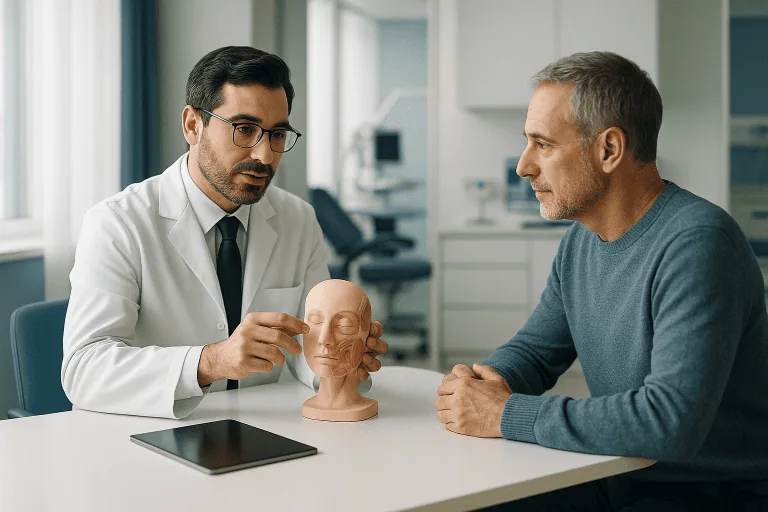
Disease reconstruction is reconstructive surgery that restores form, function and appearance after cancer, infection, trauma or tissue loss. Treatment is personalized to the affected area and patient goals.
Quick answer
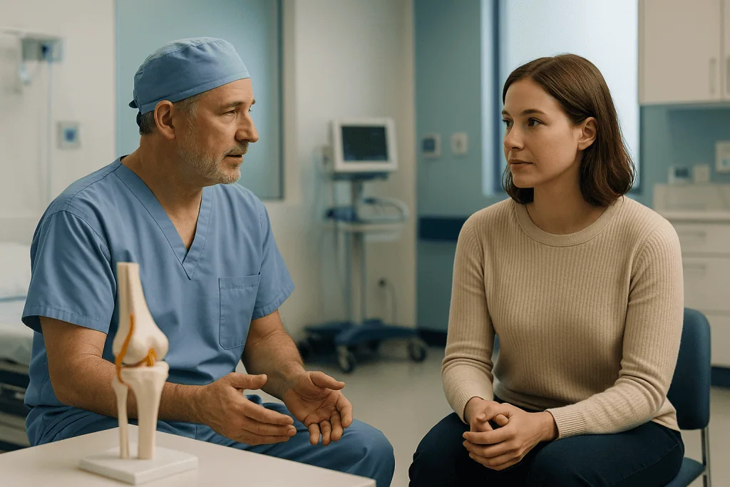
Disease reconstruction is reconstructive surgery that rebuilds areas of the body after tissue has been removed, damaged or lost through cancer surgery, severe infection, trauma, burns or chronic wounds. Surgeons use skin grafts, local or free flaps, bone reconstruction, implants and staged procedures to restore coverage, function and appearance. The plan depends on the defect, the underlying disease and the patient's overall health and goals.
Disease Reconstruction: Rebuilding After Cancer, Infection, Trauma or Tissue Loss
Disease reconstruction is a specialised area of reconstructive surgery focused on restoring form, function and appearance after tissue has been removed, damaged or lost. It may follow cancer surgery, severe infection, trauma, burns, chronic wounds, congenital or acquired deformities, or complications from earlier treatment. The goal is not simply to cover a defect. A well-planned disease reconstruction aims to return you to daily life with the best achievable balance of safety, function, durability and appearance, and it is honest, from the start, about what surgery can and cannot restore.
When illness or injury changes the way your body looks, moves or works, the impact is rarely only physical. Patients weighing up disease reconstruction usually arrive with a cluster of practical questions. Will I recognise myself after treatment? Will I be able to eat, speak, walk, work, breathe comfortably or use my hand again? Will the reconstruction be safe alongside my cancer treatment? How many operations will I need, and over what period? This page answers those questions as directly as the medicine allows.
Every reconstruction is personal. A patient who needs breast reconstruction after mastectomy has different priorities from someone requiring jaw reconstruction after oral cancer, limb reconstruction after trauma, or soft-tissue coverage after a severe infection. Your age, general health, cancer treatment plan, previous operations, smoking history, circulation, current medications and personal expectations all shape the plan. Two patients with defects of similar size may be advised to have quite different operations, because the surrounding tissue, the underlying disease and the goals of recovery are never identical. That is why disease reconstruction works best when it is designed by teams who understand both the disease being treated and the lived experience of recovering from it.
At Acibadem, disease reconstruction sits inside a broader care pathway rather than standing alone. Reconstructive surgeons work with oncology, orthopaedics, radiology, pathology, rehabilitation, wound care, anaesthesia and intensive care as the case requires, and with the infectious diseases department when infection control must come before closure. This coordination matters most when reconstruction has to be timed around chemotherapy, radiotherapy, debridement or complex wound healing.
What Is Disease Reconstruction?
Disease reconstruction refers to surgical procedures that repair or rebuild areas of the body affected by disease, trauma or tissue loss. It may involve skin, fat, muscle, bone, cartilage, nerves, blood vessels, tendons or specialised structures such as the breast, jaw, face, scalp, abdominal wall, limbs or genital region. In some cases, reconstruction is performed at the same operation as tissue removal, immediate reconstruction. In others, it is deliberately delayed until healing is complete, cancer treatment is finished, infection is controlled or you are medically stronger. Neither approach is universally better; the timing is a clinical decision made case by case.
What does “reconstruction” mean in medical terms?
In medical terms, reconstruction means surgically rebuilding a part of the body so that it looks and works as normally as possible after damage or removal. It is distinct from simple wound closure, which brings edges of tissue together, and from cosmetic surgery, which reshapes healthy tissue by choice. Reconstruction replaces or restores tissue that disease, injury or treatment has taken away. That may mean resurfacing a wound with skin, rebuilding a jaw with bone, restoring a breast mound after mastectomy, or covering exposed tendons so a hand can move again. The word covers a wide range of procedures, but the common thread is restoration rather than enhancement.
How do we reconstruct disease-damaged tissue?
Surgeons reconstruct disease-damaged tissue by moving healthy tissue, or, where appropriate, implants and grafts, into the area of loss, using a set of techniques that are often combined:
- Skin grafting transfers a thin layer of skin from one area of the body to another. The graft has no blood supply of its own and relies on the wound bed beneath it to survive, which is why grafts suit clean, well-vascularised wounds.
- Local flaps move nearby tissue into a defect while keeping its original blood supply attached, which makes them more robust than grafts in many settings.
- Regional flaps use tissue from a neighbouring area of the body, rotated or tunnelled into place on its own vessels.
- Free flaps, also called microsurgical tissue transfers, detach tissue completely from a distant part of the body and reconnect its small blood vessels, and sometimes nerves, under magnification at the new site.
- Bone reconstruction may use bone grafts, vascularised bone flaps, plates, screws or carefully planned fixation to restore alignment and strength.
- Implants, tissue expanders, biologic materials and custom prosthetic components may be used alone or alongside the patient’s own tissue, and some reconstructions are deliberately staged across more than one operation.
The “best” technique is not always the most complex one. A smaller, simpler reconstruction that heals reliably may be the right choice for a medically fragile patient. A long microsurgical reconstruction may be appropriate when a defect is large, when vital structures such as bone, vessels or hardware are exposed, or when complex function, swallowing, speech, grip, has to be rebuilt. A staged approach is often the safest strategy when infection, radiation damage or scar tissue has made the local environment hostile to healing. The plan follows the condition being treated and what matters most to you, not a fixed hierarchy of techniques.
Successful disease reconstruction also requires more than operative technique. It depends on accurate diagnosis, a precise understanding of the defect in three dimensions, careful assessment of blood supply, planning for rehabilitation from the outset, and a frank conversation about scars, sensation, symmetry, strength and the possibility of future procedures. In cancer-related reconstruction, it must respect oncologic safety above all: effective treatment of the disease comes first, and reconstruction is integrated in a way that does not interfere with necessary cancer care, radiotherapy or long-term surveillance.
Who May Need Disease Reconstruction
Patients reach a reconstructive team by several routes: after a planned operation such as tumour removal, after an emergency injury, because of a long-standing wound that will not close, or following a complication of previous treatment. Some people know in advance that reconstruction will be needed and can plan it alongside their disease treatment. Others are referred only after tissue has already been lost to infection, trauma or delayed healing. In both situations the same principle applies: the earlier a reconstructive surgeon is involved, the more options tend to remain open.
Typical situations that lead to a reconstructive consultation include an open wound that does not heal, exposed bone or tendon, a visible deformity after surgery, difficulty using a limb, problems with speech or swallowing after head and neck surgery, facial asymmetry, breast loss after mastectomy, abdominal wall weakness, chronic infection, scarring that restricts movement, or tissue damage following radiation therapy. Some patients seek reconstruction after the original disease has been fully treated, because the remaining defect still affects comfort, clothing fit, confidence or social life. Function and appearance both count as outcomes worth restoring.
Diagnosis begins with a detailed history and physical examination. Your surgeon will ask about the original disease or injury, previous operations, pathology results, infection history, current medications, smoking or nicotine use, diabetes, circulation problems and any radiotherapy or chemotherapy you have received. Photographs are often taken for documentation and planning. Depending on the region involved, imaging such as ultrasound, CT, MRI, angiographic studies or plain X-rays may be used to evaluate bone, soft tissue, blood vessels and the true depth of the defect, which is frequently larger than it looks from the surface.
For cancer patients, pathology reports and staging information are essential, and the reconstructive plan may be reviewed in a multidisciplinary tumour board where surgeons and medical specialists agree the treatment sequence together. For infection-related defects, cultures, blood tests and imaging help confirm that infection is controlled, or define what further removal of non-viable tissue is needed before any closure is attempted. For trauma, the team evaluates fracture stability, circulation, nerve function and the extent of soft-tissue loss. For chronic wounds, assessment often extends to blood flow, pressure distribution, nutrition and blood sugar, because a wound rarely stays open without a reason; conditions such as peripheral vascular disease are a common underlying factor.
The scale of what is being planned matters, too. Disease reconstruction spans an enormous range, from a relatively short day-case procedure to a complex operation requiring inpatient care, close flap monitoring and structured rehabilitation. A thorough assessment of the defect, the underlying disease and your general health comes before any surgical plan, so that expectations stay aligned with clinical reality.
Conditions and Indications Disease Reconstruction Can Address
Disease reconstruction may be considered for most regions of the body. The indications are broad, but the underlying purpose stays consistent: to restore coverage, support, contour and function when tissue has been damaged or removed. It also overlaps with the correction of some congenital conditions when a deformity present from birth affects function or development.
What are examples of reconstructive surgery?
Examples of reconstructive surgery include breast reconstruction after mastectomy, jaw and tongue reconstruction after oral cancer, skin flap or graft coverage after skin cancer removal, limb reconstruction after trauma or bone tumour surgery, abdominal wall repair after tumour resection or infection, and scar release after burns. The main categories seen in reconstructive practice are:
- Cancer-related defects: reconstruction after breast cancer surgery, skin cancer removal, sarcoma surgery, head and neck cancer surgery, bone tumour surgery or pelvic tumour surgery.
- Head and neck reconstruction: restoration of the jaw, tongue, mouth, throat, facial soft tissue, scalp or facial bones after tumour removal, trauma or infection, often microsurgical, because the region combines appearance with speech, chewing and swallowing.
- Breast reconstruction: rebuilding after mastectomy, or correction of a lumpectomy deformity, using implants, tissue expanders, the patient’s own tissue, fat grafting or combinations of these when appropriate.
- Limb and extremity reconstruction: coverage of exposed bone, tendons, nerves or orthopaedic implants; reconstruction after trauma, tumour surgery, infection, burns or chronic wounds.
- Abdominal wall and trunk reconstruction: repair after tumour removal, infection, hernia, surgical complications or loss of soft-tissue support.
- Chronic wound reconstruction: treatment of wounds related to diabetes, pressure injury, vascular disease, radiation damage, infection or previous operations.
- Post-infectious reconstruction: repair after severe soft-tissue infection, necrotising infection, osteomyelitis or tissue loss following surgical debridement.
- Burn and scar reconstruction: release of contractures, resurfacing, contour improvement and restoration of motion after burn injury or severe scarring.
- Facial and skin reconstruction: restoration after removal of skin cancers, traumatic tissue loss or deformity affecting the eyelids, nose, lips, ears or cheeks. Detail on this specific pathway is covered under reconstruction after skin disease.
Not every defect needs surgery. Some wounds heal with advanced dressings, negative pressure therapy, treatment of infection, vascular care or rehabilitation alone, and a responsible reconstructive team will say so. Surgery becomes the more durable answer when vital structures are exposed, function is impaired, the wound is unstable or recurrent, or the deformity meaningfully affects daily life.
How is disease reconstruction different from cosmetic surgery?
Disease reconstruction restores tissue that illness, injury or treatment has removed or damaged; cosmetic surgery reshapes healthy tissue by personal choice. The two fields share techniques, fat grafting, flap design, scar management, but the starting point differs. In reconstruction, the baseline is a defect, and success is measured against what was lost: coverage, movement, symmetry, the ability to eat, speak or walk. That distinction also shapes expectations. A reconstructed area is compared with the disease or wound it replaced, not with an untouched body, and honest counselling before surgery reflects that.
What do reconstructed nipples look like?
A reconstructed nipple is a small mound created from local skin flaps on the reconstructed breast, usually combined with tattooing to recreate the colour of the nipple and areola. It typically holds a modest, permanent projection, though some flattening over time is common, and it does not have the sensation or erectile response of a natural nipple. Some patients choose three-dimensional tattooing alone, which creates a realistic visual impression of a nipple on a flat surface. Nipple reconstruction is normally one of the final, smaller stages of breast reconstruction, performed after the breast mound has settled, and it is entirely optional, some patients feel complete without it.
How Disease Reconstruction Is Performed
Preparation and Planning
The process begins with the whole clinical picture, not the defect in isolation. The reconstructive team reviews your diagnosis, prior treatment and the current condition of the tissue. If cancer is involved, the surgical margins, tumour stage and any planned chemotherapy or radiotherapy are weighed before the reconstructive method is chosen, because radiation in particular changes how tissue heals. If infection is present, the team decides whether further debridement, antibiotic treatment or a period of wound preparation is needed before reconstruction is attempted. If trauma is the cause, bone stability, vascular status and nerve or tendon injury are assessed first, because soft-tissue coverage built on an unstable skeleton rarely lasts.
Before surgery you may need blood tests, imaging, cardiac or pulmonary evaluation, an anaesthesia assessment and a medication review with your treating doctor. Stopping smoking and all nicotine products is strongly encouraged, because nicotine narrows small blood vessels and increases wound-healing complications, in flap surgery, this is one of the few risk factors entirely within your control. Diabetes control, nutrition and anaemia are assessed and improved where possible. When the reconstruction will use tissue from another part of your body, the donor site is examined carefully to confirm it can heal well and that taking tissue from it will not create unacceptable weakness or functional loss. A reconstruction that fixes one problem by creating another is not a good trade.
Preparation also looks past the operation itself: where wound checks will happen, when drains are likely to come out, what rehabilitation will involve and how follow-up visits will be organised. Complex reconstructions need a period of close observation afterwards for flap monitoring, drain management, wound care and the first phase of rehabilitation; building that period into the plan from the start avoids difficult compromises later.
The Procedure Itself
The exact operation depends on the location and size of the defect, the quality of the surrounding tissue and your goals. Most significant reconstructions are performed under general anaesthesia; smaller procedures may be possible with local anaesthesia and sedation. A typical operation follows a recognisable sequence:
- Preparing the wound bed. Scarred, infected or non-viable tissue is removed to create a healthy foundation. In cancer surgery, reconstruction begins once the tumour team has completed removal and confirmed the oncologic requirements are met.
- Raising the reconstruction. A graft is harvested, or a flap is raised on its blood supply, locally, regionally or from a distant donor site.
- Transfer and fixation. The tissue is set into the defect. In free-flap surgery, the surgeon reconnects small arteries and veins under the microscope so the transferred tissue lives on its own restored circulation. Bone is fixed with plates, screws or planned fixation where the skeleton is being rebuilt.
- Closure and donor-site repair. The donor area is closed or grafted, drains are placed where needed, and dressings protect both sites.
When a skin graft is used, the surgeon takes a thin layer of skin from a donor area, commonly the thigh, and places it over the prepared wound, where it develops a blood supply from the bed beneath. When a flap is used, tissue arrives with its own circulation, which is why flaps can succeed over exposed bone, tendon, hardware or irradiated tissue where a graft would fail. Free flaps can bring healthy skin, fat, muscle or bone into an area damaged by cancer treatment, infection, radiation or trauma, effectively replacing a hostile local environment with well-vascularised tissue from elsewhere.
The functional targets differ by region. In jaw reconstruction, the aims may include facial contour, the potential for later dental rehabilitation, speech and swallowing. In limb reconstruction, priorities are stable coverage, infection control, mobility and protection of tendons, nerves or orthopaedic hardware. In breast reconstruction, the focus includes chest contour, symmetry, clothing comfort and careful coordination with ongoing cancer therapy. In abdominal wall reconstruction, the priority is durable structural support for the trunk and its contents.
Reconstruction sometimes requires more than one stage, by design. A first operation may establish safe coverage or control infection. Later procedures refine contour, improve symmetry, release scar contractures, revise scars, exchange a tissue expander for an implant, add fat grafting or support functional rehabilitation. Staging is not a sign of failure. In many complex cases it is the safest and most reliable strategy, and it is discussed openly before the first operation so the full pathway, not just the first step, is understood.
Technology and Clinical Support Used in Reconstruction
Modern disease reconstruction leans on imaging and planning before anyone enters the operating theatre. CT and MRI define bone and soft-tissue defects precisely. Vascular imaging identifies suitable blood vessels for flap surgery, both at the defect and at the donor site. In selected cases, three-dimensional planning, cutting guides or custom anatomical models assist complex bone reconstruction, facial reconstruction or jaw alignment, so that decisions about angles and fit are made in advance rather than improvised.
During microsurgery, surgeons work under magnification to join vessels a few millimetres wide, and nerves where repair is feasible. Afterwards, the transferred tissue is observed closely, its colour, temperature, swelling and circulation, because early recognition of a circulation problem is what allows it to be corrected. Around the operation itself, negative pressure dressings, specialised wound care materials and infection-control protocols support the tissue before and after transfer. For functional recovery, rehabilitation specialists guide splinting, movement, strengthening, swallowing therapy, speech therapy, gait training or hand therapy, depending on what has been rebuilt.
Duration varies widely and honestly cannot be summarised in a single figure. A limited skin graft or scar revision may take a relatively short time; a complex free flap, or a combined cancer-removal-plus-reconstruction operation, may take many hours and be followed by monitoring in a specialised postoperative unit. Length of hospital stay follows the same logic: it depends on the reconstruction performed, your overall health, the wound’s behaviour and the need for early rehabilitation. Your team explains the expected pathway before treatment, likely drains, dressings, activity restrictions and follow-up visits, so nothing about the first weeks comes as a surprise.
Recovery Process
Recovery after disease reconstruction is gradual and rarely linear. In the first days, the team concentrates on pain control, the circulation of any transferred tissue, infection prevention, wound care and safe early movement. Drains remove fluid from the operative sites. Dressings protect both the reconstruction and the donor area. If a flap has been transferred, nurses and physicians check it frequently: this close observation is normal and planned, not a sign that something is wrong.
As healing progresses, activity increases in measured steps. Some patients walk the day after surgery; others need restricted movement to protect a limb, flap, graft or bone repair for a period. Nutrition carries real weight in this phase, especially after cancer, infection or major surgery: adequate protein, steady blood sugar and complete avoidance of nicotine all support healing. Rehabilitation may begin early and continue for weeks or months, particularly after limb, hand, head and neck or abdominal wall reconstruction, and committing to it influences the final result as much as the operation did.
Scars mature over many months. Swelling settles gradually, sensation may change and often partially returns, and firm tissue tends to soften with time. It is also common to feel emotionally vulnerable during recovery, particularly when reconstruction follows cancer or trauma; the body that is healing is also the body that carried the illness. Raising this with the care team is appropriate and expected. A complete reconstructive plan covers not only the operation but the physical and psychological adjustment that follows it.
Why Acting Early Matters
Timely evaluation genuinely widens your options. When reconstruction is considered before tissue removal, the surgical team can plan incisions, preserve important structures where medically appropriate, and arrange immediate reconstruction if it suits the case. In trauma and infection, early soft-tissue coverage protects bone, tendons, nerves and implants, and can reduce the likelihood of a wound becoming chronic.
Delay tends to narrow options. Wounds can enlarge, infection can spread, scar tissue becomes more restrictive and surrounding tissue loses flexibility. Exposed bone or hardware can become infected. A chronic open wound gradually erodes mobility, sleep, nutrition and emotional wellbeing. In cancer care, poor coordination can push patients towards reconstructive choices that fit less well with radiotherapy, chemotherapy or future surveillance than an earlier, planned alternative would have.
Acting early does not always mean operating early. Sometimes the safest decision is to wait, until infection is controlled, cancer treatment is completed, nutrition improves or a medical condition stabilises. The point is early specialist assessment, so that any waiting is deliberate and medically supervised rather than the product of uncertainty. A planned delay and a drifting delay look similar on a calendar; clinically, they are very different things.
Benefits of Disease Reconstruction
The potential benefits depend on the region treated and the underlying condition, but they usually span both function and quality of life. None of them is automatic; each depends on appropriate patient selection, sound technique and completed rehabilitation.
| Benefit | What It Means for You |
|---|---|
| Restored tissue coverage | Reconstruction can protect exposed bone, tendons, nerves, vessels or implants, helping create a more stable and durable wound closure. |
| Improved function | Depending on the condition, surgery may support walking, hand use, swallowing, speech, breathing, sitting comfort or abdominal wall strength. |
| Better body contour and appearance | Reconstruction can reduce visible deformity, improve symmetry and help clothing fit more comfortably after tissue loss or surgery. |
| Support for cancer and disease care | A coordinated reconstruction can be planned around chemotherapy, radiotherapy, surveillance and the medical priorities of the underlying disease. |
| Reduced burden of chronic wounds | For selected patients, durable reconstruction may reduce prolonged dressing care, repeated infections and the daily limitations of an open wound. |
| Psychological and social recovery | Restoring form and function can help patients re-engage with work, family life, travel, intimacy and social activities with greater confidence. |
Recovery Timeline After Disease Reconstruction
Recovery varies with the procedure performed, the underlying disease and your general health. The timeline below describes the broad pattern many patients experience; your team will give you a version specific to your operation.
| Time Period | What Patients Can Expect |
|---|---|
| Day 1 | Close monitoring, pain control, wound and flap checks where applicable, early breathing exercises and cautious movement with the care team. |
| First Week | Dressings and drains are managed, walking or protected movement increases, and the team watches for swelling, bleeding, infection or circulation concerns. |
| First Month | Wounds continue to strengthen. Some activity restrictions usually remain. Rehabilitation, scar care and follow-up visits become increasingly important. |
| Two to Three Months | Swelling often decreases, function improves gradually and many patients resume more of their normal routine, depending on the reconstruction and ongoing disease treatment. |
| Longer Term | Scars mature, sensation may continue to evolve, and some patients consider refinement procedures, prosthetic planning or further rehabilitation. |
Factors That Influence Outcomes
The outcome of disease reconstruction depends on factors related to you, and factors related to the disease or injury itself. Understanding both sets before surgery is part of making an informed decision.
The defect itself. Size and location matter. Areas with good blood supply and healthy surrounding tissue heal more predictably than areas affected by radiation, infection, scarring or poor circulation. Defects involving bone, joints, nerves, tendons or mucosal surfaces demand more complex planning than superficial soft-tissue loss, and their recovery timelines are correspondingly longer.
Your general health. Diabetes, vascular disease, anaemia, kidney disease, immune suppression, malnutrition and obesity can each raise the risk of complications. Smoking and nicotine exposure are among the most important modifiable risk factors, because nicotine impairs the small-vessel blood flow that grafts and flaps depend on. Your team may recommend a period of medical optimisation before surgery, and where circulation is a concern, assessment for underlying vascular disease may come first, because closing a wound over poor blood supply rarely holds.
The underlying condition. Cancer reconstruction is planned around margins, staging, radiotherapy, chemotherapy and long-term monitoring. Infection-related reconstruction requires thorough debridement and appropriate antimicrobial treatment before durable closure. Trauma reconstruction depends on fracture stability, nerve recovery and rehabilitation. Chronic wounds usually need their cause addressed, blood flow, pressure, nutrition, blood sugar, before any closure can be expected to last.
Your goals. A good result is defined partly by you. For one person the priority is walking without an open wound; for another it is facial symmetry, breast contour, swallowing, hand function or returning to work. A successful reconstruction is not what looks good on the operating table: it is what remains stable, useful and acceptable to you months later, as you heal and resume your life.
Rehabilitation and follow-up. Physiotherapy, occupational therapy, speech and swallowing therapy, scar management, compression garments, splints, wound care and lifestyle adjustments may all form part of the plan. Patients who follow postoperative instructions, attending follow-up, reporting changes early, protecting the repair until it is ready to be loaded, give their reconstruction its best chance. This part of the outcome sits largely in your hands.
Honest limitations. Reconstructed tissue does not feel exactly like the original. Scars are permanent, though they usually fade and soften. Symmetry can improve substantially without becoming perfect. Some complex reconstructions need revisions, and sensation, strength and range of motion may recover partially rather than completely, depending on what the original disease or injury destroyed. A careful consultation covers these realities plainly, so that the expectations you carry into surgery are ones the surgery can meet.
How Acibadem Plans Disease Reconstruction
Most disease reconstruction cases involve more than one specialty. Patients may be evaluated through multidisciplinary tumour boards or specialist boards where appropriate, particularly for cancer-related reconstruction, complex wounds, orthopaedic defects and head and neck conditions, so that reconstructive decisions are aligned with the broader medical plan: surgery, chemotherapy, radiotherapy, antimicrobial treatment and rehabilitation, in the right order.
Each patient is assessed individually rather than fitted to a standard approach. Depending on your anatomy, disease status and priorities, a plan may involve local tissue rearrangement, grafting, flap surgery, microsurgical tissue transfer, implant-based reconstruction, bone reconstruction, scar revision or a staged sequence of procedures. Advanced imaging helps define the defect and surrounding structures; vascular assessment identifies suitable vessels for tissue transfer; and in selected complex cases, three-dimensional planning assists bone alignment or facial reconstruction. The purpose of the technology is practical: plan accurately, operate precisely, monitor recovery carefully.
Personalised planning is also a safety measure. Some patients are suitable candidates for single-stage reconstruction. Others are advised to complete cancer therapy first, bring an infection fully under control, improve nutrition, stop nicotine or plan a staged reconstruction over time. Sometimes the most responsible recommendation is to delay surgery or simplify the plan. A measured recommendation of this kind is not a lesser service: it is what high-quality reconstructive care looks like.
Preparing for a Consultation or Second Opinion
If you are facing tissue loss after cancer, infection, trauma or a previous operation, uncertainty about the next step is entirely reasonable. Disease reconstruction is medically complex and emotionally significant, and a sound plan should answer concrete questions: What are my options? Which is safest for my specific condition? How many stages might be involved? How will reconstruction interact with cancer treatment or infection control? What will movement, appearance and recovery realistically look like? What will follow-up involve?
Any consultation or second opinion is most useful when the surgeon can see the full picture. Pathology reports, imaging files, operative notes, recent wound photographs and a complete medication list allow a meaningful assessment of whether reconstruction is appropriate, which approach fits your anatomy and goals, and what preparation or further testing would be needed before any operation.
Disease reconstruction is ultimately about more than repairing tissue. It is about moving forward after a difficult diagnosis, injury or long illness with better function, greater stability and more confidence in your own body. With careful planning, evidence-based protocols and coordinated specialist care, many patients achieve improvement that is meaningful in exactly the terms that matter to them: the wound that finally stays closed, the meal swallowed comfortably, the hand that works, the reflection that feels like their own again.
Preparation
- Preparation begins with a detailed consultation, physical examination and review of medical history, imaging and laboratory tests. Patients may be asked to stop smoking, adjust blood-thinning medicines and optimize chronic conditions before surgery. The surgical plan is tailored to the defect, tissue quality and expected functional needs.
Aftercare
- After surgery, patients receive wound care instructions, pain control and follow-up visits to monitor healing. Activity restrictions vary by the reconstructed area, and swelling, bruising or temporary drains may occur. Physical therapy or additional staged procedures may be recommended for optimal function and appearance.
Frequently Asked Questions
What affects the cost of disease reconstruction?
The cost depends on the affected area, the size and depth of the defect, whether bone or soft tissue is involved, the need for microsurgery, hospital stay, anaesthesia, imaging, wound care, rehabilitation and any implant or graft materials. A personalised medical review is needed before a reliable quote can be prepared.
How can I get a personalised quote?
You can request a free consultation and share medical reports, photographs if appropriate, imaging, pathology results, previous operation notes and information about current symptoms. A specialist team can then review suitability, recommend a treatment plan and provide a tailored quote.
Is disease reconstruction usually performed in one operation?
Some reconstructions can be completed in a single surgical session, while others require staged treatment. This depends on tissue quality, infection control, previous cancer treatment, radiotherapy, wound healing and the desired functional or aesthetic outcome.
What is typically included in an international patient package?
Depending on the case, a package may include care coordination, specialist consultation, preoperative tests, surgery, hospital stay, standard medications during admission, interpreter support and transfer arrangements. Follow-up care, rehabilitation, additional imaging or revision procedures may be quoted separately.
Will insurance cover disease reconstruction?
Coverage varies by insurer, policy and the reason for reconstruction. Procedures related to cancer, trauma or functional restoration may be assessed differently from procedures considered aesthetic. Patients should confirm coverage directly with their insurer and request medical documentation from the treating hospital.
Medically reviewed by the Acıbadem International Medical Board September 13, 2026
See our medical review board →
Update history
- PublishedJune 8, 2026
- Medical review approvedSeptember 13, 2026
- Last content updateSeptember 12, 2026
References1
- Breast Reconstruction After Mastectomy cancer.gov
Trusted care for international patients
From the Acibadem Blog

Life After Disease Reconstruction: Activity Limits, Swelling and How the Area Settles Over Time
Life after reconstructive surgery usually unfolds in stages: several weeks of restricted lifting and reaching, months of gradual swelling reduction, and a year or…
Doctors Performing This Treatment


Aesthetic Plastic & Reconstructive Surgery
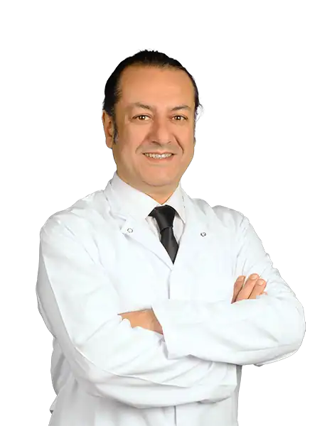
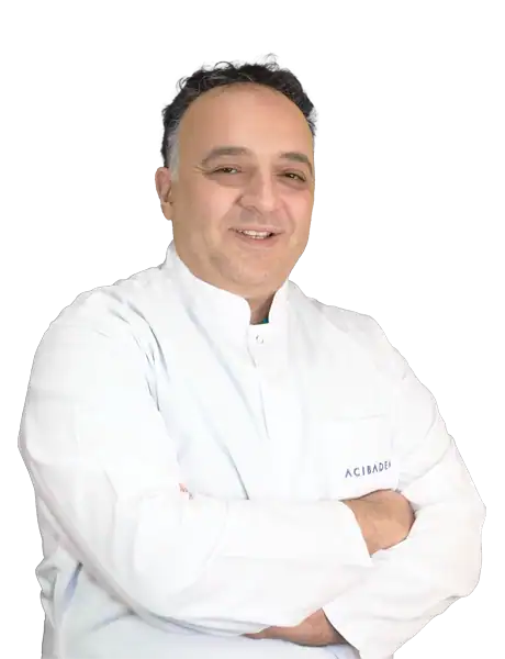
Prof. Mehmet Veli Karaaltın, MD

Aesthetic Plastic & Reconstructive Surgery
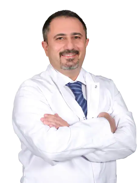
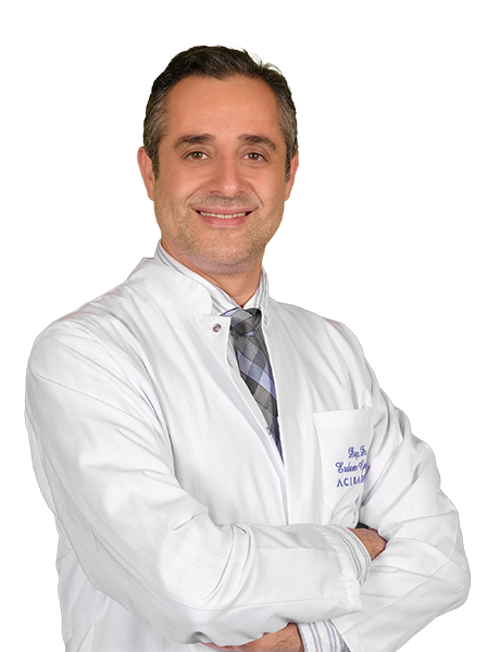
Assoc. Prof. Erdem Güven, MD

Aesthetic Plastic & Reconstructive Surgery
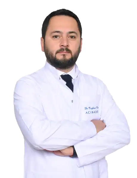
Assoc. Prof. Ahmet Küçükçelebi, MD
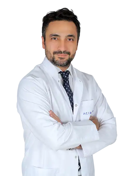
Aesthetic Plastic & Reconstructive Surgery
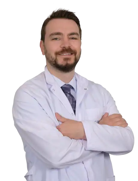
Assoc. Prof. Mehmet Altıparmak, MD

Aesthetic Plastic & Reconstructive Surgery