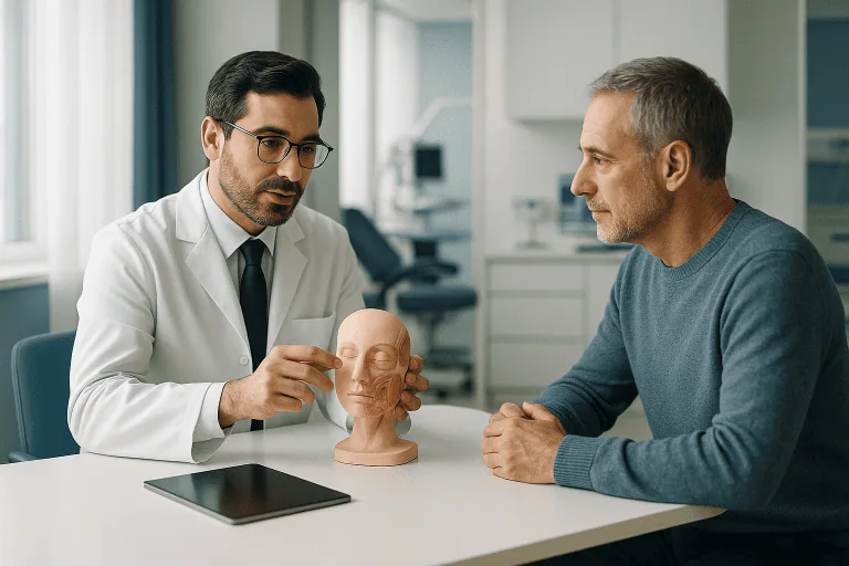
Reconstruction After Skin Disease
Reconstruction after skin disease restores skin coverage, contour, and function after tissue loss from infection, tumors, wounds, or chronic dermatologic conditions using grafts, flaps, or scar revision.
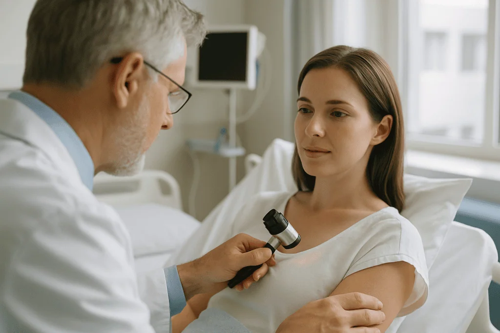
Quick answer
Reconstruction after skin disease is surgery that restores skin and soft tissue damaged or removed because of a medical condition, skin cancer, severe infection, chronic wounds or inflammatory skin disease. Depending on the size, depth and location of the defect, it may involve skin grafts, local or regional flaps, microsurgical tissue transfer or scar release, sometimes carried out in planned stages.
Reconstruction After Skin Disease: Rebuilding Skin, Function and Confidence
Reconstruction after skin disease is surgery that restores skin and soft tissue lost or damaged by a medical condition: a tumour, a severe infection, a chronic wound or an inflammatory skin disease. It rebuilds coverage, contour and movement rather than simply closing a hole. You may be considering it because a wound will not heal, because treating the disease has left a visible defect, or because scarring now restricts how you move.
Skin is not decoration. It is the body’s protective barrier: it helps control temperature, prevents fluid loss, keeps bacteria out and lets you move comfortably. When tissue is lost, scarred or distorted, everyday activities, walking, dressing, working, sleeping, using your hands, can become genuinely difficult. That is why this kind of reconstruction sits firmly in medical, not cosmetic, territory, even when appearance is one of the things it improves.
If you are weighing this up, your questions are probably practical. Will the wound finally close? Will the scar be obvious? Will movement come back? Could the disease return? These are reasonable questions, particularly if you have already been through repeated dressings, previous operations or long stretches of uncertainty. This page sets out what the surgery involves, who needs it, how it is planned and what recovery genuinely looks like, including its limits.
Reconstruction after skin disease is one branch of a wider field. It is closely related to disease reconstruction in general, and it shares many techniques with reconstruction after trauma. The difference lies in the starting point. Trauma damages otherwise healthy tissue in a single event. Disease often damages tissue gradually, leaves the surrounding skin inflamed, infected or poorly supplied with blood, and may still be active when reconstruction is being planned. That changes how surgeons think about timing, technique and safety.
At Acibadem, reconstruction is planned with careful diagnostic evaluation and multidisciplinary input where the condition demands it. Dermatologists, plastic and reconstructive surgeons, oncologic surgeons, infectious disease physicians, wound care teams, radiologists, pathologists and rehabilitation specialists may all contribute, depending on the cause. That coordination matters most when reconstruction follows skin cancer, complex infection, chronic ulceration or autoimmune-related skin damage, situations where the operation is only one part of the treatment.
What Is Reconstruction After Skin Disease?
Reconstruction after skin disease refers to surgical, and sometimes staged, treatment designed to restore damaged or missing skin and soft tissue. It may be needed after tissue has been removed to treat a tumour, destroyed by infection, lost through chronic ulceration, or distorted by scarring from an inflammatory skin condition. The goal is not simply to cover a defect. A well-planned reconstruction accounts for the cause of the disease, the quality of the surrounding tissue, blood supply, infection control, cancer safety where relevant, future mobility, appearance and your overall health.
There is no single standard operation. The plan is individual. Some patients need a modest scar revision or a local rearrangement of nearby tissue. Others need a skin graft, a flap of adjacent tissue, or a complex reconstruction that transfers tissue from another part of the body. The right choice depends on the size, depth and location of the defect, whether structures such as tendon, nerve, bone or cartilage are exposed, and, critically, whether the underlying disease is under control. The main techniques are described below.
Skin Grafts
Skin grafts are thin or thicker layers of skin taken from another area of your body and placed over a prepared wound. A graft has no blood supply of its own at first: it survives by drawing nourishment from the wound bed beneath it and then developing new blood vessels over the following days. That is why grafts work well on broad, shallow wounds with a healthy, well-vascularised base, and why they are unreliable over bare bone, exposed tendon, infected tissue or previously irradiated areas. Thinner split-thickness grafts take more readily and can cover larger areas, while thicker full-thickness grafts contract less and blend better in visible sites, at the cost of a donor wound that must be closed directly. Grafts are the workhorse of durable skin coverage, but they need the right foundation.
Local Flaps
Local flaps move nearby skin and soft tissue into the defect while keeping the tissue attached to its own blood supply. Because the transferred tissue comes from the immediate area, a local flap usually offers a better match in colour, thickness and contour than a graft, which matters on the face, hands and other visible or functionally sensitive areas. Local flaps are often the preferred option for small and medium defects after tumour removal, provided enough healthy tissue lies nearby.
Regional Flaps
Regional flaps bring tissue from an adjacent anatomical region, for example, from the forehead to the nose, or from the chest to the neck, to reconstruct larger or deeper defects that local tissue alone cannot fill. A regional flap may include skin, fat, fascia and sometimes muscle. Because it carries its own robust blood supply, it can succeed where a graft would fail, including over exposed deep structures.
Free Tissue Transfer (Microsurgery)
Free tissue transfer moves tissue from a distant part of the body and reconnects its blood vessels to vessels near the defect using microsurgical techniques, performed under magnification. It is reserved for complex situations: very large defects, wounds in areas with poor local tissue, sites damaged by radiation, or reconstructions that need substantial bulk or specialised tissue. Microsurgery demands longer operating times and closer postoperative monitoring, but for the right patient it can achieve coverage that no simpler method could provide.
Scar Revision and Contracture Release
Scar revision and contracture release address scars that have become tight, painful, raised or restrictive. Skin disease and its treatment can leave scars that shorten over time and pull on joints, eyelids, lips or fingers. Surgical techniques such as Z-plasty, local flap rearrangement, grafting after release, or staged correction can reduce tension, restore movement and improve comfort and appearance. The choice depends on the direction, depth and tightness of the scar and what lies beneath it.
Dermal Substitutes and Staged Reconstruction
Dermal substitutes and staged reconstruction use specialised biologic or synthetic materials to support tissue regeneration before definitive coverage. In some wounds, a dermal regeneration template or temporary wound covering is applied first; once a new vascularised layer has formed, a skin graft or final reconstruction follows. Staging can also mean deliberately separating disease removal from reconstruction, for example, waiting for final pathology results before committing healthy tissue to the repair. These materials and strategies do not replace surgical judgement; they extend it.
Is Reconstruction After Skin Disease One Operation or Several?
For some patients it is a single procedure; for many it is a planned sequence. Reconstruction is best understood as a decision-making process rather than one fixed operation. The surgeon balances reliable healing, function, appearance, safety and the risk of recurrence or infection. Where the disease is fully treated and the tissue is healthy, one operation may achieve everything needed. Where infection must first be cleared, pathology confirmed or the wound bed prepared, a staged pathway is often safer and more predictable. Neither approach is inherently better: the right one is the one that matches the biology of your wound.
Who May Need Reconstruction After Skin Disease?
You may need reconstruction after skin disease when the skin cannot heal on its own, when surgical removal of diseased tissue leaves a defect, or when scarring causes pain, deformity or loss of function. Sometimes the need is obvious: a large excision or a severe infection leaves a wound that clearly will not close by itself. Just as often, the need develops gradually, as a chronic skin condition slowly scars and distorts an area over months or years.
Typical problems that bring patients to reconstructive assessment include:
- A wound that does not close despite regular dressings or previous procedures
- Exposed tendon, bone, cartilage, implant or other deep structures
- Repeated skin breakdown over a scarred or fragile area
- Painful, tight or raised scars that limit movement
- Distortion of facial features, eyelids, lips, nose, ears, hands or genital skin after disease or surgery
- Drainage, odour, recurrent infection or inflammation in chronic skin wounds
- Open areas after removal of skin cancer or other tumours
- Functional difficulties such as trouble closing the eye, opening the mouth, walking, gripping or straightening a joint
How Is the Need for Reconstruction Diagnosed?
Diagnosis starts with the original disease, not with the wound. A detailed medical history is essential: when the lesion started, how it changed, what treatments were tried, whether infection occurred, and whether conditions such as diabetes, vascular disease, immune suppression, autoimmune disease or a smoking history could be interfering with healing. A wound that has not closed usually has a reason, and reconstruction is far more likely to hold if that reason is identified and addressed first.
The clinical examination assesses wound depth, tissue quality, circulation, sensation, movement and the condition of nearby skin. If the cause is uncertain, a biopsy may be needed before any reconstruction, chronic wounds can occasionally harbour tumour tissue, and a repair placed over undiagnosed disease helps no one. When cancer is involved, pathology reports, margin status and imaging are reviewed to confirm that reconstruction will not compromise oncologic safety. For deep or complex wounds, imaging can clarify bone involvement, abscesses, sinus tracts and the relationship of the wound to important structures.
Much of this groundwork can be organised before the first surgical consultation. Previous pathology reports, operative notes, culture results and photographs of the wound over time give the assessing team a picture of how the disease has behaved, not just how it looks today. Where earlier repairs have failed, understanding exactly what was done, and why it broke down, often changes the plan more than any single test.
Conditions and Indications Reconstruction Can Address
This field of reconstruction covers a broad spectrum of conditions. The common thread is tissue loss, scarring or deformity caused by a medical problem rather than a purely elective cosmetic concern. Indications range from urgent situations, such as infected wounds after debridement, to carefully scheduled reconstruction after planned tumour removal.
Reconstructive treatment may be considered for:
- Skin cancer and soft tissue tumour defects: Reconstruction is frequently needed after removal of basal cell carcinoma, squamous cell carcinoma and other tumours treated within skin cancer care, and after excision of melanoma skin cancer or rarer tumours such as dermatofibrosarcoma protuberans, particularly on the face, scalp, hands, limbs and trunk, where every millimetre of tissue matters.
- Severe skin and soft tissue infections: After debridement for necrotising infections, abscesses or extensive cellulitis-related tissue loss, reconstruction restores coverage once the infection is controlled, often in coordination with the infectious diseases department.
- Chronic wounds and ulcers: Diabetic foot ulcers, venous ulcers, pressure injuries and wounds linked to poor circulation may need reconstruction when conservative wound care is not enough on its own.
- Inflammatory dermatologic disease: Conditions such as hidradenitis suppurativa cause repeated abscesses, tunnels and scarring. Once the diseased tissue is removed, reconstructive closure can improve comfort, hygiene and daily function.
- Autoimmune or vascular skin damage: Some patients with vasculitis, scleroderma-related wounds or other immune-mediated conditions need careful wound coverage after their medical treatment has stabilised the disease.
- Scar contractures: Scars from chronic inflammation, infection or previous surgery can tighten over time, restricting joint movement or distorting nearby anatomy.
- Radiation-related skin injury: Skin previously treated with radiation heals poorly and breaks down easily; these areas sometimes need flap reconstruction that imports healthier tissue with its own blood supply.
- Complex postoperative wounds: When a surgical wound opens or fails to heal, reconstruction may be needed to achieve durable closure.
Because the causes are so diverse, the right timing varies. In some cases reconstruction can be performed immediately after the diseased tissue is removed. In others it is safer to wait until infection has cleared, inflammation is controlled, blood flow has been improved or pathology results are complete. A delay chosen for good clinical reasons is not lost time; it is part of the treatment.
Can Reconstruction Be Done at the Same Time as Skin Cancer Removal?
Sometimes, yes, but not always, and the decision rests on cancer safety. When the tumour type is well understood and margins can be confirmed reliably, immediate reconstruction in the same operation is often reasonable and spares you a second procedure. When margin status is uncertain, or when further treatment such as radiotherapy may follow, surgeons may prefer a delayed or staged reconstruction so that the repair never obscures residual disease. Complex cases are typically reviewed with pathology and oncology input before the sequence is fixed. The guiding principle is simple: reconstruction supports cancer treatment; it must never get in its way.
How Reconstruction After Skin Disease Is Performed
Preparation and Planning
Preparation begins with a clear diagnosis and a realistic plan. Your physician assesses the wound or defect, reviews prior treatments, evaluates your general health and identifies anything that could work against healing. Blood tests, wound cultures, biopsy, vascular studies or imaging may be requested depending on the situation.
For tumour-related reconstruction, pathology is central. The team needs to understand the tumour type, depth and margins, and whether additional treatment, radiotherapy, systemic therapy or further excision, may be needed. Complex cancer cases may be discussed in a multidisciplinary tumour board so that the reconstruction is coordinated with the overall oncologic plan rather than bolted onto it.
For infected or chronic wounds, preparation may involve debridement, targeted antibiotics, better dressings, pressure relief, glucose control, vascular assessment, nutritional support and management of swelling. A reconstruction performed before the wound is ready carries a higher risk of breaking down, so this preparation phase is often as important as the operation itself. It is unglamorous work, and it is where many previously failed wounds are finally turned around.
You will also be prepared for anaesthesia and recovery. The surgical team reviews your medications, allergies, blood-thinning drugs, smoking or nicotine exposure and any previous reactions to anaesthesia; decisions about medication belong to your treating doctors and are made individually. Good planning also covers the practicalities after discharge: who will help you at home, how dressing changes will be managed, when follow-up visits and any drain removal are expected, and when it is realistic to return to work, driving and exercise.
The Procedure, Step by Step
The operation varies with the disease and the defect, but most reconstructions follow a recognisable sequence:
- Removal of unhealthy tissue. The surgeon excises scarred or inflamed skin, removes residual tumour tissue, or cleans an infected wound. Nothing durable can be built on diseased tissue.
- Confirmation that the field is safe. In tumour cases, timing may be coordinated with pathology so that cancer clearance is confirmed before healthy tissue is committed to the repair. In infected wounds, cultures and the appearance of the wound bed guide readiness.
- Preparation of the wound bed. Bleeding is controlled, the base is refreshed to healthy tissue, and the defect is measured against the reconstructive options.
- The reconstruction itself. A graft is harvested and secured, a flap is raised and moved, a scar is released and resurfaced, or a dermal substitute is applied as the first stage, whichever the plan specifies.
- Care of the donor site. Wherever tissue was borrowed, that area is closed and dressed with the same attention as the reconstruction.
- Protection and monitoring. Dressings, splints, drains or negative pressure devices are placed as needed, and the early monitoring plan is set.
If a skin graft is used, skin is harvested from a donor site such as the thigh, groin or another suitable area, placed over the prepared wound and secured with sutures, staples or specialised dressings. The graft first survives on nourishment from the wound bed and then grows a new blood supply, which is why the dressing over a fresh graft is often left undisturbed for several days, and why movement at the site is restricted early on.
If a flap is used, tissue arrives with its own circulation, which must be protected. Local and regional flaps suit deeper defects, exposed bone or tendon, and areas where contour match matters. In microsurgical free tissue transfer, vessels are joined under magnification and the flap is then watched closely for colour, warmth, swelling and blood flow in the first days: the period when circulation problems, if they occur, usually declare themselves.
If scar revision or contracture release is performed, the surgeon removes or rearranges scar tissue to reduce tension and restore movement, using Z-plasty, local flap rearrangement, grafting after release or staged correction depending on the scar’s direction and tightness.
Some reconstructions use negative pressure wound therapy before or after surgery to manage fluid, support graft adherence or prepare the wound bed. Others use dermal regeneration templates or temporary coverings as part of a staged plan. These technologies support healing when applied to the right patient and the right wound; they do not replace surgical judgement.
Technology Used to Support Safety and Precision
Modern reconstructive care uses technology at every stage. Before surgery, imaging defines the depth and extent of disease, identifies fluid collections, evaluates bone involvement and maps blood vessels; in selected cases, vascular imaging guides the choice of flap and donor site. During surgery, magnification and microsurgical instruments allow delicate vessel and tissue work, and advanced energy devices support precise dissection and bleeding control. Afterwards, specialised dressings, negative pressure systems and biologic or synthetic tissue matrices can support graft and flap healing, while digital photography and structured wound documentation let the team track progress objectively over time.
The most important technology, though, is the pathway itself: accurate diagnosis, appropriate timing, careful tissue handling and coordinated follow-up. Reconstruction succeeds when the surgical method matches the biology of the wound, not when the newest device is used for its own sake.
How Long Does Surgery Take, and How Long Is the Hospital Stay?
It varies widely, and honest planning reflects that. A small scar revision or a straightforward graft may take relatively little operating time and, in selected patients, can be performed as day surgery. Larger flap reconstructions, infected wounds requiring debridement, microsurgical procedures and operations in medically complex patients can take several hours and usually involve a hospital stay. After surgery, the team monitors circulation, pain control, bleeding, drainage and early healing. Grafts may sit under protective dressings that stay untouched for several days. Flaps are observed for colour, temperature, swelling and blood flow. Drains, where placed, come out once their output falls to an appropriate level. Your surgeon should be able to give you a realistic estimate for your specific plan before you commit to it.
The Recovery Process
Recovery depends on the site and extent of the reconstruction. You may need to limit movement, elevate the area, keep pressure off the wound, wear splints or compression garments and attend scheduled dressing changes. Where joints, tendons or mobility are involved, physical therapy or hand therapy is often part of the plan, and skipping it can undo good surgery.
Donor sites need care too. A skin graft donor site can feel like a deep graze and stays sensitive while it heals. Flap donor sites are monitored for wound healing and scar maturation. Over time, scars usually soften and fade, although they never disappear completely: a point worth being clear about from the start. Sensation over grafted or flap-covered skin is often reduced at first and typically improves gradually, though it may never be identical to the original skin.
Surgical teams typically ask patients to watch for changes such as increasing redness, worsening pain, fever, unusual drainage, foul odour, sudden swelling, bleeding, darkening of a graft or flap, or separation of wound edges, because reviewing these changes early keeps small issues small. Follow-up appointments exist precisely so that healing is checked rather than assumed.
Why Acting Early Matters
Delaying evaluation of a non-healing wound, an infected area, a suspicious skin lesion or a tightening scar tends to make reconstruction harder, not easier. Skin disease can progress into deeper tissue, involve tendons or bone, increase the bacterial burden or create wider zones of scarring. In cancer-related cases, delay may allow the tumour to grow and require a larger excision, and therefore a larger reconstruction.
Chronic wounds also wear down general health. Persistent inflammation, drainage and infection risk reduce mobility, disturb sleep and drive repeated courses of antibiotics or minor procedures. In patients with diabetes, vascular disease or immune suppression, small wounds can deteriorate quickly and end up demanding far more complex care than they would have a few months earlier.
Early assessment does not automatically mean early surgery. Sometimes the right first step is a biopsy, infection control, vascular evaluation, dermatologic treatment or general medical optimisation. But early assessment preserves options. A defect that can be reconstructed with a local flap today may need a larger flap later if it expands or the surrounding tissue scars. In reconstructive surgery, healthy neighbouring tissue is capital: the longer a wound is left, the more of that capital is spent.
Early planning is also simply practical. It leaves time to clarify the diagnosis with biopsy or imaging where needed, treat infection, improve nutrition and blood sugar control, and complete the relevant specialist assessments, so that when surgery does happen, it happens once, under the best possible conditions, rather than as a rescue procedure on a wound that has been allowed to worsen.
Benefits of Reconstruction After Skin Disease
What reconstruction can achieve depends on the underlying condition, but the core aims are consistent: stable coverage, comfort and function, delivered in a way that supports the broader medical treatment plan rather than competing with it.
| Benefit | What It Means for You |
|---|---|
| Durable wound closure | Reconstruction can close wounds that have not healed with dressings alone, reducing fluid loss, contamination and the daily burden of ongoing wound care. |
| Improved function | By releasing tight scars or covering exposed structures, treatment may help restore movement, grip, walking ability, facial expression or eyelid and mouth function. |
| Better protection of deeper tissues | Healthy coverage over tendon, bone, cartilage or hardware can reduce the risk of further breakdown and infection. |
| More natural contour and appearance | Flaps, grafts and scar revision can improve shape, symmetry and skin surface quality, especially in visible or functionally sensitive areas. |
| Support for cancer and disease management | When coordinated with dermatology, oncology or infectious disease care, reconstruction fits safely into the overall treatment strategy. |
| Reduced long-term care burden | A stable reconstruction may decrease the need for frequent dressings, repeated minor procedures and ongoing wound-related limitations. |
Set against these benefits are real limits. Reconstruction cannot return diseased tissue to exactly what it was before. Every technique leaves a scar somewhere, including at the donor site. And no reconstruction protects against the underlying disease returning if that disease remains active, which is why the surgical plan is always paired with continuing medical care for the original condition.
Recovery Timeline After Reconstruction
Recovery varies with the procedure type and the complexity of the wound, but the following timeline gives a general sense of what many patients experience. Your own plan may run faster or slower, and your surgical team will adjust the milestones to your operation.
| Time Period | What Patients Can Expect |
|---|---|
| Day 1 | Monitoring focuses on pain control, bleeding, swelling, circulation of any flap, and protection of the reconstruction. The treated area may be elevated or immobilised. |
| First week | Dressings are checked or changed according to the surgical plan. Grafts and flaps are monitored closely. Activity is limited to protect healing tissue. |
| First month | Most early healing occurs. Sutures may be removed when appropriate. Physical therapy, compression, scar care or gradual movement may begin depending on the site. |
| Two to three months | Strength, comfort and mobility often improve. Scars remain active and may be red, firm or sensitive. Further scar management may be recommended. |
| Longer term | Scar maturation continues for many months. Some patients need secondary refinement, contour adjustment or ongoing dermatologic care to reduce recurrence risk. |
What Influences Outcomes and a Good Result?
A good reconstructive result is not defined by how the area looks the day after surgery. It is measured by stable healing, restored function, comfort, control of the original disease and a result that fits your body and your priorities. Several factors shape that outcome, and it is worth understanding them before you decide.
The underlying disease matters more than anything else. Reconstruction after a clean tumour excision is a different proposition from reconstruction in a chronically infected or inflamed wound. If the original disease remains active, the repair is more likely to break down or the problem to recur. That is why dermatologic, oncologic or infectious disease treatment often continues before and after surgery: the operation treats the defect, not the disease.
Blood supply determines what will heal. Areas with poor circulation, previous radiation, dense scarring or vascular disease may need robust flap coverage rather than a simple graft. Patients with diabetes, peripheral vascular disease or smoking-related vascular changes may need additional evaluation and risk reduction before the operation is scheduled.
Wound preparation is non-negotiable. A graft will not reliably survive on dead tissue, uncontrolled infection or a poorly vascularised bed. Debridement, cultures, antibiotics, negative pressure therapy, pressure offloading or vascular treatment may all be needed to create the conditions in which a reconstruction can take. When previous repairs have failed, inadequate preparation is one of the most common reasons.
Location matters. The face, eyelids, nose, lips, hands, feet, scalp and genital area each carry particular functional and aesthetic demands. Reconstruction there requires refined planning to preserve movement, sensation, drainage, contour and symmetry. A technique that performs well on the trunk may be entirely wrong for an eyelid or a fingertip.
Your health and habits influence healing directly. Nutrition, protein intake, anaemia, immune suppression, steroid use, blood sugar control and nicotine exposure all affect outcomes. Surgeons often strongly advise stopping smoking and all nicotine products before and after surgery, because nicotine narrows blood vessels and increases the risk of wound complications: a graft or flap depends on exactly the circulation nicotine restricts.
Postoperative care is part of the operation. Even a technically excellent reconstruction can fail if the area is exposed too early to pressure, excessive movement, trauma or missed dressing care. Following instructions on elevation, splinting, hygiene, compression, weight-bearing and follow-up visits is not an optional extra; it is half the treatment.
Realistic expectations protect you from disappointment and from poor decisions. Reconstruction can improve coverage, shape and function, but it cannot make diseased tissue exactly as it was. Scars are permanent, although they usually mature and fade over time. Some patients benefit from staged procedures or later refinement once the tissue has settled, and a surgeon who tells you this before the first operation is doing their job properly.
How Acibadem Approaches Reconstruction After Skin Disease
Complex skin reconstruction is rarely a single-specialty problem, and Acibadem organises it accordingly. The operation is usually one step in a longer pathway that may include dermatologic diagnosis, oncologic assessment, infection management, vascular evaluation, wound care, rehabilitation and long-term surveillance. Coordinating those elements under one roof reduces fragmented decision-making: the situation where each specialist treats their piece of the problem and nobody owns the whole.
When cancer is involved, cases may be reviewed through multidisciplinary tumour boards, where surgeons, oncologists, radiologists, pathologists and other specialists consider the timing of excision, reconstruction and any additional therapy together. For chronic wounds and infections, plastic and reconstructive surgeons work with infectious disease physicians, endocrinologists, vascular specialists, radiologists, wound care nurses and rehabilitation teams to answer the question that matters most: why has this wound not healed, and what must change before reconstruction is attempted?
Diagnostic and surgical technology is applied according to clinical need. Imaging defines the problem, modern operating theatres support complex procedures, microsurgical capability is available for selected large or difficult defects, and specialised wound care tools support healing before and after surgery. The value of these resources lies in how they are integrated into an individual plan, not in their existence.
Experience shapes the judgement calls. This kind of reconstruction demands knowing when a simple method is sufficient and when a more complex technique is safer, and when staged care, with time between operations for pathology results or wound preparation, is the wiser path. Some patients also use a specialist review as a second opinion: to understand alternatives to a major operation they have been offered, to get a fresh evaluation after repeated wound breakdown, or to plan reconstruction alongside a forthcoming tumour removal. A sound plan explains not only what will be done, but why that approach fits your disease, your anatomy and your goals.
Preparing for a Reconstructive Consultation
Living with an open wound, a disfiguring scar or tissue loss after skin disease is physically and emotionally draining, and it is easy to arrive at a consultation with a long history and scattered paperwork. Whichever team you consult, a specialist review is far more productive when the documentation is complete. Useful items include your medical records, photographs of the area over time, biopsy or pathology reports, imaging results, culture reports, a current medication list and details of previous surgeries or wound treatments. Together, these let a reconstructive surgeon understand the disease behind the defect, judge whether further testing or specialist review is needed, and set out the realistic options.
It also helps to arrive with your own questions written down. Which technique is proposed, and why that one rather than a simpler or more complex alternative? Is the underlying disease fully treated, and how will that be confirmed? Will the reconstruction be single-stage or staged? What will the donor site look like? How long is recovery, what restrictions apply, and what follow-up will be needed at home? Reconstruction after skin disease works best as a shared decision, one built on a clear diagnosis, honest expectations and a plan that treats the disease and the defect together rather than the defect alone.
Preparation
- A plastic and reconstructive surgeon evaluates the skin defect, scarring, blood supply, and any active disease before planning surgery. Patients may need blood tests, imaging, medication review, and optimization of conditions such as diabetes or infection. Smoking is usually stopped before surgery to improve wound healing.
Aftercare
- The surgical area is protected with dressings, and patients receive instructions on wound care, hygiene, and activity limits. Follow-up visits monitor healing, graft or flap survival, and scar maturation. Sun protection and scar management may be recommended for several months.
Frequently Asked Questions
What affects the cost of reconstruction after skin disease?
Cost is influenced by the size and depth of the defect, the reconstruction method, wound condition, surgeon expertise, anaesthesia, hospital stay, tests, dressings, and follow-up needs. A specialist assessment is required to estimate the most appropriate plan.
How can I get a personalised quote?
You can request a free consultation by sharing medical photographs, diagnosis details, previous biopsy or pathology reports if available, current medications, and information about any infection or chronic condition. The clinical team can then advise on likely treatment options and provide a personalised quote.
Does a package usually include aftercare?
Packages may include hospital care, dressings during admission, medications used in hospital, interpreter support, and planned follow-up. Items such as long-term scar care, extra dressings, rehabilitation, or additional procedures may be assessed separately depending on the case.
Is the cheapest option always suitable?
Not necessarily. Reconstruction must restore safe skin coverage, protect function, and reduce the risk of wound problems. The most appropriate option depends on blood supply, infection status, tissue quality, and the specialist’s examination.
Can international patients plan surgery and travel together?
In many cases, international patient teams can help coordinate consultation, testing, interpreter services, airport transfer, accommodation guidance, surgery scheduling, and follow-up planning. Travel timing should be confirmed by the treating specialist, especially if the wound is active or infected.
Medically reviewed by the Acıbadem International Medical Board September 1, 2026
See our medical review board →
Update history
- PublishedJune 8, 2026
- Medical review approvedSeptember 1, 2026
- Last content updateSeptember 1, 2026
References1
- Skin Graft medlineplus.gov
Trusted care for international patients
From the Acibadem Blog
Timing Reconstruction After Skin Disease: Why Active Infection or Tumor Must Settle First
Reconstruction after skin cancer is usually timed to two conditions rather than a calendar date: the pathology report must confirm the tumor has been…
Healing After Reconstruction for Skin Disease: Graft Survival Checks and Scar Maturation
After reconstruction for skin cancer, a skin graft usually begins forming its own blood supply within about 36 hours and looks settled within 2–3…
Doctors Performing This Treatment


Aesthetic Plastic & Reconstructive Surgery
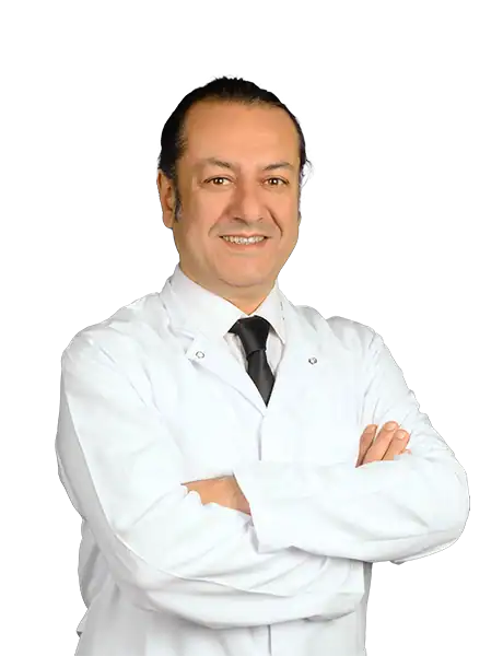
Prof. Mehmet Veli Karaaltın, MD

Aesthetic Plastic & Reconstructive Surgery
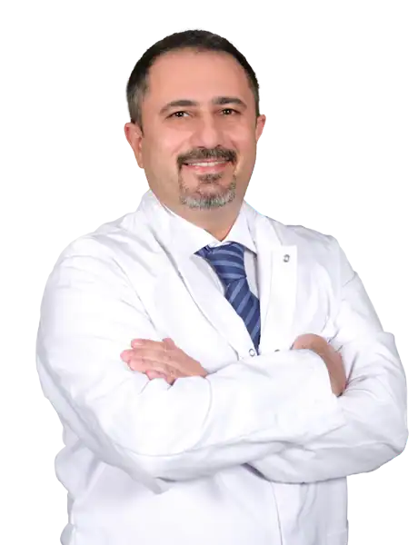
Assoc. Prof. Erdem Güven, MD

Aesthetic Plastic & Reconstructive Surgery
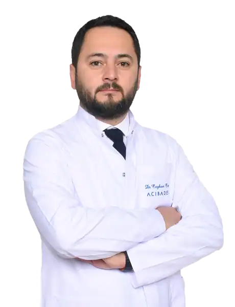
Assoc. Prof. Ahmet Küçükçelebi, MD
Aesthetic Plastic & Reconstructive Surgery
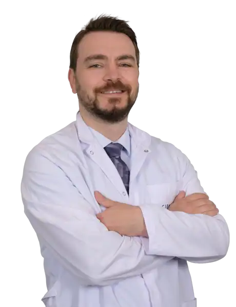
Assoc. Prof. Mehmet Altıparmak, MD

Aesthetic Plastic & Reconstructive Surgery