Macular Diseases Treatment
Macular diseases affect the central retina, causing blurred, distorted, or reduced central vision. Care may include retinal imaging, injections, laser therapy, or surgery depending on the condition.
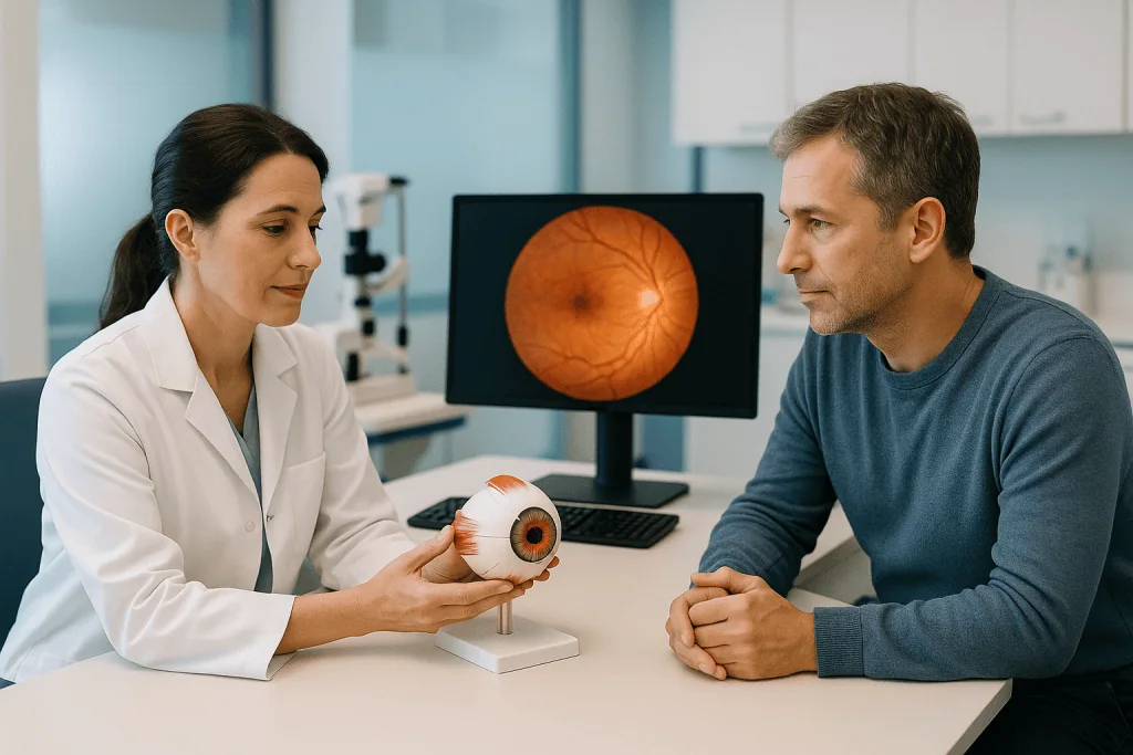
Quick answer
Macular diseases are disorders of the macula, the central part of the retina responsible for sharp, detailed vision, and they can cause blurred, distorted, or reduced central sight. At Acibadem in Turkey, evaluation typically includes detailed retinal examination and imaging, with treatment tailored to the specific condition and may involve eye injections, laser procedures, or surgery.
When Central Vision Changes, Timely Retinal Care Matters
Macular disease can be unsettling because it affects the part of vision people rely on most: reading, recognizing faces, driving, using a phone, seeing details, and noticing contrast. Many patients describe a gray spot in the center of vision, straight lines that appear wavy, words that seem to disappear on the page, or a gradual blur that does not improve with new glasses. Others experience a sudden change and fear they may be losing sight permanently.
The macula is a small but highly specialized area in the center of the retina, the light-sensitive tissue at the back of the eye. Although it is only a tiny part of the eye, it is responsible for sharp central vision. When the macula is damaged by swelling, bleeding, scar tissue, abnormal blood vessels, traction, holes, or age-related changes, vision can become blurred, distorted, or reduced. Peripheral vision is often preserved, but the loss of central detail can significantly affect independence and quality of life.
Modern retinal medicine has changed the outlook for many macular diseases. Conditions that once led to progressive central vision loss can often be stabilized, slowed, or treated when diagnosed early. Some conditions are managed with careful monitoring and lifestyle measures. Others require intravitreal injections, laser therapy, retinal surgery, or a combination of treatments. The best approach depends on the exact diagnosis, the stage of disease, the condition of the other eye, general health, and the patient’s visual goals.
For international patients, the decision to seek treatment abroad often comes with additional questions: How urgent is the condition? Which tests are needed? Will treatment require repeated visits? Is surgery necessary? What recovery time should be expected before flying home? At Acibadem, care for macular diseases is organized around accurate diagnosis, evidence-based treatment planning, and coordinated support for patients traveling from outside Turkey.
What Macular Disease Treatment Is
Macular disease treatment refers to the full medical and surgical management of disorders affecting the macula. It begins with a detailed retinal evaluation and high-resolution imaging to identify the cause of visual symptoms. Treatment may then aim to reduce fluid, control abnormal blood vessel growth, repair structural problems, close a macular hole, relieve traction on the retina, or monitor stable disease safely over time.
There is no single treatment for all macular diseases. The retina specialist first determines the mechanism causing vision loss. For example, wet age-related macular degeneration is often treated with medicines injected into the eye to control abnormal blood vessel leakage. Diabetic macular edema may require injections, laser therapy, improved systemic disease control, or a combination of approaches. A macular hole or epiretinal membrane may require vitrectomy surgery, a microsurgical operation performed inside the eye. Certain inflammatory or vascular macular conditions may need additional medical evaluation and coordination with other specialties.
In many cases, the goal is to preserve existing vision and prevent further deterioration. In some situations, vision can improve when swelling decreases, bleeding resolves, or traction is relieved. The degree of improvement varies widely. It depends on how long the macula has been affected, whether retinal cells are still functioning, the underlying disease, and how consistently treatment can be maintained.
Macular disease care often includes long-term follow-up. Some patients need only periodic monitoring. Others require a structured treatment schedule with repeat imaging and injections over months or years. A clear plan is especially important for international patients, because some treatments must be continued after returning home or coordinated with an ophthalmologist in the patient’s country.
Who May Need Evaluation and Treatment for Macular Disease
Patients may need macular evaluation when central vision changes are persistent, progressive, or sudden. Symptoms can affect one eye or both eyes. Because the brain may compensate when one eye is stronger, a macular problem can go unnoticed until the patient covers one eye or has a routine eye examination.
Common symptoms of macular disease include:
- Blurred central vision, especially when reading or looking at fine detail
- Straight lines appearing bent, wavy, or distorted
- A dark, gray, or empty area in the center of vision
- Difficulty recognizing faces or reading small print
- Reduced color intensity or contrast sensitivity
- Visual distortion after cataract surgery or retinal disease
- Sudden or gradual worsening of vision in one eye
- Needing brighter light for reading or close work
Diagnosis begins with a complete eye examination, including visual acuity testing, eye pressure measurement, pupil dilation, and detailed assessment of the retina. Retinal imaging is central to decision-making. Optical coherence tomography, often called OCT, creates cross-sectional images of the macula and can detect fluid, swelling, thinning, holes, traction, or scar tissue. Fundus photography documents the appearance of the retina over time. Angiography studies may be used to evaluate blood flow, leakage, abnormal vessels, or areas of poor circulation. In selected cases, additional imaging helps distinguish between inflammatory, vascular, degenerative, and inherited retinal disorders.
Some patients are referred after an eye screening detects diabetic changes, macular degeneration, or retinal vein occlusion. Others come after receiving injections elsewhere and seeking a second opinion about treatment intervals, persistent fluid, or whether surgery is appropriate. Patients with a family history of retinal disease, diabetes, high blood pressure, cardiovascular disease, high myopia, or previous retinal problems may need earlier or more frequent evaluation.
A prompt examination is particularly important when symptoms appear suddenly, such as a new central blind spot, sudden distortion, or rapid loss of vision. These signs may indicate bleeding, retinal vascular blockage, active wet macular degeneration, or another urgent retinal condition.
Conditions and Indications Addressed by Macular Disease Care
Macular disease care covers a broad group of retinal conditions. Each has a different cause and treatment pathway, so accurate diagnosis is essential before starting therapy.
Age-related macular degeneration is one of the most common causes of central vision loss in older adults. The dry form may progress slowly and is often monitored with imaging, risk assessment, nutritional guidance when appropriate, and lifestyle recommendations such as smoking cessation. The wet form involves abnormal blood vessels that leak fluid or blood under or within the retina. It commonly requires intravitreal medication to control leakage and reduce the risk of further vision loss.
Diabetic macular edema occurs when diabetes damages small retinal blood vessels, allowing fluid to accumulate in the macula. Treatment may include intravitreal injections, laser therapy in selected cases, and close coordination with diabetes care to improve blood sugar, blood pressure, and kidney-related risk factors. Stable systemic control is an important part of protecting vision.
Retinal vein occlusion with macular edema develops when a retinal vein becomes blocked, leading to hemorrhage, swelling, and reduced central vision. Treatment often focuses on reducing macular fluid and monitoring for abnormal blood vessel growth. Patients may also need evaluation for blood pressure, vascular risk factors, and other medical contributors.
Epiretinal membrane, sometimes called macular pucker, is a thin layer of scar-like tissue that forms on the macular surface. It can cause distortion, blurred vision, and difficulty reading. Mild cases may be monitored. More significant cases may be treated with vitrectomy surgery to remove the membrane and reduce traction.
Macular hole is a small full-thickness opening in the central macula. It often causes a central blind spot, distortion, or difficulty seeing fine detail. Many macular holes are treated surgically using vitrectomy, membrane peeling, and a gas bubble to support closure while the eye heals.
Central serous chorioretinopathy involves fluid accumulation under the retina, often associated with stress physiology, steroid exposure, or choroidal circulation changes. Some cases resolve with observation and risk-factor modification. Persistent or recurrent cases may require targeted treatment depending on imaging findings.
Myopic macular disease can occur in patients with high myopia, where stretching of the eye contributes to degenerative changes, macular traction, bleeding, or choroidal neovascularization. Management depends on the specific complication and may involve injections, surgery, or monitoring.
Inflammatory, hereditary, and medication-related macular conditions may require a more specialized diagnostic pathway. These cases may involve additional laboratory testing, genetic counseling, medication review, or collaboration with other medical specialists.
How Macular Disease Treatment Is Performed
Step 1: Comprehensive Retinal Assessment
The treatment process begins with a careful review of symptoms, prior eye treatments, medical history, medications, and family history. Patients who have already had imaging or injections are encouraged to bring previous reports, OCT scans, angiography images, and surgical notes if available. Comparing old and new images can help determine whether the disease is active, stable, or progressing.
A dilated retinal examination is performed to evaluate the macula, optic nerve, retinal blood vessels, and peripheral retina. Visual acuity and other functional assessments help measure how the disease is affecting daily life. In some cases, the retina specialist may also assess cataract, glaucoma, corneal conditions, or optic nerve disease, because these can influence vision and treatment decisions.
Step 2: Advanced Retinal Imaging and Diagnosis
Retinal imaging guides both diagnosis and treatment. OCT is one of the most important tools because it shows the retinal layers in fine detail. It can identify macular edema, subretinal fluid, drusen, atrophy, vitreomacular traction, epiretinal membrane, or macular hole features. OCT angiography may help assess blood flow and abnormal vascular networks without dye in selected cases. Dye-based angiography may be recommended when leakage patterns, blocked vessels, or choroidal circulation need closer evaluation.
These technologies help the physician determine whether treatment is urgent, what type of treatment is most appropriate, and how response should be monitored. For many macular diseases, small changes on OCT can influence whether an injection is given, whether an interval is adjusted, or whether surgery should be considered.
Step 3: Personalized Treatment Planning
After diagnosis, the retina specialist explains the condition, the expected course, and the treatment options. For international patients, this conversation also includes practical planning: how many visits may be needed, whether same-trip treatment is appropriate, whether follow-up can be done locally, and when it is safe to travel after a procedure.
Some patients are managed with observation and scheduled imaging, particularly when the condition is mild, stable, or likely to resolve. Others require active treatment. In complex cases, decisions may be discussed within specialist boards or multidisciplinary meetings, especially when diabetes, vascular disease, oncology-related eye problems, inflammatory disease, or inherited conditions affect management.
Intravitreal Injections
Intravitreal injections deliver medication directly into the vitreous cavity of the eye, allowing high concentration at the retina with limited exposure to the rest of the body. They are commonly used for wet macular degeneration, diabetic macular edema, retinal vein occlusion-related swelling, and certain abnormal blood vessel conditions.
The injection is usually performed as an outpatient procedure. The eye is cleaned carefully with antiseptic solution, numbing drops are applied, and a small instrument may be used to keep the eyelids open. The medication is injected through the white part of the eye using a very fine needle. The procedure itself is brief, although preparation and post-injection checks take longer. Patients may feel pressure, mild irritation, or watering afterward. Temporary floaters or a small red spot on the white of the eye can occur.
Many injection-based treatments require a series of visits. The interval may be monthly at first and then adjusted according to disease activity, imaging response, and the medication used. Regular follow-up is important because fluid can recur even when vision feels stable.
Laser and Light-Based Retinal Treatments
Laser therapy may be used in selected macular and retinal vascular diseases. Modern retinal lasers can target leaking microaneurysms, areas of retinal ischemia, or specific lesions depending on the diagnosis. In some conditions, photodynamic therapy or other light-based treatment may be considered to address abnormal choroidal circulation or persistent fluid. The choice depends on imaging findings and the location of the abnormality relative to the central fovea, the most sensitive part of the macula.
Laser treatment is usually outpatient and does not require general anesthesia. The eye is numbed with drops, and a contact lens may be placed on the eye to focus treatment accurately. Some patients notice brief flashes of light or mild discomfort. Recovery is generally quick, but vision may be temporarily blurred from the examination drops or contact lens.
Vitrectomy Surgery for Structural Macular Problems
Vitrectomy is a microsurgical procedure used for conditions such as macular hole, epiretinal membrane, vitreomacular traction, some complications of diabetic retinopathy, and selected cases of macular detachment or hemorrhage. During vitrectomy, the surgeon works through tiny openings in the eye to remove the vitreous gel, release traction, peel delicate membranes when needed, and stabilize the macula. A gas bubble, air bubble, or other internal support may be placed depending on the condition.
Vitrectomy is typically performed in an operating room under local anesthesia with sedation or, in selected cases, general anesthesia. The procedure duration varies according to complexity. Some macular surgeries may be completed in under an hour, while more complex cases take longer. Patients usually go home the same day or after short observation, depending on the medical situation and travel arrangements.
If a gas bubble is placed, patients may need to follow specific head-positioning instructions for a period of time. Flying is not allowed while a significant gas bubble remains in the eye, because changes in cabin pressure can dangerously increase eye pressure. This is a crucial point for international patients and is discussed before surgery so travel plans can be adjusted safely.
Recovery and Follow-Up
Recovery depends on the condition and treatment type. After injections or laser, patients often resume many normal activities within a short time, while avoiding eye rubbing and following medication instructions. After vitrectomy, recovery is more structured and may include eye drops, activity limitations, positioning, protective eyewear, and several follow-up examinations.
Vision may fluctuate during recovery. After injections, improvement can occur gradually as fluid decreases, although some patients primarily achieve stabilization rather than improvement. After macular hole or membrane surgery, vision may continue to change over months as the retina remodels. The final visual result depends on the health of the macular cells and how long the disease was present before treatment.
Why Acting Early Matters
Macular diseases often respond best when treated before prolonged damage has occurred. The macula contains highly specialized nerve tissue. When swelling, bleeding, traction, or abnormal blood vessels persist, retinal cells may become permanently damaged. Early diagnosis gives physicians a better opportunity to reduce fluid, control leakage, repair mechanical problems, and preserve central vision.
Delay can allow a treatable condition to become more advanced. In wet macular degeneration, ongoing leakage or bleeding can lead to scar formation. In diabetic macular edema, chronic swelling can damage retinal layers and reduce the chance of visual recovery. In macular hole, long-standing holes may become larger and less likely to regain fine central vision after repair. In epiretinal membrane, prolonged traction can distort the macula and limit postoperative improvement.
Some patients delay care because vision changes are mild or affect only one eye. Others assume the problem is cataract, tiredness, or the need for new glasses. Any new distortion, central blur, or dark spot should be evaluated by an eye specialist. Prompt assessment does not always mean urgent treatment is needed, but it helps determine whether observation is safe or whether intervention should begin.
Benefits of Macular Disease Treatment
The potential benefits depend on the diagnosis, but appropriate treatment can help protect central vision and support daily visual function.
| Benefit | What It Means for You |
|---|---|
| More accurate diagnosis | High-resolution retinal imaging helps identify the specific cause of blurred or distorted central vision, allowing treatment to be tailored to the disease mechanism. |
| Stabilization of vision | Many macular diseases can be slowed or controlled, reducing the risk of further central vision loss when treatment is started and monitored appropriately. |
| Reduction of macular fluid | In conditions such as wet macular degeneration, diabetic macular edema, and retinal vein occlusion, treatment may reduce swelling and improve retinal structure. |
| Repair of traction or macular holes | Microsurgical treatment can relieve mechanical pulling on the macula or close selected macular holes, helping the retina return to a more functional shape. |
| Personalized follow-up plan | Patients receive guidance on visit intervals, imaging, medications, travel timing, and coordination of care after returning home. |
Recovery Timeline After Macular Disease Treatment
Recovery varies by treatment type, but the following timeline gives a general sense of what many patients can expect.
| Time Period | What Patients Can Expect |
|---|---|
| Day 1 | After injections or laser, mild irritation, watering, light sensitivity, or temporary blur may occur. After surgery, the eye is usually protected, and drops are started as instructed. |
| First Week | Injection and laser patients often return to routine activities quickly, with precautions. Surgical patients attend follow-up visits, use prescribed drops, and may need positioning or activity restrictions. |
| First Month | Retinal imaging may be repeated to assess response. Swelling may decrease gradually. After vitrectomy, vision can remain blurred while the eye heals or while a gas bubble is present. |
| Longer Term | Some patients need ongoing injections or monitoring. Surgical vision recovery may continue for several months. Long-term results depend on the underlying disease and macular health. |
Factors That Influence Outcomes
Outcomes in macular disease care depend on multiple medical and practical factors. The first is the exact diagnosis. A patient with fluid from wet macular degeneration has a different prognosis and treatment pathway than a patient with a long-standing macular hole or advanced dry macular degeneration. Even within the same diagnosis, severity varies from mild and reversible changes to advanced damage.
The duration of symptoms is also important. The longer the macula has been swollen, detached, distorted, or affected by abnormal blood vessels, the greater the risk that photoreceptor cells have been damaged. Early treatment often gives the retina a better chance to recover structure and function.
Baseline vision and retinal imaging findings help guide expectations. OCT can show whether the retinal layers are preserved, whether fluid is active, whether scar tissue is present, and whether atrophy has developed. These details are often more informative than vision testing alone.
Consistency of follow-up is critical, especially for injection-based treatment. Some macular diseases require repeated therapy to maintain control. Skipping visits can allow fluid or bleeding to recur. For international patients, a practical follow-up plan may include treatment at Acibadem during the visit and coordination with a trusted retina specialist after returning home.
General health also influences outcomes. Diabetes control, blood pressure management, kidney function, cholesterol levels, smoking status, and cardiovascular risk factors can affect retinal vascular disease. In inflammatory conditions, systemic disease activity may need to be addressed. Patients taking steroid medications may need a review if central serous chorioretinopathy is suspected, but medication changes should always be made under medical supervision.
For surgical macular conditions, outcomes depend on surgical timing, the size and chronicity of the problem, lens status, postoperative positioning when required, and careful adherence to instructions. Cataract progression can occur after vitrectomy in some patients, particularly older adults, and may later affect vision even when the macula has improved structurally.
Finally, realistic expectations are important. Treatment may improve vision, stabilize vision, reduce distortion, or prevent further decline, but results vary. A thoughtful consultation should include not only what can be done technically, but also what the patient is likely to experience in everyday life.
Why International Patients Choose Acibadem for Macular Disease Care
International patients seeking retinal care need more than a single procedure. They need a reliable diagnosis, a clear explanation, coordinated scheduling, and a treatment plan that fits both medical needs and travel realities. Acibadem Hospitals provide care within JCI-accredited hospital environments, with ophthalmology teams experienced in diagnosing and managing complex retinal conditions.
Macular disease care at Acibadem is supported by modern diagnostic pathways, including detailed retinal examination and advanced imaging used to evaluate macular structure, fluid, circulation, and treatment response. These tools help physicians distinguish between conditions that may look similar to patients but require very different treatment. A patient with central blur may have cataract, macular edema, epiretinal membrane, optic nerve disease, or more than one condition at the same time. Careful assessment helps avoid incomplete or misdirected treatment.
Experienced ophthalmologists and retina specialists develop personalized treatment plans based on international and evidence-based protocols. When cases involve diabetes, vascular disease, inflammatory disease, oncology history, neurological concerns, or surgical complexity, care may be coordinated with other specialties. This multidisciplinary approach is particularly valuable for patients whose eye condition is connected to broader health issues.
For international patients, Acibadem International provides dedicated support before, during, and after the visit. Services may include assistance with medical record review, appointment coordination, language support in more than 20 languages, travel-related planning, and communication with clinical teams. This is especially important for retinal disease, where timing matters and some treatments may require repeat visits or restrictions on flying after surgery.
Patients also benefit from access to a broad hospital infrastructure. If a patient needs medical clearance before surgery, management of diabetes or hypertension, laboratory testing, or consultation with another specialist, these services can be coordinated within the same healthcare group. This reduces fragmentation and helps the retina team understand the full clinical picture.
Technology plays a central role, but it is not used in isolation. Retinal imaging, microsurgical visualization systems, fine-gauge vitrectomy instruments, laser platforms, and injection protocols are valuable because they help physicians diagnose precisely, treat carefully, and monitor response over time. The most appropriate technology is selected according to the patient’s condition rather than applying the same pathway to every case.
Second opinions are also a common reason patients seek care. A second opinion may be helpful if injections are not producing the expected response, if surgery has been recommended and the patient wants to understand alternatives, if imaging findings are unclear, or if there is concern about rapid progression. Reviewing previous imaging alongside new tests can clarify whether the disease is active, chronic, or changing.
Taking the Next Step
Changes in central vision should be taken seriously, but a diagnosis of macular disease does not mean that vision loss is inevitable. Many macular conditions can be monitored, controlled, or treated when evaluated carefully and addressed at the right time. The first step is to understand exactly what is affecting the macula and whether treatment is needed now, soon, or only if changes occur.
If you have blurred central vision, distortion, a central dark spot, diabetic retinal changes, age-related macular degeneration, a macular hole, epiretinal membrane, or persistent macular edema, a retina consultation can help define your options. International patients may request an evaluation or second opinion and share existing eye reports and retinal images before traveling whenever possible. This allows the clinical team to advise on urgency, likely testing, treatment planning, and travel considerations.
With accurate diagnosis, evidence-based treatment, and careful follow-up, macular disease care can help protect the vision that supports reading, work, mobility, and daily independence.
This information is general and is not a substitute for professional medical advice. Diagnosis and treatment decisions should be made after consultation with a qualified ophthalmologist or retina specialist.
Preparation
- Before treatment, an ophthalmologist evaluates vision and performs retinal imaging such as OCT and fundus examination. Patients should share current medications, eye history, diabetes or hypertension status, and any allergies. Blood thinners or eye drops should only be adjusted if the doctor recommends it.
Aftercare
- After intravitreal injection or laser treatment, patients may need prescribed eye drops and should avoid rubbing the eye. Mild irritation or floaters can occur, but severe pain, sudden vision loss, or increasing redness should be reported immediately. Regular follow-up visits are important because many macular diseases require ongoing monitoring.
Frequently Asked Questions
What affects the cost of macular disease treatment?
The main factors are the diagnosis, required imaging, whether treatment involves injections, laser or surgery, medication choice, hospital setting and the number of follow-up visits needed. A specialist review is required to estimate the most appropriate pathway.
How can I get a personalised quote for treatment in Turkey?
You can request a free consultation by sharing recent eye reports, retinal scans if available, your diagnosis and current symptoms. The medical team can then advise what further assessment may be needed and prepare a personalised cost estimate.
Are macular injections included in a treatment package?
Package contents vary by case. Some packages may include consultation, imaging and coordination services, while medication and repeat injections may be quoted separately depending on the treatment plan.
Will I need to stay in Turkey for follow-up?
This depends on the condition and treatment. Some patients need short-term review after a procedure, while others require ongoing monitoring that may be continued with a local ophthalmologist after the treating specialist confirms it is appropriate.
Is surgery always required for macular diseases?
No. Many macular conditions are managed with monitoring, injections or laser therapy. Surgery is usually considered for specific problems such as macular hole, epiretinal membrane or vitreomacular traction, and suitability is decided by a retina specialist.
Medically reviewed by the Acıbadem International Medical Board June 20, 2026
See our medical review board →
Update history
- PublishedJune 8, 2026
- Medical review approvedJune 20, 2026
- Last content updateSeptember 8, 2026
References3
- Macular Degeneration medlineplus.gov
- Age-related macular degeneration (AMD) nhs.uk
- Macular Degeneration my.clevelandclinic.org
Trusted care for international patients
From the Acibadem Blog

Injections, Laser or Surgery for Macular Diseases: How the Retina Team Picks a Path
Macular disease treatment depends on what is happening in the macula. Leaking new blood vessels and swelling are usually treated with repeated anti-VEGF eye…