Ocular Immunology
Ocular immunology focuses on diagnosis and medical treatment of immune-related eye inflammation, such as uveitis and scleritis, aiming to protect vision and control recurrence.
Quick answer
Ocular immunology is the medical specialty that diagnoses and treats immune-related eye inflammation, including uveitis, scleritis and retinal vasculitis. Evaluation combines detailed eye examination, imaging and targeted laboratory tests to identify the cause. Treatment is usually medical — corticosteroid drops, injections around or inside the eye, or steroid-sparing immune therapy — and is chosen according to the type, location and severity of inflammation and any underlying systemic disease.
When Eye Inflammation Threatens Vision, the Diagnosis Comes First
Eye inflammation driven by the immune system is the territory of ocular immunology — the specialty that diagnoses and treats conditions such as uveitis, scleritis and retinal vasculitis, in which the body’s own defence system inflames the delicate structures of the eye. It exists for people whose inflammation is severe, recurrent, unexplained or linked to a wider disease, and its purpose is straightforward: identify why the eye is inflamed, control that inflammation, and protect vision over the long term. Treatment is usually medical rather than surgical, and it works best when it starts from an accurate diagnosis.
Immune-related eye inflammation can be frightening because it often begins suddenly, affects vision quickly, and can return without warning. A red, painful, light-sensitive eye may look like a simple infection or irritation at first. Blurred vision, floaters or a dull ache around the eye are easy to dismiss as tiredness. Yet in uveitis, scleritis, retinal vasculitis and inflammation linked to autoimmune disease, early and accurate treatment can make a meaningful difference to how well sight is preserved.
Ocular immunology sits at the intersection of several disciplines. It draws on ophthalmology, immunology, rheumatology, infectious disease, internal medicine, and sometimes neurology or oncology, because inflammation in the eye is frequently one part of a larger picture. The goal is never just to calm the current flare. It is to reduce the chance of recurrence, protect the eye’s structures from cumulative damage, and identify any underlying systemic condition that also needs attention.
If you are considering travelling for this kind of care, you probably have specific questions. Is the inflammation serious? Can vision recover? Is steroid treatment safe over time? Will you need long-term immune therapy? Many patients arrive after repeated attacks of uveitis, unclear test results, or treatment that has not held the disease in check. A structured ocular immunology evaluation exists to answer exactly these questions — with evidence-based diagnosis, careful risk assessment, and a plan tailored to both the eye and the whole person.
What Is Ocular Immunology?
Ocular immunology is the diagnosis and medical management of inflammatory eye diseases caused by an overactive, misdirected or dysregulated immune response. These conditions can affect the front of the eye, its middle layers, the retina, the optic nerve, the sclera, or the tissues around the eye. Some are confined to the eye alone. Others are linked to systemic inflammatory or autoimmune disorders such as Behçet’s disease, ankylosing spondylitis, rheumatoid arthritis, inflammatory bowel disease, sarcoidosis, lupus or vasculitis — diseases in which the eye may be the first organ to show trouble.
Treatment in this field is usually medical. Depending on the diagnosis, it may involve corticosteroid eye drops, injections around or inside the eye, oral medication, steroid-sparing immunomodulatory therapy, biologic therapies, or targeted treatment for an infection if one is identified. The right approach depends on the type, location, severity, cause and recurrence pattern of the inflammation. Because several of these treatments suppress the immune system, careful diagnosis and ongoing monitoring are not optional extras — they are the foundation of safe care.
A central principle of ocular immunology is precision. Not every red eye is uveitis. Not every uveitis is autoimmune. And no two patients should automatically receive the same medication. Before any immune-suppressing therapy is used, physicians must rule out or account for infectious causes such as tuberculosis, syphilis, herpes-family viruses, toxoplasmosis, and other regional or travel-related diseases — an area where ocular microbiology testing plays a decisive role. In some cases, inflammation can also mimic, or be associated with, malignancy. That is why this specialty relies on detailed examination, imaging, laboratory testing and multidisciplinary interpretation rather than a single test or a standard prescription.
What is uveitis?
What is uveitis, exactly? It is inflammation of the uvea — the vascular middle layer of the eye that includes the iris at the front, the ciliary body behind it, and the choroid, which lines the back of the eye beneath the retina. Uveitis is the most widely recognised condition in ocular immunology, and depending on where it sits, it can involve the iris, the ciliary body, the choroid, the retina, or all compartments of the eye at once. You may occasionally see it spelled “uvitis” in searches and informal writing; the correct medical term is uveitis. Whatever the spelling, the condition deserves careful attention, because the uvea supplies much of the eye’s blood flow and sits directly against the tissues responsible for sight.
Is uveitis the same as scleritis?
No — scleritis is a separate condition, though the two belong to the same specialty. Scleritis is a deeper, often intensely painful inflammation of the sclera, the white wall of the eye. Its pain is typically boring in character, may radiate to the temple, jaw or head, and can worsen at night. Ocular immunology also covers inflammatory diseases of the retina and choroid, optic nerve inflammation related to immune conditions, and certain orbital inflammatory disorders. Each behaves differently, and each has its own diagnostic pathway.
Who May Need Ocular Immunology Care?
You may need evaluation by an ocular immunology specialist if you have eye inflammation that is severe, recurrent, unexplained, associated with systemic symptoms, or not responding as expected to standard treatment. Some patients are referred after an emergency visit for a painful red eye. Others arrive after months or years of repeated flares, steroid dependence, or slowly progressive changes in vision. Both routes are common, and neither means it is too late to establish a clear diagnosis.
What are the symptoms of uveitis?
Uveitis symptoms depend on which part of the eye is inflamed. In anterior uveitis, the most common pattern, you typically notice redness, eye pain, light sensitivity, tearing and blurred vision. Intermediate or posterior uveitis behaves differently: floaters, hazy vision, reduced central vision or distortion may develop with little or no pain, which is one reason inflammation deeper in the eye is sometimes recognised late. Retinal vasculitis or choroiditis may cause blind spots, dim vision or a sudden decline in sight. Scleritis, by contrast, announces itself with deep, persistent pain rather than visual haze. Because these patterns overlap with other eye problems, the pattern of symptoms is a starting point for diagnosis, never the whole answer.
What causes uveitis?
Uveitis has many possible causes, and identifying the right one shapes everything that follows. Broadly, the causes fall into three groups: autoimmune and inflammatory conditions in which the immune system attacks eye tissue, sometimes as part of a systemic disease; infections, including tuberculosis, syphilis, viral infections and toxoplasmosis; and masquerade processes, in which something that is not primarily inflammatory — such as lymphoma — imitates uveitis. In a proportion of patients, no systemic cause is found even after thorough testing, and the inflammation is managed as an eye-limited disease. This range of possibilities is precisely why treatment should not begin with assumptions.
How is uveitis diagnosed?
Diagnosis begins with a detailed history, and this conversation is often as important as the examination itself. Your ophthalmologist will ask about the timing of symptoms, previous episodes, response to medications, past eye surgery, infections, travel, current medicines, family history, and systemic symptoms such as joint pain, back stiffness, mouth or genital ulcers, skin rashes, bowel symptoms, breathing problems, fever or neurological complaints. Questions may seem far removed from the eye. They are not — immune-related eye inflammation can be the first sign of a systemic disease, and the history is where that link is usually first suspected.
The eye examination usually includes visual acuity testing, eye pressure measurement, slit-lamp examination, dilated retinal examination, and grading of inflammatory cells inside the eye. Imaging then documents where the inflammation sits and how severe it is. Optical coherence tomography shows swelling in the macula, fluid under the retina or damage within retinal layers. Wide-field retinal photography helps track peripheral inflammation over time. Fluorescein or indocyanine green angiography can reveal leakage from retinal vessels, choroidal inflammation, or areas of poor circulation that ordinary examination misses. Ultrasound is useful when inflammation or opacity prevents a clear view into the eye.
Laboratory testing is selected according to the clinical pattern rather than ordered indiscriminately. Blood tests, chest imaging, infectious disease screening, autoimmune markers and rheumatological evaluation may all be recommended — but only where the pattern justifies them. In selected cases, a sample of eye fluid or tissue is needed to distinguish infection, masquerade syndromes or rare inflammatory diseases. The aim is to avoid two opposite mistakes: undertreating a disease that threatens vision, and exposing you to medication that was never appropriate for your condition.
Conditions and Indications Addressed by Ocular Immunology
Ocular immunology covers a wide range of inflammatory eye conditions, and they do not all behave the same way. Some are one-time episodes that settle with short-term therapy. Others are chronic diseases that need long-term control and regular monitoring. The treatment plan is shaped by the condition’s location, severity, whether one or both eyes are involved, its systemic associations, and the risk it poses to vision.
What is anterior uveitis?
Anterior uveitis is inflammation in the front chamber of the eye, involving the iris and often the ciliary body, and it is the form of uveitis most people encounter first. It typically causes redness, pain, light sensitivity and blurred vision, and it may be associated with HLA-B27-related disease, arthritis or inflammatory bowel disease — or occur with no identifiable systemic condition at all. Anterior uveitis often responds well to local treatment, but recurrent episodes are a signal that systemic evaluation is worthwhile, because repeat attacks sometimes point to an underlying disease that has not yet declared itself elsewhere.
The broader list of conditions managed within ocular immunology includes:
- Anterior uveitis: Inflammation in the front chamber of the eye, often causing redness, pain, light sensitivity and blurred vision, with or without an associated systemic disease.
- Intermediate uveitis: Inflammation centred in the vitreous and peripheral retina, usually presenting with floaters and hazy vision. In selected cases it is associated with systemic inflammatory or neurological conditions, which is one reason evaluation sometimes overlaps with neuroimmunology.
- Posterior uveitis and panuveitis: Inflammation involving the retina, the choroid, or multiple compartments of the eye at once. These forms can threaten vision and call for prompt imaging and systemic work-up.
- Scleritis and episcleritis: Inflammation of the sclera or of the superficial tissues over it. Scleritis can be severely painful and may be associated with rheumatoid arthritis, vasculitis, inflammatory bowel disease or infection.
- Retinal vasculitis: Inflammation of retinal blood vessels leading to leakage, swelling, bleeding or ischaemia. It occurs in Behçet’s disease, sarcoidosis, infectious disease and other immune-mediated conditions.
- Behçet’s-related eye disease: A potentially recurrent form of ocular inflammation that may involve the retina and its vessels, requiring coordinated and rapid control to limit visual risk.
- Sarcoidosis-related eye inflammation: Ocular inflammation associated with granulomatous disease that may also involve the lungs, lymph nodes, skin or nervous system.
- Inflammation associated with systemic autoimmune disease: Eye disease linked to lupus, rheumatoid arthritis, ankylosing spondylitis, inflammatory bowel disease, vasculitis or other immune disorders. Where these diseases affect other organs — the kidneys, for example — care is coordinated with the relevant specialty, such as renal immunology.
- Inflammatory complications after surgery or trauma: Persistent or unusual inflammation after cataract surgery, retinal procedures, injury or other intraocular interventions, which must be carefully distinguished from infection.
- Masquerade syndromes: Conditions that look inflammatory but are actually driven by infection, lymphoma or other non-immune disease. These require specialised investigation before any treatment decision is made.
How Ocular Immunology Evaluation and Treatment Are Performed
Ocular immunology care is a process, not a single procedure. It moves through a logical sequence: establish the correct diagnosis, assess the risk to vision, identify any underlying cause, and select treatment that controls inflammation while limiting avoidable side effects. For patients travelling from another country, the first visit usually also includes a review of prior records, imaging, laboratory reports and medication history, so the team starts with the full story rather than a fragment of it.
In practice, the pathway typically follows these steps:
- Detailed history and review of all prior records, treatments and flare patterns.
- Comprehensive eye examination, including slit-lamp and dilated retinal assessment.
- Imaging selected to the clinical picture — tomography, photography, angiography or ultrasound.
- Targeted laboratory and systemic testing, with specialist referral where indicated.
- Diagnosis and risk assessment, discussed with you in plain terms.
- Treatment initiation, followed by monitoring and adjustment over subsequent visits.
Preparation Before Your Visit
Preparation matters most if you are seeking a second opinion or have had multiple previous episodes. It helps to gather prior eye examination notes, imaging, laboratory results, surgical reports, complete medication lists, and the dates of past flares. Documenting how your eye responded to steroid drops, oral steroids, injections, immune medications, antibiotics or antiviral therapy is genuinely useful — patterns of response and relapse are diagnostic information in their own right. If you already carry a systemic diagnosis, records from your rheumatologist, internist, infectious disease specialist or neurologist help the team see the whole picture from the start.
During the initial evaluation, expect questions that range well beyond the eye. Back pain, mouth ulcers, skin lesions, cough, digestive symptoms, neurological symptoms, fever, weight change and previous infections can all be relevant, because ocular inflammation frequently connects to immune, infectious and systemic disease. Answering these questions fully — even where they seem unrelated — shortens the road to a correct diagnosis.
Diagnostic Examination and Imaging
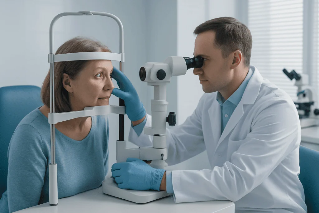
The examination itself uses magnified biomicroscopy and dilated retinal assessment. The physician looks for the type of inflammatory cells present, deposits on the cornea, iris changes, adhesions of the pupil, vitreous haze, retinal lesions, vascular leakage, optic nerve involvement and macular swelling. Eye pressure is measured at every stage, because both inflammation and steroid therapy can raise pressure and increase glaucoma risk — a complication that is far easier to manage when it is caught early.
Modern imaging makes inflammation visible and measurable rather than a matter of impression. Optical coherence tomography produces cross-sectional images of the retina and macula, revealing swelling or structural damage in fine detail. Retinal angiography shows leakage from inflamed vessels or choroidal disease that routine examination cannot see. Fundus photography documents disease activity over time, so the appearance of the eye can be compared visit to visit rather than remembered. Where inflammation or opacity blocks the view, ultrasound assesses the back of the eye instead. Together, these tools do two jobs: they locate the disease precisely, and they create an objective baseline against which treatment response is judged.
Laboratory and Systemic Evaluation
Laboratory testing is tailored to the pattern of disease and framed around specific questions. Is there evidence of infection? Is the inflammation linked to an autoimmune disease? Is any organ outside the eye involved? Are there safety concerns to resolve before immune therapy begins? Testing may include blood counts, liver and kidney function, inflammatory markers, autoimmune panels, infectious disease screening, and chest imaging where indicated.
Some patients are referred onward to rheumatology, infectious disease, pulmonology, neurology or internal medicine. In a centre where these specialties collaborate routinely and complex cases can be reviewed by multidisciplinary boards, a difficult diagnosis is examined from several angles at once — and eye treatment stays aligned with the rest of your medical care rather than running parallel to it.
How Is Uveitis Treated?
Uveitis treatment depends on the diagnosis, the location of inflammation and its severity — there is no single protocol. Mild anterior inflammation is often treated with corticosteroid eye drops together with pupil-dilating drops, which reduce pain and prevent the iris from sticking to the lens. More severe inflammation may require steroid injections around the eye or inside it, particularly when swelling at the back of the eye threatens vision. Oral corticosteroids come into play when disease is bilateral, posterior, severe, or tied to a systemic condition.
Because corticosteroids carry side effects with long-term use, chronic or recurrent disease frequently calls for steroid-sparing therapy. These medications regulate the immune response over time and may include conventional immunomodulatory drugs or biologic therapies, chosen according to the condition and your broader health profile. Selection weighs disease severity, pregnancy plans, infection risk, liver and kidney function, other medications and prior response to therapy. These are decisions your treating physician makes with you, and any change to dosing or tapering happens under that physician’s supervision — immune and steroid medicines are adjusted to a plan, not stopped abruptly.
If an infectious cause is suspected or confirmed, treatment targets the infection rather than relying on immune suppression alone. Antiviral, antibiotic, antiparasitic or anti-tuberculosis therapy may be required. In some situations anti-inflammatory treatment runs carefully alongside antimicrobial therapy — but only once the infectious risk is properly understood, because suppressing the immune system in the face of an untreated infection can make matters worse.
Typical Duration of Care and Follow-Up
A diagnostic visit can take several hours when imaging, blood testing and specialist consultation all happen in sequence. Treatment visits vary with the plan — eye drops, injections, systemic medication decisions or monitoring tests each take different amounts of time. Some patients improve over days to weeks. Others need months of therapy and long-term follow-up to keep relapse at bay, and knowing which group you fall into is itself part of the diagnostic work.
Follow-up frequency tracks disease activity and treatment risk. Active inflammation is monitored at short intervals. Once the eye is quiet and medication is stable, visits stretch out. For international patients, the team can coordinate ongoing follow-up with your ophthalmologist or physician at home, providing reports and treatment plans that support continuity of care after you return.
Why Acting Early Matters
Inflammation inside or around the eye damages delicate structures even when symptoms come and go. Delayed diagnosis or undertreatment leaves room for complications: cataract, glaucoma, macular oedema, retinal scarring, optic nerve damage, retinal detachment, or permanent loss of vision. Some of these complications then need treatment of their own — inflammation-related cataract, for example, may eventually require surgery with an intraocular lens once the eye is quiet. In scleritis, untreated severe inflammation can thin the wall of the eye and may signal serious systemic disease.
Is uveitis serious?
It can be — uveitis ranges from a single, mild anterior episode that settles with drops to a chronic, sight-threatening disease involving the retina and optic nerve. Seriousness depends on where the inflammation sits, how long it has been active, whether it recurs, and what is driving it. That variability is exactly why an early, structured evaluation matters: it distinguishes the mild, self-limited case from the one that needs systemic therapy before damage accumulates.
There is a second reason to act early. Immune-related eye disease can be the opening sign of a broader health condition. Eye inflammation sometimes precedes the diagnosis of Behçet’s disease, sarcoidosis, inflammatory bowel disease, rheumatological disease or infection — which means identifying the cause protects more than vision.
Early evaluation also reduces the risk of the wrong treatment. Treating an infection as if it were autoimmune inflammation can make the infection worse. Treating recurrent autoimmune inflammation with repeated short steroid courses alone may fail to prevent relapse while exposing you to avoidable steroid effects. A structured ocular immunology approach determines whether the disease needs local treatment, systemic therapy, infectious disease management, or a combination — before those mistakes can happen.
Potential Benefits of Ocular Immunology Treatment
When treatment rests on an accurate diagnosis and is monitored carefully, many patients achieve better control of inflammation and reduce the risk of vision-threatening complications. The benefits are practical and specific:
| Benefit | What It Means for You |
|---|---|
| Control of active inflammation | Redness, pain, light sensitivity, floaters and blurred vision may improve as inflammation settles, depending on the cause and how much of the eye is involved. |
| Protection of vision | Timely treatment can reduce the risk of complications such as macular swelling, retinal damage, glaucoma, cataract progression or optic nerve injury. |
| Reduced recurrence risk | For chronic or relapsing disease, long-term strategies may decrease the frequency and severity of flares. |
| Lower reliance on long-term steroids | Steroid-sparing medications may control disease while reducing cumulative steroid exposure in appropriate patients. |
| Identification of systemic disease | Eye inflammation may reveal an underlying autoimmune, inflammatory or infectious condition that also needs medical care. |
| Coordinated treatment planning | Ophthalmology and the relevant specialties can align eye treatment with your overall health, medication safety and follow-up needs. |
Recovery and Follow-Up Timeline
Recovery varies with the type of inflammation, how long it has been present, whether the retina or optic nerve is involved, and whether systemic therapy is needed. The timeline below describes a typical course, not a promise — your own pattern may run faster or slower.
| Time Period | What Patients Can Expect |
|---|---|
| Day 1 | Evaluation may include a detailed eye examination, imaging, eye pressure measurement and initial treatment decisions. If inflammation is active, drops or other therapy may begin the same day. |
| First Week | Pain, redness and light sensitivity may start to improve in many anterior inflammation cases. More severe or posterior disease may require further tests, systemic evaluation or treatment adjustment. |
| First Month | The physician assesses treatment response, monitors eye pressure and macular swelling, and may taper steroids or introduce longer-term immune therapy where disease is recurrent or severe. |
| Several Months | Chronic conditions may need ongoing medication monitoring, blood tests, imaging and coordination with other specialists. The goal is stable, inactive inflammation on the safest effective plan. |
| Longer Term | Some patients need periodic follow-up to catch recurrence early. Those on immunomodulatory or biologic therapy require continued safety monitoring and communication between eye and systemic care teams. |
Factors That Influence Outcomes
The outlook for immune-related eye inflammation depends on several factors, and it is worth understanding them honestly. In general, patients do better when inflammation is diagnosed early, the correct cause is identified, and treatment is adjusted before structural damage sets in. Many inflammatory eye diseases can be controlled effectively — but the course can be unpredictable, and some conditions are prone to recurrence. Anyone promising a fixed result is not describing this field accurately.
Location of inflammation. Anterior uveitis often responds well to local treatment, though repeated episodes justify systemic evaluation. Posterior uveitis, retinal vasculitis, optic nerve involvement and panuveitis carry higher visual risk because they affect the tissues directly responsible for sight. Macular oedema, retinal ischaemia, scarring or optic nerve damage can limit visual recovery even after the inflammation itself improves.
Duration of disease. Inflammation that has been active for a long time may leave behind cataract, glaucoma, retinal changes or adhesions that need their own management. Repeated flares add cumulative damage. That is why long-term disease control — not just short-term symptom relief — is the central aim of ocular immunology.
The underlying cause. Infectious uveitis needs targeted antimicrobial therapy. Behçet’s-related eye disease may demand rapid systemic control. Sarcoidosis-related inflammation may require evaluation for lung or neurological involvement. Scleritis linked to systemic vasculitis or rheumatoid disease calls for close medical coordination. In every case, the eye findings are read in the context of your full health picture, not in isolation.
Medication response and tolerability. Some patients respond quickly to corticosteroids and taper without incident. Others relapse as steroids reduce, which signals the need for steroid-sparing treatment. Immune-modulating medicines can take time to reach full effect and require regular blood testing and safety monitoring. A good result is not a quiet eye at a single visit — it is a durable plan that controls inflammation, protects vision, and fits your medical circumstances.
Your own participation. Taking medication exactly as prescribed, attending follow-up visits, and reporting side effects promptly all reduce risk. Steroid and immune therapies follow a tapering plan set by your treating doctor, and adjustments belong in that doctor’s hands. If you travel internationally for care, clear documentation and coordination with your physicians at home keep treatment continuous rather than fragmented.
Can uveitis come back?
Yes — uveitis can recur, and some forms are defined by their tendency to relapse. Recurrence does not mean earlier treatment failed; it reflects the underlying biology of the disease. What changes the picture is planning for it: recognising your relapse pattern, choosing a long-term strategy where flares are frequent, and monitoring so that a recurrence is caught while it is small. For chronic and relapsing forms, prevention of the next flare is as much a treatment goal as calming the current one.
How Acibadem Approaches Ocular Immunology
Patients who travel for ocular immunology care usually need more than a routine eye appointment. They arrive with recurrent inflammation, conflicting diagnoses, systemic symptoms, or the worry that vision is at risk. At Acibadem, the evaluation is organised around careful diagnosis, multidisciplinary collaboration, and treatment planning that considers the whole patient rather than the eye alone.
Acibadem’s hospitals provide an environment where ophthalmologists work closely with rheumatology, infectious disease, internal medicine, radiology, neurology and other specialties when the case requires it. This matters in ocular immunology specifically, because the eye is often one part of a wider inflammatory or infectious process. Complex cases can be reviewed through specialist discussion or multidisciplinary boards, so diagnostic possibilities and treatment risks are weighed from several perspectives rather than one.
Diagnostic pathways draw on retinal tomography, angiographic imaging, fundus documentation, ultrasound where needed, and detailed slit-lamp and retinal examination — the tools that locate inflammation, track response, and detect complications. Laboratory and systemic investigations are chosen according to the clinical picture, avoiding unnecessary testing while making sure important causes are not missed.
Treatment plans are personalised rather than protocol-driven. A patient with a first episode of mild anterior uveitis may need local therapy and observation. A patient with recurrent bilateral inflammation, macular oedema or retinal vasculitis may need systemic treatment and coordinated follow-up. A patient with suspected infection needs a different pathway entirely. Keeping those distinctions clear is what makes the care safe.
For international patients, Acibadem International supports the practical side of care before, during and after the visit — coordination of medical record review, appointment planning, interpretation services, help with hospital processes, and communication of medical reports. The purpose is to make navigating care in another country manageable while keeping medical decision-making clear and physician-led.
Moving Forward With Clarity
Immune-related eye inflammation is complex, but it is understandable. With the right evaluation, most patients come away knowing the cause of their symptoms, the reasoning behind their treatment, and the plan for reducing future risk. The essential step is always the same: identify the exact pattern of disease, then build a plan appropriate to your eyes and your overall health.
What a thorough ocular immunology work-up ultimately delivers is not just a prescription — it is answers. Whether the inflammation is limited to the eye or part of something systemic. Whether it is autoimmune, infectious, or a masquerade. Whether it needs weeks of local therapy or a long-term immune strategy with monitoring built in. In a field where uveitis, scleritis and retinal vasculitis can each look like several other things, that clarity is what protects vision — and it is what a structured, multidisciplinary evaluation is designed to provide.
Preparation
- Patients should bring previous eye reports, imaging, laboratory results, and a list of current medications. The ophthalmologist may request blood tests or systemic evaluation to identify autoimmune or infectious causes. Contact lenses may need to be removed before examination.
Aftercare
- Treatment may include eye drops, oral medication, injections, or coordination with other specialties for systemic immune disease. Regular follow-up is important to monitor inflammation, eye pressure, and medication side effects. Patients should seek urgent care if vision worsens, pain increases, or new floaters appear.
Turkey vs UK, Germany & USA
Ocular immunology costs vary because diagnosis and treatment often involve specialist eye examinations, laboratory work, imaging, and sometimes long-term medication monitoring. Comparing destinations can help international patients understand the main factors that influence cost and the overall care experience.
For immune-related eye inflammation such as uveitis or scleritis, the overall cost is shaped by the complexity of diagnosis, the treatment plan, and the need for coordinated follow-up.
| Factor | Turkey | UK | Germany | USA |
|---|---|---|---|---|
| Care pathway | Private hospital access with coordinated ophthalmology, imaging, laboratory, and related specialties when needed. | Public and private pathways; private care may offer more scheduling flexibility. | Structured specialist care with public or private insurance pathways. | Specialist care commonly linked to insurance networks and pre-authorization processes. |
| Hospital and quality factors | International hospitals may offer JCI-accredited services, multilingual coordination, and bundled international patient support. | Quality standards vary by provider and pathway; academic eye units may manage complex inflammatory disease. | Strong specialist and university hospital networks; standards vary by centre and insurance route. | Wide range of academic and private specialist centres; costs and coverage vary substantially by provider and plan. |
| Specialist factors | Cost may depend on ocular immunology expertise, retina involvement, and collaboration with rheumatology or infectious disease specialists. | Cost and timing depend on whether care is public or private and on access to subspecialists. | Specialist availability and referral requirements can influence timing and cost. | Subspecialist fees, facility charges, and insurance rules can strongly affect final out-of-pocket cost. |
| Waiting times | Private scheduling can often be arranged around international travel plans, subject to medical urgency. | Public waiting times may vary; private appointments may be quicker. | Waiting times depend on referral pathway, region, and insurance status. | Appointments may be quick in some private networks, but insurance authorization can affect timing. |
| Travel and language logistics | International patient teams may assist with appointment planning, translation, airport transfers, and local logistics. | English-language care is standard; travel planning depends on provider location and pathway. | Language support may be available in larger centres; international coordination varies. | English-language care is standard; travel and accommodation can add significantly to the overall journey. |
| Typical package scope | May include consultation, ocular imaging, laboratory coordination, treatment planning, translation, and follow-up guidance. | Private packages may include selected consultations and tests; medications and follow-up may be separate. | Packages are less common in standard insurance pathways; itemized billing may apply. | Billing is often itemized across physician, facility, imaging, laboratory, and medication services. |
- What affects your final cost:
- The underlying diagnosis and whether inflammation is infectious or immune-mediated.
- The need for imaging, blood tests, systemic evaluation, or multidisciplinary consultation.
- Medication type, including topical therapy, systemic steroids, immunomodulatory medicines, or biologic treatment.
- Whether injections, implants, or procedures are required for associated complications.
- Follow-up frequency, monitoring tests, and management of recurrence or side effects.
- Travel, accommodation, translation, and international patient coordination needs.
Compare your options
Ocular immunology treatment is individualized according to the cause, severity, location of inflammation, recurrence risk, and general health. Suitability for any option is decided by a specialist after examination and diagnostic workup.
| Option | What it is | Typical use | Key considerations |
|---|---|---|---|
| Diagnostic and systemic workup | Eye examination, imaging, laboratory tests, and assessment for autoimmune, infectious, or systemic disease links. | Used before or during treatment to identify the cause of uveitis, scleritis, or related inflammation. | Helps avoid inappropriate therapy; may require coordination with rheumatology, infectious disease, or internal medicine. |
| Topical eye treatment | Anti-inflammatory eye drops and supportive drops prescribed for inflammation control and comfort. | Often used for anterior eye inflammation or as part of a broader treatment plan. | Requires monitoring for pressure changes, cataract risk, and response to treatment. |
| Systemic corticosteroids | Oral or intravenous anti-inflammatory medication used to rapidly suppress active inflammation. | May be used for more severe or sight-threatening inflammation, or when topical treatment is not enough. | Usually requires careful tapering and monitoring for systemic side effects. |
| Immunomodulatory therapy | Longer-term medicines that regulate immune activity and reduce dependence on steroids. | Considered for recurrent, chronic, bilateral, or systemic immune-related eye inflammation. | Needs regular blood monitoring and coordination with a specialist familiar with these medicines. |
| Biologic therapy | Targeted immune therapy used for selected inflammatory eye diseases. | May be considered when inflammation is severe, recurrent, or not controlled with standard medicines. | Requires infection screening, ongoing monitoring, and review of access or insurance coverage. |
| Local steroid injection or implant | Medication delivered near or inside the eye to control localized inflammation. | Used in selected cases, especially when inflammation is concentrated in the back of the eye or systemic therapy is unsuitable. | May increase risk of eye pressure elevation or cataract and requires specialist follow-up. |
| Management of complications | Treatment for inflammation-related issues such as macular swelling, cataract, glaucoma, or retinal involvement. | Used when inflammation has affected eye structures or vision. | May involve additional medications, imaging, procedures, or surgical planning. |
General information only — not medical or financial advice. Final costs depend on the factors above and your individual case; request a free, personalised quote.
Frequently Asked Questions
What affects the cost of ocular immunology treatment?
The main factors are the diagnosis, severity and location of inflammation, need for imaging and laboratory tests, medication type, follow-up schedule, and whether other specialists are involved. Costs also vary if injections, implants, or treatment for complications are needed.
How can I get a personalised quote for ocular immunology care in Turkey?
You can request a free consultation by sharing your diagnosis, symptoms, previous eye reports, imaging, laboratory results, current medicines, and any systemic disease history. A specialist review helps estimate the likely examinations, tests, and treatment options before travel.
Are medicines included in an ocular immunology package?
Package content varies by case. Some packages may include consultation, eye imaging, selected tests, translation, and care coordination, while medicines, biologic therapy, laboratory monitoring, or long-term follow-up may be billed separately depending on the plan.
Why can the cost change after the first examination?
Immune-related eye inflammation can have different causes and complications. After examination, the specialist may recommend additional imaging, blood tests, systemic evaluation, or a different treatment approach, which can change the final cost.
Is ocular immunology treatment a one-time visit?
Often it is not. Conditions such as uveitis and scleritis may require monitoring to control recurrence, adjust medicines, and check for side effects. Your specialist will advise whether follow-up can be coordinated locally, remotely, or during return visits.
Does insurance usually cover ocular immunology treatment abroad?
Coverage depends on your insurer, policy, destination rules, and the medical reason for care. It is best to request written confirmation from your insurer and obtain a personalised treatment plan and estimate before making travel arrangements.
Medically reviewed by the Acıbadem International Medical Board — August 31, 2026
See our medical review board →
Update history
- PublishedJune 6, 2026
- Medical review approvedAugust 31, 2026
- Last content updateAugust 31, 2026
Trusted care for international patients
Doctors Performing This Treatment

Prof. Dr. Müslime Akbaba
Ophthalmology
Prof. Dr. Dilek Güven
Ophthalmology
Prof. Dr. Mehdi S.Öğüt
Ophthalmology
Prof. Dr. Banu Coşar
Ophthalmology
Prof. Dr. Dilaver Erşanlı
Ophthalmology
Prof. Dr. Haluk Esgin
Ophthalmology
Prof. Dr. Özgül Altıntaş
Ophthalmology
Prof. Dr. Berna Özkan
Ophthalmology
Prof. Dr. Muhsin Eraslan
Ophthalmology
Prof. Dr. Nazan Bengüdeniz Erda
Ophthalmology
Prof. Dr. G. Ertuğrul Mirza
Ophthalmology
Prof. Dr. Solmaz Balcı Akar
Ophthalmology
Prof. Dr. Sarper Karaküçük
Ophthalmology
Prof. Dr. Altan Göktaş
Ophthalmology
Prof. Dr. Özlem Şahin
Ophthalmology
Prof. Dr. Selçuk Sızmaz
Ophthalmology
Prof. Dr. Ayşe Öner
Ophthalmology
Prof. Dr. Gökhan Pekel
Ophthalmology
Prof. Dr. Seyhan Topbaş
Ophthalmology
Assoc. Prof. Dr. Özgür Çakıcı
Ophthalmology
Dr. Nezih Özdemir
Ophthalmology
Dr. Mürüvvet Ayten Tüzünalp
Ophthalmology
Dr. Safiye Küçükgül
Ophthalmology
Dr. Öznur İşcan
OphthalmologyMedical Units
Available at These Hospitals