Chambers Are in the Heart: A Complete Medical Overview

The heart has four chambers: right atrium, right ventricle, left atrium, and left ventricle. Each chamber has a specific job in moving blood in one direction through the heart and body.
Key Takeaways
- The heart has four chambers: right atrium, right ventricle, left atrium, and left ventricle.
- Each chamber has a specific job in moving blood in one direction through the heart and body.
- Heart valves, electrical signals, and the heart muscle help the chambers work together efficiently.
- Problems affecting heart chambers may cause chest discomfort, shortness of breath, palpitations, fatigue, or swelling.
- Doctors use tests such as physical examination, ECG, echocardiogram, and imaging to assess heart chamber health.
- Early medical evaluation can help identify conditions such as valve disease, heart failure, rhythm disorders, or congenital heart defects.
Chambers are in the heart as four connected spaces: two upper atria and two lower ventricles. Together, they receive blood, send it to the lungs for oxygen, and pump oxygen-rich blood to the rest of the body.
Overview: What it means that chambers are in the heart
Chambers are in the heart as four hollow spaces that work in sequence to keep blood moving. The two upper chambers are called the right atrium and left atrium, and the two lower chambers are the right ventricle and left ventricle. This arrangement allows the heart to collect blood returning from the body, send it to the lungs, receive oxygen-rich blood back, and then pump it out again to the entire body.
Rather than acting as one simple pump, the heart functions as a coordinated double pump. The right side manages blood flow to the lungs, where oxygen is picked up and carbon dioxide is released. The left side then receives that oxygen-rich blood and sends it under higher pressure to organs, muscles, and tissues.
This four-chamber design is important because it keeps oxygen-poor and oxygen-rich blood largely separated. That separation supports efficient circulation and helps the body meet changing needs during rest, exercise, sleep, illness, and recovery.
The four chambers and what each one does
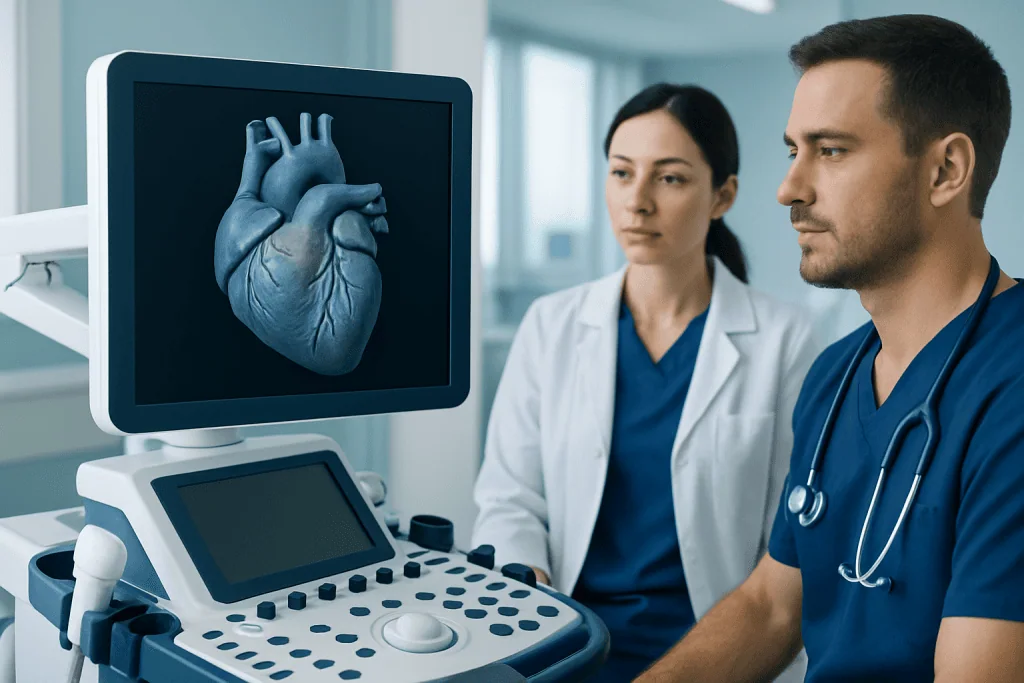
The right atrium is the chamber that receives oxygen-poor blood returning from the body through large veins. It acts as a collecting room, passing blood through the tricuspid valve into the right ventricle. The right ventricle then pumps that blood through the pulmonary valve and into the pulmonary arteries, which carry it to the lungs.
After blood has picked up oxygen in the lungs, it returns to the left atrium through the pulmonary veins. The left atrium passes blood through the mitral valve into the left ventricle. The left ventricle is the heart’s strongest pumping chamber because it must push blood through the aortic valve into the aorta and out to the whole body.
Each chamber has a different wall thickness based on its task. The atria have thinner walls because they mainly receive blood and move it a short distance into the ventricles. The ventricles, especially the left ventricle, have thicker muscular walls because they generate more force.
- Right atrium: receives blood returning from the body
- Right ventricle: pumps blood to the lungs
- Left atrium: receives oxygen-rich blood from the lungs
- Left ventricle: pumps oxygen-rich blood to the body
How the chambers work together with valves and electrical signals

The chambers do not work alone. Four heart valves help blood move in the correct direction and reduce backward flow. These are the tricuspid, pulmonary, mitral, and aortic valves. When they open and close properly, blood passes smoothly from one chamber to the next and then out to the lungs or body.
The heart’s electrical system also guides the chambers. A natural pacemaker in the right atrium starts an electrical impulse that travels through the atria, causing them to contract. The signal then passes to the ventricles, which contract slightly afterward. This timing helps the ventricles fill before they pump.
When the valves, muscle, and electrical system are coordinated, circulation is efficient. If one part is affected by disease, the chambers may enlarge, weaken, stiffen, or beat out of rhythm. This can reduce how well the heart fills or pumps and may eventually lead to symptoms or complications.
Some conditions involve the heart’s pumping ability directly, such as heart failure, while others affect the muscle itself, including cardiomyopathy. In both cases, understanding the role of each chamber helps explain why symptoms develop and why different tests or treatments may be needed.
Common problems that can affect the heart chambers
Heart chambers can be affected by many medical conditions. High blood pressure can make the left ventricle work harder over time, causing thickening or stiffness. Coronary artery disease can reduce blood flow to the heart muscle, weakening a chamber’s pumping function. Valve disorders may overload a chamber because blood leaks backward or faces resistance moving forward.
Heart rhythm disorders can also affect chamber function. For example, atrial fibrillation changes the normal rhythm of the atria and may reduce how effectively they move blood. Congenital heart defects present from birth may alter the structure of one or more chambers or the walls between them. Infections, inflammation, and certain systemic diseases may also affect the heart muscle.
When a chamber becomes enlarged, weakened, or unusually thick, the heart may not pump or fill efficiently. This may lead to shortness of breath, reduced exercise tolerance, tiredness, swelling in the legs, palpitations, or dizziness. Some people have mild abnormalities without symptoms, while others need ongoing care.
Examples include enlargement of the atria from longstanding pressure changes, or reduced ventricular pumping strength in conditions such as arrhythmia-related heart disease or cardiomyopathy. In selected cases, doctors may recommend advanced imaging, medication, catheter-based procedures, or surgery depending on the cause.
Symptoms that may suggest a chamber-related heart problem
Many people asking about heart chambers want to know when anatomy becomes a health concern. A problem affecting one or more chambers may cause symptoms, but symptoms alone cannot identify the exact cause. That is why evaluation by a qualified clinician is important, especially if symptoms are new, persistent, or worsening.
Possible symptoms include breathlessness during activity or when lying flat, chest pressure or discomfort, unusual fatigue, swelling in the ankles or abdomen, rapid or irregular heartbeat, fainting, or reduced ability to exercise. Some people notice only subtle changes, such as becoming more easily tired than usual or feeling their heart race.
In children, signs may be different and can include poor feeding, sweating with feeds, bluish lips, poor growth, or tiring quickly during activity. Older adults may present with weakness, confusion, dizziness, or swelling rather than classic chest symptoms.
- Shortness of breath
- Palpitations or irregular pulse
- Fatigue or reduced stamina
- Swelling in the legs, feet, or abdomen
- Chest discomfort
- Dizziness or fainting
How doctors diagnose problems involving heart chambers
Diagnosis usually begins with a medical history and physical examination. A doctor may ask about symptoms, blood pressure, exercise tolerance, family history, previous infections, and any known heart disease. During the examination, they may listen for murmurs, irregular rhythm, or signs of fluid retention.
One of the most useful tests for assessing heart chambers is an echocardiogram, an ultrasound scan that shows chamber size, wall thickness, valve movement, and pumping function. This can help identify enlargement, weakness, stiffness, congenital abnormalities, or valve-related strain. An electrocardiogram may also be used to evaluate heart rhythm and signs of chamber enlargement.
Additional testing depends on the situation. Blood tests may look for markers of heart strain or related medical issues. Chest X-ray, stress testing, CT, or MRI may provide more detail. If a rhythm problem is suspected, doctors may recommend longer-term monitoring. In some cases, invasive studies or coronary imaging may be needed to guide treatment.
When symptoms point to structural or blood flow issues, care may involve cardiology evaluation and noninvasive imaging such as echocardiography. If a blocked artery is suspected as a cause of weakened chamber function, a doctor may consider coronary angiography in appropriate patients.
Treatment options and long-term care
Treatment depends on why a chamber is not working normally. The goal may be to improve pumping, reduce pressure on the heart, control rhythm, repair a valve problem, treat blocked arteries, or manage a congenital condition. Some people need only monitoring, while others need medications or procedures.
Medicines may help the heart pump more effectively, reduce excess fluid, lower blood pressure, slow an abnormal rhythm, or decrease strain on the chambers. Procedures can include catheter-based treatments for rhythm problems or narrowed valves, and surgery may be needed for some valve disorders, congenital defects, or advanced coronary disease. Severe cases may require specialized heart failure care.
Lifestyle measures remain important even when treatment is medical or surgical. Doctors often recommend stopping smoking, limiting excess salt, maintaining a healthy weight, staying physically active as advised, controlling diabetes, and treating high blood pressure or sleep apnea. Follow-up care helps monitor whether chambers are stable, improving, or under new strain.
For some patients, treatment may involve heart valve surgery or coordinated care from multiple specialists. Near the end of the care pathway, some international patients choose centers such as Acibadem International, where multidisciplinary specialists in JCI-accredited hospitals diagnose and treat heart conditions affecting the chambers.
Prevention, self-care, and when to seek medical care
People cannot usually prevent every disorder that affects the heart chambers, especially congenital conditions or inherited cardiomyopathies. However, many risks can be reduced by protecting overall heart health. This includes managing blood pressure, cholesterol, and blood sugar; avoiding tobacco; limiting excess alcohol; staying active; and following a balanced eating pattern.
Self-care also means paying attention to symptoms and keeping regular checkups if there is known heart disease, a strong family history, or previous valve or rhythm problems. People with prescribed heart medicines should take them as directed and ask a doctor before starting supplements, decongestants, or exercise programs that may affect the heart.
Medical care should be sought promptly for new or worsening shortness of breath, chest pain, fainting, a very fast or irregular heartbeat, unexplained swelling, or sudden severe weakness. Emergency care is especially important for chest pressure, severe breathing difficulty, bluish lips, or symptoms of stroke such as facial droop, speech difficulty, or weakness on one side.
If symptoms are mild but persistent, such as palpitations, fatigue, or reduced exercise tolerance, arranging a nonurgent medical evaluation is still a sensible step. Early assessment can identify whether the issue involves the heart chambers, valves, rhythm, lungs, or another cause and can support timely treatment.
Frequently asked questions
How many chambers are in the heart?
There are four chambers in the heart. The right and left atria are the two upper chambers, and the right and left ventricles are the two lower chambers.
What is the difference between the atria and ventricles?
The atria mainly receive blood returning to the heart, while the ventricles pump blood out of the heart. Ventricles have thicker muscle walls because they must generate more force, especially the left ventricle.
Why is the left ventricle thicker than the other chambers?
The left ventricle pumps blood to the entire body, so it needs more muscular strength than the other chambers. Its thicker wall helps create the pressure needed for this larger workload.
Can a person have a problem in one heart chamber without knowing it?
Yes, some chamber abnormalities cause no symptoms at first and may be found during a routine examination or imaging test. Over time, however, they can lead to symptoms such as fatigue, palpitations, breathlessness, or swelling.
Which test best shows the heart chambers?
An echocardiogram is one of the most common and useful tests for viewing the heart chambers. It can show chamber size, wall motion, valve function, and how well the heart pumps blood.
Do all heart chamber problems need surgery?
No, treatment depends on the cause and severity of the problem. Many conditions can be managed with monitoring, lifestyle measures, or medication, while others may need catheter-based procedures or surgery.
References
- American Heart Association
- National Heart, Lung, and Blood Institute
- Centers for Disease Control and Prevention
- European Society of Cardiology
- Mayo Clinic
This article is for general information only and is not a substitute for professional medical advice. Please consult a qualified doctor about your individual situation.
Explore treatments in Turkey — costs, top hospitals & a free quote
JCI-accredited · board-certified surgeons · reply within 24h
Add us as a Preferred Source to see more of our trusted health content across Google Search, AI Overviews and Discover.
Check your numbers in seconds
BMI, calories, due date, blood pressure and 30+ more clinical calculators — free, instant, doctor-reviewed ranges.