

Glaucoma
Glaucoma treatment aims to lower eye pressure and protect the optic nerve to slow or prevent vision loss. Care may include eye drops, laser therapy, or surgery depending on disease severity.
Quick answer
Glaucoma treatment lowers pressure inside the eye to protect the optic nerve and help slow or prevent vision loss. At Acibadem in Turkey, care is based on the type and stage of glaucoma and may include eye drops, laser procedures, or surgery, with ophthalmology teams monitoring the condition over time.
Glaucoma Care Begins With Protecting the Vision You Still Have
Glaucoma is often described as a “silent” eye disease because many people do not notice symptoms until vision has already been affected. For patients and families, this can be unsettling. You may have been told that your eye pressure is high, that your optic nerve looks damaged, or that a visual field test shows changes you cannot yet perceive in daily life. You may also be wondering whether eye drops will be enough, whether laser treatment is painful, or whether surgery is necessary.
The most important point is that glaucoma can usually be managed, especially when it is diagnosed and treated early. Treatment cannot restore vision that has already been permanently lost, but it can lower the risk of further damage by reducing pressure inside the eye and protecting the optic nerve. This makes timely, carefully planned care essential. The goal is not only to lower a number on an eye pressure test, but to preserve functional vision for reading, driving, working, recognizing faces, moving safely, and maintaining independence.
For international patients considering glaucoma treatment abroad, the decision involves both medical and practical questions. You need a precise diagnosis, clear explanation of your treatment options, access to modern diagnostic and surgical technology, and a care team that can coordinate appointments efficiently. At Acibadem, glaucoma care is planned around the individual patient: the type of glaucoma, the level of optic nerve damage, eye pressure trends, previous treatments, general health, lifestyle, and the patient’s ability to continue follow-up after returning home.
What Is Glaucoma Treatment?
Glaucoma treatment is medical, laser, or surgical care designed to reduce intraocular pressure, commonly called eye pressure, and slow damage to the optic nerve. The optic nerve carries visual information from the eye to the brain. In glaucoma, this nerve becomes damaged, most often because the pressure inside the eye is higher than the nerve can tolerate. Some patients develop glaucoma even with eye pressure that appears to be within the statistically normal range, which is why optic nerve evaluation is as important as pressure measurement.
Eye pressure is related to the production and drainage of a clear fluid inside the eye called aqueous humor. This fluid is not the same as tears. It is produced inside the eye and normally drains through small channels. If drainage is reduced or blocked, pressure can rise. Over time, increased or poorly tolerated pressure may damage optic nerve fibers, leading first to loss of peripheral vision and, in advanced cases, central vision.
Treatment may include prescription eye drops that reduce fluid production or improve drainage, laser procedures that help fluid exit the eye more effectively, or surgery that creates new drainage pathways or implants tiny drainage devices. In some cases, treatment may combine these approaches. The best option depends on the form of glaucoma, the severity of disease, the target eye pressure needed to protect the optic nerve, and how the eye has responded to previous therapy.
Glaucoma treatment is usually long-term. Even after successful pressure reduction, patients require monitoring because glaucoma can progress silently. A well-structured treatment plan includes regular measurement of eye pressure, optic nerve imaging, visual field testing, and adjustment of therapy when needed.
Who May Need Glaucoma Treatment?
Patients may need glaucoma treatment when tests show elevated eye pressure, optic nerve damage, visual field loss, or an eye structure that places them at risk for pressure spikes. Some people are diagnosed during a routine eye examination before they notice any symptoms. Others seek care because they have blurred vision, halos around lights, eye pain, headache, nausea, or sudden vision disturbance, especially in acute forms of glaucoma that require urgent treatment.
Open-angle glaucoma, the most common form in many adult populations, often develops gradually. Patients may feel completely well. Reading vision can remain normal for a long time while peripheral vision slowly narrows. This is why glaucoma screening is especially important for people over 40, those with a family history of glaucoma, people with diabetes or high myopia, patients who use steroid medications, and individuals with previous eye injury or eye surgery.
Angle-closure glaucoma is different. It can occur when the drainage angle inside the eye becomes narrow or blocked. Some patients have intermittent symptoms such as eye discomfort, temporary blurry vision, colored halos, or headache in dim light. A sudden angle-closure attack can cause severe eye pain, redness, nausea, vomiting, and rapid vision loss. This is an emergency because high pressure can damage the optic nerve quickly.
Diagnosis begins with a comprehensive ophthalmologic examination. The ophthalmologist measures intraocular pressure, examines the optic nerve, evaluates the drainage angle, checks corneal thickness, and assesses the retina and visual function. Common diagnostic tests may include optic nerve imaging, visual field testing, optical coherence tomography, gonioscopy to view the drainage angle, pachymetry to measure corneal thickness, and photography to document structural changes over time.
Patients who already have a diagnosis may seek treatment because their eye pressure remains above target despite medication, they cannot tolerate drops, they have difficulty using drops consistently, or test results show progression. Others come for a second opinion before laser therapy or surgery, particularly if both eyes are involved or if prior treatments have not achieved stable control.
Conditions and Indications Addressed by Glaucoma Treatment
Glaucoma treatment is not a single procedure for a single disease. It covers a group of conditions that damage the optic nerve through pressure-related mechanisms or impaired fluid drainage. Identifying the exact type of glaucoma is essential because treatment choices differ.
Primary open-angle glaucoma is usually chronic and progressive. The drainage angle appears open, but fluid does not drain efficiently enough. Treatment often begins with eye drops or laser therapy, with surgery considered when pressure control is insufficient or the disease is advanced.
Normal-tension glaucoma occurs when optic nerve damage progresses even though eye pressure readings are not very high. These patients still benefit from pressure lowering, but the ophthalmologist may also evaluate circulation, blood pressure patterns, sleep apnea risk, migraine history, and other factors that may affect optic nerve health.
Angle-closure glaucoma involves a narrow or blocked drainage angle. Laser treatment to create a small opening in the iris may be recommended to prevent or treat angle closure. In some cases, cataract surgery or other procedures may be part of the treatment plan because lens position can contribute to angle narrowing.
Secondary glaucoma develops because of another eye or medical condition. Causes may include eye trauma, inflammation, steroid use, advanced cataract, diabetes-related eye disease, pigment dispersion, pseudoexfoliation syndrome, tumors, or previous eye surgery. Treatment must address both pressure control and the underlying cause when possible.
Congenital or childhood glaucoma is less common but requires specialized evaluation. Infants or children may have enlarged eyes, cloudy corneas, light sensitivity, tearing, or vision concerns. Surgical treatment is often needed, and early diagnosis is especially important for visual development.
Advanced glaucoma requires careful planning because the remaining optic nerve reserve may be limited. In these cases, the target pressure is often lower, and treatment may need to be more intensive. The physician balances the need for stronger pressure reduction with the safety of the eye and the patient’s overall condition.
How Glaucoma Treatment Is Performed
Glaucoma care begins with confirming the diagnosis and defining the target pressure for each eye. This target is not the same for every patient. An eye with early glaucoma may require moderate pressure reduction, while an eye with advanced optic nerve damage may need lower pressure to reduce the risk of progression. The treatment plan is adjusted as new test results become available.
Preparation and Evaluation
Before treatment, the ophthalmologist reviews your medical history, current medications, allergies, previous eye surgery, and any family history of glaucoma. If you are using glaucoma drops, the care team will review the exact names, dosing schedule, and whether you experience redness, burning, dry eye, shortness of breath, fatigue, or other side effects. This is especially important for patients with asthma, heart rhythm problems, low blood pressure, or certain neurologic conditions, because some eye drops may not be appropriate.
Diagnostic imaging helps the physician evaluate the optic nerve and retinal nerve fiber layer. Visual field testing measures how well you see in different areas of your field of vision. Gonioscopy allows the doctor to examine the eye’s drainage angle directly. These tests are generally noninvasive. They provide a baseline for future comparison, which is crucial because glaucoma management depends on detecting change over time.
Medication-Based Treatment
Many patients begin with prescription eye drops. These medications work by reducing fluid production inside the eye, increasing fluid outflow, or both. Some are used once daily; others require more frequent dosing. Combination drops may reduce the number of bottles needed. For some patients, oral medication may be used temporarily, especially when eye pressure must be lowered quickly, but long-term oral therapy is less common because of potential systemic side effects.
Correct use of drops matters. Patients are often taught how to place drops without touching the bottle tip to the eye, how to wait between different medications, and how to gently close the eye or press near the inner corner after application to reduce medication absorption into the bloodstream. If drops are difficult to use, expensive in the patient’s home country, or causing side effects, laser or surgical options may be considered earlier.
Laser Treatment
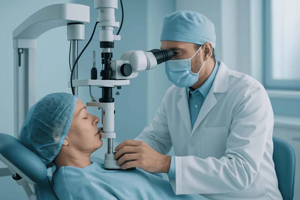
Laser therapy can be used in several glaucoma situations. In open-angle glaucoma, laser treatment may improve drainage through the natural drainage tissue. It is typically performed in an outpatient setting with numbing drops. Patients sit at a laser microscope similar to the equipment used during an eye examination. The procedure usually takes a short time, and most patients return to normal light activities soon afterward.
In narrow-angle or angle-closure glaucoma, a laser may be used to create a tiny opening in the iris, allowing fluid to move more freely and reducing the risk of angle closure. This procedure may be recommended preventively in an eye with a dangerously narrow angle or urgently in certain angle-closure situations.
Laser treatment is not the same as eye surgery in an operating room, and it does not involve removing the eye or making a large incision. Some patients feel brief discomfort or see flashes of light. Anti-inflammatory drops may be prescribed afterward. Eye pressure is usually checked after the procedure, and the physician explains whether current glaucoma drops should be continued, reduced, or adjusted.
Surgical Treatment
Surgery is considered when medications and laser therapy do not lower pressure enough, when glaucoma is progressing, when pressure is very high, or when a patient cannot safely or reliably continue medication. The purpose of glaucoma surgery is to create a controlled way for fluid to leave the eye, lowering pressure on the optic nerve.
Several surgical approaches may be used. Traditional filtering surgery creates a new drainage pathway under the surface of the eye. Tube or shunt procedures use a small drainage implant to help fluid exit the eye in a controlled manner. Minimally invasive glaucoma procedures may be appropriate for selected patients, often those with mild to moderate disease or those also undergoing cataract surgery. The choice depends on the type and severity of glaucoma, the anatomy of the eye, previous surgeries, cataract status, and the degree of pressure reduction needed.
Glaucoma surgery is usually performed with local anesthesia and sedation, although the anesthesia plan may vary. The procedure may take less than an hour in some cases, while more complex eyes require longer. Patients generally go home the same day, but close follow-up is necessary, especially in the early healing period. Postoperative visits allow the surgeon to check eye pressure, inflammation, healing of the drainage area, and signs of complications.
Technology Used in Modern Glaucoma Care
Modern glaucoma management relies on precise measurement and long-term comparison. Diagnostic technologies may include high-resolution optic nerve and retinal nerve fiber imaging, computerized visual field analysis, corneal thickness measurement, drainage angle assessment, and retinal photography. These tools help the ophthalmologist distinguish stable disease from progression and select treatment intensity appropriately.
During procedures, microscopes, microsurgical instruments, laser platforms, and fine drainage devices may be used depending on the treatment. The practical value for the patient is accuracy: better visualization of delicate eye structures, more controlled treatment delivery, and more individualized planning. Technology supports decision-making, but it does not replace physician judgment or careful follow-up.
Recovery After Treatment
Recovery depends on the treatment type. After medication changes, patients may notice mild irritation, redness, or changes in eyelash growth or eye color depending on the medication. Follow-up is needed to confirm that pressure has reached the intended level.
After laser therapy, mild irritation, light sensitivity, or blurred vision may occur for a short period. Many patients resume routine activities quickly, although they may be advised to avoid strenuous activity for a brief time. After surgery, recovery is more structured. Patients usually use antibiotic and anti-inflammatory drops, avoid rubbing the eye, and limit heavy lifting, swimming, or dusty environments until cleared by the surgeon. Vision may fluctuate during healing, and eye pressure may need careful adjustment.
Why Acting Early Matters
Glaucoma-related vision loss is usually permanent. Once optic nerve fibers are damaged, current treatments cannot fully regenerate them. This is why early diagnosis and consistent treatment are central to preserving vision. Waiting until symptoms appear can be risky because many forms of glaucoma progress silently for years before noticeable vision loss develops.
Delay can allow peripheral vision loss to advance. Patients may not realize what has been lost because the brain adapts and the unaffected areas of vision compensate. Over time, tasks such as driving, walking on stairs, navigating crowded places, and seeing objects from the side may become more difficult. In advanced glaucoma, central vision can also be threatened.
In acute angle-closure glaucoma, delay is particularly dangerous. Very high eye pressure can cause rapid optic nerve injury and damage to other eye structures. Severe eye pain, redness, blurred vision, halos, headache, nausea, or vomiting should be treated as urgent symptoms.
Acting early does not always mean having surgery immediately. It means completing a proper evaluation, understanding your risk, starting appropriate therapy, and monitoring whether the disease is stable. For many patients, timely drops or laser therapy may delay or reduce the need for more invasive treatment. For others, early surgery may be the safer choice if the optic nerve is already vulnerable or pressure is dangerously high.
Benefits of Glaucoma Treatment
The main benefits of glaucoma treatment are related to protecting the optic nerve, preserving daily function, and reducing the risk of future vision loss.
| Benefit | What It Means for You |
|---|---|
| Lower eye pressure | Reducing intraocular pressure decreases stress on the optic nerve and is the most proven way to slow glaucoma progression. |
| Protection of remaining vision | Treatment aims to preserve the vision you still have, including peripheral vision that is important for mobility and safety. |
| Individualized treatment options | Care may include drops, laser therapy, surgery, or a combination, depending on your diagnosis, severity, and response to treatment. |
| Reduced medication burden in selected patients | Laser or surgical treatment may help some patients reduce reliance on multiple eye drops, although ongoing monitoring remains necessary. |
| Better long-term disease monitoring | Regular imaging and visual field testing help detect progression earlier and guide timely changes in treatment. |
Recovery Timeline After Glaucoma Treatment
Recovery varies according to whether treatment involves eye drops, laser therapy, or surgery, but the following timeline gives a general sense of what many patients can expect.
| Time Period | What Patients Can Expect |
|---|---|
| Day 1 | After laser treatment, patients may have mild irritation or blurred vision. After surgery, the eye is usually protected, drops are started, and activity restrictions are explained. |
| First Week | Follow-up visits are important to check eye pressure and healing. Patients may need to avoid heavy lifting, swimming, eye rubbing, and dusty environments, especially after surgery. |
| First Month | Vision may fluctuate after surgery as the eye heals and inflammation improves. Medication schedules may be adjusted based on pressure readings and clinical findings. |
| Longer Term | Ongoing monitoring with pressure checks, optic nerve imaging, and visual field testing remains essential because glaucoma is a chronic condition. |
Factors That Influence Outcomes
A good glaucoma result depends on more than one pressure reading. The most important factors include the type of glaucoma, the amount of optic nerve damage present at diagnosis, baseline eye pressure, the target pressure needed, the patient’s age, corneal thickness, drainage angle anatomy, and whether other eye diseases are present.
Severity at the time treatment begins is especially important. Patients diagnosed early often have more optic nerve reserve, which may make long-term vision preservation more achievable. Advanced glaucoma can still be treated, but the margin for additional damage is smaller. In these cases, follow-up must be particularly careful.
Adherence to treatment strongly affects outcomes. Eye drops must be used consistently and correctly. If a patient forgets doses, cannot tolerate side effects, or has difficulty obtaining medication after returning home, the treatment plan may need to be changed. For international patients, it is useful to discuss which medications are available in the home country and whether a written treatment plan can be shared with the local ophthalmologist.
The eye’s healing response also matters after surgery. Some eyes scar more aggressively, which can reduce the effectiveness of a drainage procedure. Previous eye surgery, inflammation, trauma, diabetes-related changes, or certain secondary glaucomas may make surgical planning more complex. The surgeon may adjust technique, postoperative medications, and visit frequency based on these risks.
Other medical conditions can influence glaucoma management. Low blood pressure at night, vascular disease, sleep apnea, migraine, diabetes, and steroid use may affect optic nerve vulnerability or eye pressure. A careful medical history helps the ophthalmologist select safer medications and identify factors that may need coordination with other physicians.
Finally, outcomes depend on monitoring over time. Glaucoma is assessed through patterns: pressure trends, optic nerve appearance, imaging changes, and visual field progression. A treatment that works well initially may need modification later. This is not a failure of care; it is part of managing a chronic optic nerve disease responsibly.
Why International Patients Choose Acibadem for Glaucoma Care
International patients seeking glaucoma treatment often need more than a single appointment. They need a coordinated diagnostic pathway, clear communication, efficient scheduling, and a treatment plan that can continue safely after they return home. Acibadem’s approach is designed for this reality.
Care is provided in JCI-accredited hospitals with established quality and safety processes. Ophthalmologists evaluate glaucoma using modern diagnostic tools that support detailed assessment of eye pressure, optic nerve structure, visual field function, and drainage angle anatomy. When cases are complex, care may involve discussion among relevant specialists, such as glaucoma surgeons, retinal specialists, cataract surgeons, cornea specialists, anesthesiology teams, and internal medicine physicians when systemic conditions affect treatment choices.
For patients traveling from the United States or other countries, Acibadem International supports the practical side of care. Services may include appointment coordination, medical record transfer, language interpretation in more than 20 languages, assistance with hospital processes, and communication of treatment plans. This support is particularly valuable for glaucoma patients because prior test results, medication lists, and follow-up needs must be clearly understood.
Personalized treatment planning is central. Some patients are best managed with optimized eye drops and follow-up testing. Others may benefit from laser therapy during the same travel period, if clinically appropriate. Patients with advanced or uncontrolled glaucoma may require surgery and a longer stay for early postoperative monitoring. The care team explains the expected timeline before treatment so patients can plan travel, accommodation, and return visits realistically.
Acibadem’s physicians follow evidence-based international treatment principles while adapting care to the individual eye. This includes defining a target pressure, choosing therapy based on disease severity and anatomy, monitoring for progression, and discussing benefits and risks in understandable terms. For many international patients, a second opinion can be especially helpful when they have been advised to undergo surgery, when they have conflicting test results, or when glaucoma is progressing despite treatment.
Technology is used to support precision and continuity. Imaging and visual field results help document the condition before and after treatment. Surgical microscopes, laser systems, and microsurgical techniques allow ophthalmologists to work within very delicate eye structures. Just as important, patients receive practical instructions: how to use drops, what symptoms require urgent attention, what activities to avoid after procedures, and how to continue care with an ophthalmologist at home.
The experience of receiving care abroad should feel medically rigorous and personally organized. For glaucoma, this means that diagnosis, treatment, recovery, and follow-up planning are connected. The patient should leave not only with treatment completed, when appropriate, but also with a clear understanding of the condition and the next steps needed to protect vision over time.
Taking the Next Step
If you have been diagnosed with glaucoma, have elevated eye pressure, or are concerned about optic nerve changes, a timely specialist evaluation can help clarify your risk and treatment options. Glaucoma care is most effective when it is proactive, measured, and individualized. Whether you need confirmation of a diagnosis, adjustment of medications, laser treatment, surgery, or a second opinion, the first step is a detailed review of your eye health and previous test results.
International patients may request a consultation or second opinion by sharing available records, including eye pressure measurements, visual field tests, optic nerve imaging, medication lists, previous surgery details, and any relevant medical history. The care team can then advise what additional testing may be needed and what treatment pathways may be appropriate.
This information is general and is not a substitute for professional medical advice. Diagnosis and treatment recommendations should always be made by a qualified ophthalmologist after an individual examination.
Preparation
- Before treatment, an ophthalmologist performs a comprehensive eye examination, including eye pressure measurement, optic nerve evaluation, and visual field testing. Patients should share all medications, allergies, and previous eye surgeries. Contact lenses may need to be removed before laser or surgical procedures.
Aftercare
- After treatment, prescribed eye drops should be used exactly as instructed to control pressure and reduce inflammation. Patients should avoid rubbing the eye and attend follow-up visits to monitor eye pressure and optic nerve health. Temporary blurred vision or mild discomfort may occur after laser or surgical care.
Turkey vs UK, Germany & USA
Glaucoma care is planned to lower eye pressure and protect the optic nerve, with the treatment choice depending on the type and severity of disease. Comparing destinations can help patients understand how hospital factors, specialist expertise, waiting time, and travel logistics may influence the overall experience and cost.
The overall cost of glaucoma care can vary by country because treatment may involve ongoing diagnostics, medicines, laser procedures, or surgery, as well as follow-up monitoring.
| Factor | Turkey | UK | Germany | USA |
|---|---|---|---|---|
| Price drivers | Often shaped by whether care includes consultation, imaging, laser or surgery, medication, and follow-up in an international patient package. | Private care costs vary by clinic and procedure; public pathways may involve referral and eligibility processes. | Costs depend on specialist centre, diagnostic workup, procedure type, and whether care is public or private. | Costs are strongly influenced by provider, insurance status, facility fees, imaging, procedure choice, and medication plans. |
| Hospital and surgeon factors | International hospitals may offer ophthalmology teams experienced in glaucoma assessment, laser, and surgical care. | Care may be delivered in private eye hospitals, NHS-affiliated services, or consultant-led clinics. | Care is commonly provided through university hospitals, private clinics, and specialist ophthalmology centres. | Care may be provided by academic eye centres, private ophthalmology groups, or hospital-based surgical units. |
| Accreditation and quality | JCI-accredited hospitals are available, with structured international patient pathways and documented clinical protocols. | Quality oversight depends on the provider type and national healthcare regulation. | Providers follow national healthcare standards, with quality systems varying by hospital and clinic. | Accreditation and quality frameworks vary by hospital, clinic, and surgical centre. |
| Typical waiting time | Private international scheduling may allow coordinated appointments for diagnosis and treatment planning. | Public pathways may involve waiting lists; private appointments may be faster depending on availability. | Waiting time varies by region, provider, and whether the pathway is public or private. | Scheduling can be prompt in private settings, but insurance approvals and specialist availability may affect timing. |
| Travel and language logistics | International patient teams may help with appointment planning, translation, airport transfers, and hotel coordination. | English-speaking care is standard; international patients usually arrange travel and accommodation separately. | Interpreter support may be available, but arrangements vary by centre and language. | English-speaking care is standard; travel, accommodation, and insurance administration can add complexity. |
| Package inclusions | Packages may combine consultation, diagnostic tests, procedure planning, hospital coordination, and selected support services. | Private quotes may be itemised, with diagnostics, procedure fees, medicines, and follow-up billed separately. | Quotes may separate medical tests, physician fees, facility charges, procedure fees, and aftercare. | Billing may be highly itemised, including specialist fees, facility charges, imaging, medicines, and follow-up visits. |
- What affects your final cost: glaucoma type and severity, eye pressure level, optic nerve status, visual field findings, diagnostic imaging, need for laser or surgery, type of implant or device if used, anaesthesia requirements, hospital stay if needed, medication plan, follow-up schedule, translation support, and travel arrangements.
Compare your options
Glaucoma treatment options aim to reduce eye pressure and preserve remaining vision. Suitability is decided by a specialist after eye pressure measurement, optic nerve assessment, visual field testing, and evaluation of overall eye health.
| Option | What it is | Typical use | Key considerations |
|---|---|---|---|
| Prescription eye drops | Medicines used regularly to lower eye pressure by reducing fluid production or improving fluid drainage. | Commonly used as initial or long-term treatment for many forms of open-angle glaucoma. | Requires consistent use, monitoring for side effects, and periodic assessment to confirm pressure control. |
| Laser treatment | A procedure that improves fluid drainage or treats the drainage angle depending on glaucoma type. | May be used when drops are not enough, not tolerated, or when a specialist recommends laser as part of the care plan. | Usually performed as an outpatient procedure, but pressure and optic nerve status still require ongoing monitoring. |
| Trabeculectomy | A filtering surgery that creates a new drainage pathway to lower eye pressure. | Often considered for more advanced glaucoma or cases where medicines and laser do not provide adequate control. | Requires close follow-up, wound healing management, and careful discussion of benefits and risks. |
| Glaucoma drainage device | A small implant used to help drain fluid from the eye and reduce pressure. | May be considered in complex glaucoma, eyes with previous surgery, or when other options are less suitable. | Choice depends on eye anatomy, prior treatments, target pressure, and surgeon assessment. |
| Minimally invasive glaucoma surgery | Smaller-incision procedures designed to improve drainage with a less invasive approach than traditional filtering surgery. | May be considered in selected patients, sometimes alongside cataract surgery. | Not suitable for every glaucoma stage; expected pressure reduction and long-term plan should be discussed with the specialist. |
| Cataract and glaucoma combined care | Cataract surgery performed with a glaucoma procedure when clinically appropriate. | May be used when cataract affects vision and glaucoma also needs pressure management. | Planning depends on lens status, glaucoma severity, target pressure, and expected recovery needs. |
Trusted care for international patients
General information only — not medical or financial advice. Final costs depend on the factors above and your individual case; request a free, personalised quote.
Doctors Performing This Treatment

Prof. Dr. Altan Göktaş
Ophthalmology
Prof. Dr. Ayşe Öner
Ophthalmology
Prof. Dr. Banu Coşar
Ophthalmology
Prof. Dr. Berna Özkan
Ophthalmology
Prof. Dr. Dilaver Erşanlı
Ophthalmology
Prof. Dr. Dilek Güven
Ophthalmology
Prof. Dr. G. Ertuğrul Mirza
Ophthalmology
Prof. Dr. Gökhan Pekel
Ophthalmology
Prof. Dr. Haluk Esgin
Ophthalmology
Prof. Dr. Mehdi S. Öğüt
Ophthalmology
Prof. Dr. Muhsin Eraslan
Ophthalmology
Prof. Dr. Müslime Akbaba
Ophthalmology
Prof. Dr. Nazan Bengüdeniz Erda
Ophthalmology
Prof. Dr. Sarper Karaküçük
Ophthalmology
Prof. Dr. Selçuk Sızmaz
Ophthalmology
Prof. Dr. Seyhan Topbaş
Ophthalmology
Prof. Dr. Solmaz Balcı Akar
Ophthalmology
Prof. Dr. Özgül Altıntaş
Ophthalmology
Prof. Dr. Özlem Şahin
Ophthalmology
Assoc. Prof. Dr. Özgür Çakıcı
Ophthalmology
Dr. Akın Banaz
Ophthalmology
Dr. Alpaslan Koç
Ophthalmology
Dr. Buket Ayoğlu
Ophthalmology
Dr. Ercan Paşaoğlu
OphthalmologyMedical Units
Available at These Hospitals












Frequently Asked Questions
What affects the cost of glaucoma treatment?
Cost is influenced by the type and severity of glaucoma, diagnostic tests, whether treatment involves drops, laser, or surgery, the need for implants or devices, anaesthesia, follow-up visits, and travel or language support for international patients.
How can I get a personalised quote for glaucoma care in Turkey?
You can request a free consultation and share recent eye pressure measurements, visual field tests, optic nerve imaging, medication history, and any prior surgery details. A specialist review helps determine the likely treatment pathway and a personalised quote.
Is glaucoma treatment usually a single procedure or ongoing care?
Glaucoma is usually a long-term condition that needs monitoring even after successful laser or surgery. The care plan may include regular pressure checks, optic nerve assessment, visual field testing, and medication adjustments if needed.
Does a package usually include glaucoma medicines and follow-up?
Package content varies by hospital and treatment plan. Some packages may include consultation, diagnostic testing, procedure coordination, and selected follow-up, while medicines, additional tests, or extended monitoring may be quoted separately.
How does the treatment option affect the quote?
Eye drops, laser procedures, filtering surgery, drainage devices, and minimally invasive glaucoma procedures involve different facility needs, equipment, surgeon time, and follow-up requirements. Suitability and cost can only be confirmed after specialist assessment.