Liver Cysts and Benign Liver Lesions: When to Monitor or Treat

Most simple liver cysts and benign liver lesions are non-cancerous and are found incidentally during imaging for another reason. Ultrasound, CT, and MRI help doctors distinguish simple cysts, hemangiomas, focal nodular hyperplasia, and liver adenomas from lesions that need further evaluation.
Key Takeaways
- Most simple liver cysts and benign liver lesions are non-cancerous and are found incidentally during imaging for another reason.
- Ultrasound, CT, and MRI help doctors distinguish simple cysts, hemangiomas, focal nodular hyperplasia, and liver adenomas from lesions that need further evaluation.
- Monitoring is often enough when a lesion has typical benign features and the patient has no symptoms.
- Treatment may be considered for large or symptomatic cysts, uncertain imaging findings, or lesions such as some liver adenomas that carry specific risks.
- New abdominal pain, fever, jaundice, unexplained weight loss, or a known liver lesion that changes over time should be discussed with a qualified doctor.
Liver cysts and many benign liver lesions are common findings on ultrasound, CT, or MRI and often do not require treatment. The key is confirming the diagnosis, identifying features that need closer follow-up, and treating only when symptoms or specific risks are present.
Overview
Liver cysts and benign liver lesions are non-cancerous changes within the liver. They are frequently discovered by chance during an abdominal ultrasound, computed tomography, or magnetic resonance imaging scan performed for another reason. For many people, this unexpected finding can feel worrying, but most simple cysts and many benign liver lesions remain stable and never cause health problems.
A liver cyst is a fluid-filled space in the liver. The most common type is a simple liver cyst, which usually has a thin wall, clear fluid, and no solid parts. Benign solid liver lesions include hemangioma, focal nodular hyperplasia, and hepatocellular adenoma, also called liver adenoma. Each has different imaging features, typical patient patterns, and management considerations.
The main goal after a liver cyst or lesion is found is to confirm what it is. Doctors assess its appearance, size, location, symptoms, liver health, medical history, and sometimes blood test results. If the lesion has classic benign features, reassurance or periodic monitoring may be all that is needed. If the diagnosis is unclear, additional imaging or specialist review helps guide safe next steps.
Common Types of Benign Liver Lesions
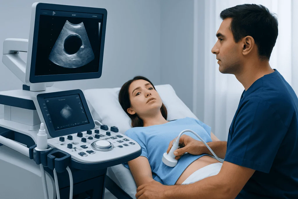
Simple liver cysts are among the most frequent benign liver findings. They are usually round or oval and filled with fluid. Most are small and silent. Larger cysts may occasionally create pressure symptoms, especially if they are located near the liver capsule or close to the stomach, bile ducts, or other nearby structures.
Hemangiomas are benign collections of blood vessels and are the most common benign solid liver tumors. They typically have a very characteristic pattern on contrast-enhanced imaging. In most cases, hemangiomas do not need treatment unless they are very large, symptomatic, or the diagnosis is uncertain.
Focal nodular hyperplasia is a benign overgrowth of normal liver cells and supporting tissue. It is often found in people with otherwise healthy livers and usually does not become cancerous. When imaging findings are typical, focal nodular hyperplasia is generally managed with observation rather than intervention.
Liver adenomas are benign tumors linked in some cases to hormonal factors, certain metabolic conditions, or specific medications. Unlike many other benign liver lesions, some adenomas can bleed or rarely develop concerning changes, so they usually require more individualized assessment. Management may involve stopping contributing medications when appropriate, monitoring, or treatment depending on size, growth, subtype, symptoms, and patient-specific factors.
Symptoms

Most liver cysts and benign liver lesions cause no symptoms. They are often described as incidental findings because the person feels well and the lesion is found during imaging for indigestion, kidney stones, back pain, routine check-ups, or another unrelated reason. Normal liver blood tests are common, especially when the lesion is small and the rest of the liver is healthy.
When symptoms occur, they are usually related to the size or position of the lesion rather than to liver failure. A large cyst or mass may press on surrounding tissues and cause a feeling of fullness in the upper right abdomen, early satiety, bloating, dull discomfort, or a sensation of pressure under the ribs. Symptoms may come and go and can overlap with gallbladder, stomach, or bowel conditions.
Less commonly, a cyst or lesion may become complicated. A cyst can bleed, become infected, rupture, or press on bile ducts, although these events are not typical for simple cysts. Some liver adenomas can bleed, especially when larger or growing. Symptoms that deserve prompt medical attention include sudden severe abdominal pain, fever, yellowing of the skin or eyes, dark urine, fainting, or persistent vomiting.
Causes and Risk Factors
Simple liver cysts are usually congenital, meaning they develop from small bile duct structures before birth, although they may not be detected until adulthood. They are not caused by diet, stress, or ordinary physical activity. Most people with a simple cyst have only one or a few cysts and do not have an inherited disease.
Some people have multiple liver cysts as part of polycystic liver disease, which can occur alone or with polycystic kidney disease. In these situations, cyst number and liver size can increase over time, and care may involve both gastroenterology and nephrology specialists. Family history and kidney imaging may be relevant when many cysts are present.
Benign solid lesions have different associations. Hemangiomas are vascular lesions that may be present for many years without being noticed. Focal nodular hyperplasia is thought to relate to changes in local blood flow within the liver. Liver adenomas may be associated with estrogen-containing medications, anabolic steroid use, certain inherited metabolic disorders, obesity, and metabolic syndrome, although risk factors vary by individual.
Not every cystic liver lesion is a simple cyst. In some regions or travel histories, parasitic hydatid disease may be considered. Cystic tumors and abscesses can also mimic benign cysts on early imaging. This is why doctors interpret imaging together with symptoms, blood tests, exposure history, and the exact appearance of the lesion.
Diagnosis and Imaging
Diagnosis usually begins with imaging. Ultrasound can often identify a simple liver cyst by showing a thin-walled, fluid-filled structure with no internal solid component. It may also detect solid lesions, but contrast-enhanced CT or MRI is often more informative when the type of lesion needs confirmation.
MRI is especially useful for characterizing benign liver lesions because it provides detailed information about tissue structure and blood flow patterns after contrast injection. Hemangiomas, focal nodular hyperplasia, and adenomas often have distinct enhancement patterns. In many cases, high-quality imaging can provide a confident diagnosis without biopsy.
Blood tests may include liver enzymes, bilirubin, markers of inflammation, and tests for viral hepatitis or other liver diseases when clinically relevant. Tumor markers are not used alone to diagnose or exclude cancer, but they may be part of a broader evaluation in selected patients. If infection or parasitic disease is suspected, additional blood tests may be requested.
Biopsy is not routinely needed for classic simple cysts, typical hemangiomas, or typical focal nodular hyperplasia. In fact, biopsy may be avoided for some vascular lesions due to bleeding risk and because imaging is usually sufficient. A multidisciplinary review is helpful when imaging is indeterminate, the patient has a known cancer history, or the lesion changes over time.
When to Monitor
Monitoring is appropriate for many liver cysts and benign liver lesions when imaging features are typical and the person has no concerning symptoms. A simple liver cyst with classic ultrasound findings may not need repeated follow-up. For other benign lesions, follow-up plans depend on the certainty of the diagnosis, lesion size, medical history, and whether the lesion is stable.
Monitoring may include repeat ultrasound, CT, or MRI after an interval chosen by the treating doctor. The purpose is to confirm stability, not to treat the lesion. If the lesion remains unchanged and has benign characteristics, follow-up can often be reduced or stopped according to specialist guidance.
Doctors may recommend closer observation when the lesion is large, newly discovered without prior imaging for comparison, atypical in appearance, or associated with symptoms. Liver adenomas are usually followed more carefully than hemangiomas or focal nodular hyperplasia because management may change if they grow or if risk factors are present.
Patients can support safe monitoring by keeping copies of imaging reports and, when possible, actual scan images. Comparing current and previous images is often more useful than looking at a single report. It helps doctors determine whether a lesion is stable, slowly changing, or in need of additional evaluation.
Treatment Options
Treatment is not needed for most asymptomatic simple cysts, hemangiomas, or focal nodular hyperplasia. When treatment is considered, it is usually because the lesion is causing symptoms, growing, at risk of complications, or cannot be confidently classified as benign. The choice of treatment depends on the lesion type and the patient’s overall health.
For symptomatic simple liver cysts, options may include drainage with sclerotherapy or laparoscopic fenestration, a procedure that opens part of the cyst wall so fluid does not reaccumulate in the same way. Simple aspiration alone is less commonly used as a definitive treatment because fluid may return. If infection, bleeding, or bile duct compression is present, management is tailored to the specific complication.
Hemangiomas are usually observed. Treatment for hemangiomas is uncommon but may be discussed for persistent symptoms clearly related to the lesion, very large lesions with complications, or uncertain diagnosis. Options can include surgery or interventional radiology procedures in carefully selected cases.
Focal nodular hyperplasia rarely requires treatment when the diagnosis is secure. Liver adenomas require individualized care. A doctor may review hormonal medications, metabolic risk factors, pregnancy plans, lesion size, growth, and imaging subtype. Some adenomas are monitored, while others are treated with surgical removal or image-guided therapies when the risk profile suggests that intervention is safer than observation.
Prevention, Self-Care, and When to See a Doctor
There is no proven way to prevent simple liver cysts, hemangiomas, or focal nodular hyperplasia. However, maintaining general liver health is helpful. This includes avoiding excessive alcohol, maintaining a healthy weight, managing diabetes or cholesterol when present, using medications and supplements only as directed, and being vaccinated or screened for viral hepatitis when recommended by a doctor.
People with liver adenomas may be advised to review hormone-related medications or anabolic steroid exposure with their physician. No medication should be stopped without medical guidance, especially if it is used for contraception, hormone therapy, or another health condition. Lifestyle measures that support metabolic health can also be part of long-term care.
A medical review is recommended if a person has persistent upper abdominal discomfort, a sense of fullness, unexplained weight loss, fever, jaundice, sudden severe abdominal pain, or abnormal liver blood tests. Follow-up is also important if a lesion is described as complex, atypical, growing, or indeterminate on the imaging report.
International patients who need evaluation of liver cysts or benign liver lesions can be assessed by multidisciplinary specialists at Acibadem International’s JCI-accredited hospitals, where gastroenterology, radiology, hepatobiliary surgery, and interventional radiology teams can coordinate diagnosis and treatment planning. Care decisions should always be based on the individual patient’s imaging findings, symptoms, and medical history.
Frequently asked questions
Are liver cysts cancer?
Most simple liver cysts are not cancer and do not turn into cancer. They have a typical fluid-filled appearance on imaging. If a cyst has solid areas, thick walls, septations, or other complex features, doctors may recommend further imaging to confirm the diagnosis.
Do all benign liver lesions need follow-up scans?
No. Some lesions with classic benign imaging features may not need ongoing follow-up, especially if the patient has no symptoms. Others, such as uncertain lesions or liver adenomas, may require repeat imaging to confirm stability or guide treatment decisions.
Can a liver cyst cause pain?
A small simple cyst usually does not cause pain. Larger cysts can sometimes create pressure, fullness, or dull discomfort in the upper right abdomen. Sudden severe pain, fever, or jaundice should be assessed promptly because these symptoms may suggest a complication or another abdominal condition.
What is the difference between a liver cyst and a liver hemangioma?
A liver cyst is a fluid-filled space, while a hemangioma is a benign cluster of blood vessels. They look different on ultrasound, CT, and MRI. Both are commonly benign, but management depends on size, symptoms, and how confidently the diagnosis is made.
Is biopsy needed for benign liver lesions?
Biopsy is often not needed when imaging findings are typical for a simple cyst, hemangioma, or focal nodular hyperplasia. MRI or contrast-enhanced CT can usually provide enough information. Biopsy may be considered only when imaging and clinical information do not provide a clear diagnosis.
Can diet or supplements shrink liver cysts?
There is no reliable evidence that a specific diet or supplement can shrink simple liver cysts. A balanced diet, healthy weight, and avoidance of unnecessary alcohol support overall liver health. Patients should discuss supplements with a doctor because some products can affect the liver or interact with medicines.
When is surgery considered for a benign liver lesion?
Surgery may be considered when a lesion causes significant symptoms, grows, bleeds, compresses nearby structures, or cannot be confidently diagnosed as benign. Some liver adenomas may also be removed depending on their size, subtype, growth pattern, and patient-specific risks. The decision is made after specialist review of imaging and overall health.
References
- European Association for the Study of the Liver
- American College of Gastroenterology
- American College of Radiology
- Radiological Society of North America
- Merck Manual Professional Edition
This article is for general information only and is not a substitute for professional medical advice. Please consult a qualified doctor about your individual situation.
Explore treatments in Turkey — costs, top hospitals & a free quote
JCI-accredited · board-certified surgeons · reply within 24h
Add us as a Preferred Source to see more of our trusted health content across Google Search, AI Overviews and Discover.

Gastroenterology
Diagnosis and treatment of the digestive system and liver, with advanced endoscopy.
46 specialists in this unit