

How Doctors Choose the Right Imaging Test for Symptoms

Different imaging tests show different body structures and problems. Symptoms, age, pregnancy status, and medical history all affect scan choice.
Key Takeaways
- Different imaging tests show different body structures and problems.
- Symptoms, age, pregnancy status, and medical history all affect scan choice.
- Doctors balance accuracy, speed, safety, and availability when selecting imaging.
- Not every symptom needs imaging, and sometimes monitoring or blood tests come first.
- Radiologists and referring doctors often work together to choose the most useful test.
Doctors choose imaging tests by matching a person’s symptoms, medical history, and urgency with the strengths and limits of each scan. The goal is to get the clearest useful answer while avoiding unnecessary delay, radiation, or invasive testing.
Overview: Why the “right” imaging test matters
Imaging tests help doctors look inside the body without surgery. They are used to investigate pain, swelling, weakness, shortness of breath, injuries, unusual bleeding, neurological symptoms, and many other concerns. However, there is no single scan that is best for every situation. Each type of imaging provides different information, and choosing the right test can make diagnosis faster and more accurate.
Doctors usually begin with the patient’s symptoms, how long they have been present, where they are located, and whether they suggest an urgent problem. A sudden severe headache, for example, may require a different approach from long-standing mild knee pain. The doctor also considers physical examination findings, blood test results, past medical problems, and whether the person has already had imaging.
The goal is not simply to order the most advanced scan available. Instead, doctors try to select the test that is most likely to answer the clinical question clearly and safely. In some cases, that may be a simple X-ray. In others, it may be an ultrasound, CT scan, MRI, or a more specialized test in radiology.
Imaging decisions are often made collaboratively. The referring physician may consult a radiologist, especially when symptoms are complex or when several scans are possible. This teamwork helps reduce unnecessary testing and improves the chances of finding the real cause of symptoms.
What doctors consider before ordering a scan
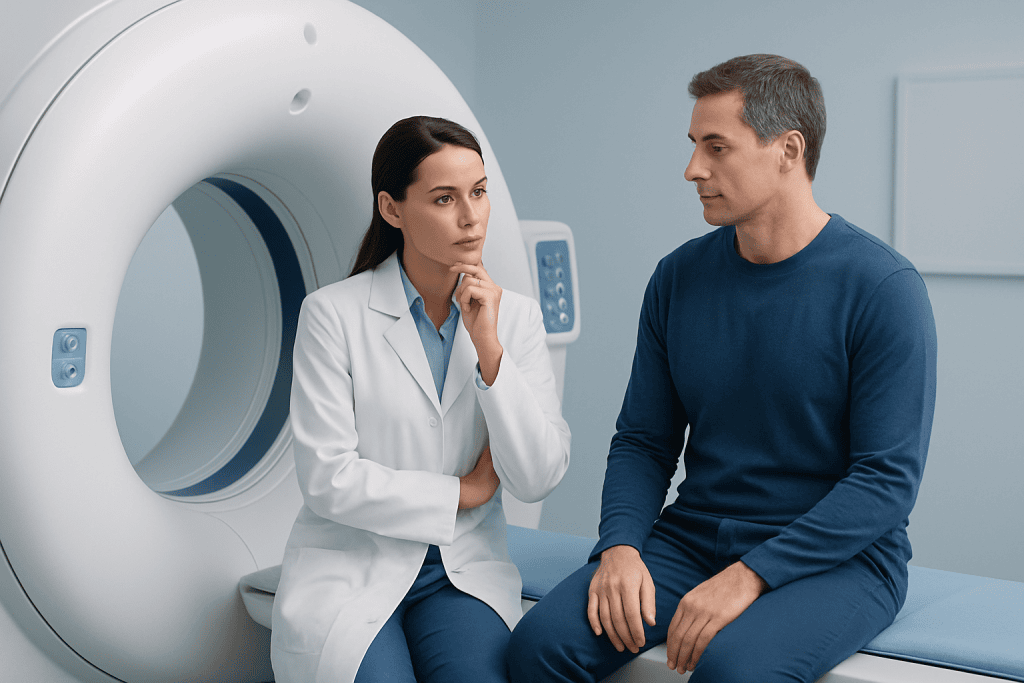
The first step is understanding the symptom itself. Doctors ask where it started, whether it is getting worse, what makes it better or worse, and whether there are warning signs such as fever, weakness, confusion, chest pain, or unexplained weight loss. They are trying to identify the most likely causes and rule out any problem that needs rapid treatment.
Age and personal medical history also matter. A child, a pregnant person, and an older adult may need different imaging strategies because safety and likely diagnoses differ. Previous surgeries, metal implants, kidney disease, allergies to contrast dye, and a history of cancer can all influence which scan is safest and most informative.
Another important factor is what the doctor needs to see. Bones, lungs, soft tissues, blood vessels, abdominal organs, and the brain are not all shown equally well on the same test. Some scans are better for detecting fractures, while others are better for inflammation, bleeding, tumors, or problems involving nerves and discs.
Practical issues are considered too. In an emergency, a fast test may be preferred over one that takes longer, even if both could be useful. Doctors also think about whether contrast material is needed, whether the patient can stay still, and whether the scan is readily available. The best test is often the one that answers the question accurately in the safest and most timely way.
How common imaging tests differ

X-rays are often the first imaging test for suspected bone injuries, some chest symptoms, and certain dental or joint problems. They are quick and widely available, and they are especially useful for fractures, some infections, and lung findings such as pneumonia. However, X-rays do not show soft tissues in as much detail as MRI or CT.
Ultrasound uses sound waves rather than radiation. It is commonly used to assess the abdomen, pelvis, gallbladder, kidneys, thyroid, blood flow in vessels, and pregnancy. It can also help evaluate lumps, swelling, and some causes of pain. Ultrasound is especially useful when doctors want real-time imaging, but image quality can depend on body area and operator technique.
CT scans provide detailed cross-sectional images and are often chosen when speed is important. They are very useful for trauma, suspected bleeding, many chest and abdominal emergencies, kidney stones, and some cancers. CT can show more detail than a plain X-ray, but it uses more radiation, so doctors use it thoughtfully.
MRI is particularly helpful for soft tissues, the brain, spinal cord, joints, ligaments, and some internal organs. It can provide excellent detail for conditions affecting nerves, discs, cartilage, and certain tumors. MRI does not use ionizing radiation, but it takes longer than CT and may not be suitable for some patients with specific implants or severe claustrophobia. In selected situations, a doctor may also recommend MRI or CT scanning as the most direct way to clarify symptoms.
Examples of how symptoms guide imaging choices
When a person has sudden chest pain or shortness of breath, imaging may be chosen to quickly look for serious causes such as pneumonia, fluid around the lungs, or blood clots in the lungs. A chest X-ray may be the starting point, but a CT scan may be needed if more detailed information is required. If the concern is related to the heart, doctors may turn to other heart-specific tests as well.
For headaches or neurological symptoms, the choice depends on urgency and accompanying signs. A sudden severe headache after trauma or with new confusion may lead to emergency CT imaging because it is fast and can detect acute bleeding. Persistent headaches, seizures, weakness, numbness, or suspected nerve or spinal problems may be better evaluated with MRI, especially if the doctor is concerned about the brain or spinal cord, including conditions such as brain tumors.
Abdominal pain is another common example. Pain in the upper right abdomen may prompt ultrasound to look at the gallbladder. Flank pain with suspected kidney stones may lead to CT. Pelvic pain may be assessed with ultrasound first, particularly in women and in younger patients. The exact location of pain and associated symptoms such as fever, vomiting, or urinary changes help guide the choice.
Joint or back pain also requires tailored imaging. After an injury, an X-ray may be enough to detect a fracture. But if symptoms suggest a ligament tear, disc problem, or nerve compression, MRI may be more useful. For long-term back pain, imaging is not always needed immediately unless there are warning signs such as trauma, weakness, bowel or bladder changes, fever, or a history that raises concern for more serious disease.
Safety, radiation, and contrast: how risks are balanced
Doctors try to get the information they need while minimizing risk. One of the main considerations is radiation exposure. X-rays and CT scans use ionizing radiation, while ultrasound and MRI do not. This does not mean CT or X-ray should be avoided when needed, but it does mean doctors think carefully about whether a lower-radiation or no-radiation option could answer the same question.
Contrast materials can improve image detail, but they are not always necessary. CT contrast is commonly used to better see blood vessels, inflammation, and some tumors. MRI contrast may help characterize certain lesions or areas of abnormal tissue. Before giving contrast, doctors review kidney function, past reactions, and other medical factors to reduce the chance of complications.
Special situations require extra care. In pregnancy, ultrasound is often preferred when appropriate, and MRI may also be considered in some circumstances. In children, doctors are especially mindful of radiation and often choose ultrasound or MRI if those tests can provide the needed answer. People with pacemakers, metal fragments, severe kidney disease, or implanted devices may need modifications to the imaging plan.
Comfort and practicality matter as well. Some patients have difficulty lying flat, staying still, or being in an enclosed scanner. Sedation may occasionally be needed, especially for children or for longer studies such as MRI. Doctors balance all of these issues to keep testing both safe and useful.
Why one scan may lead to another
Sometimes the first imaging test answers the question completely. In other cases, it narrows the possibilities but does not provide a final diagnosis. This is not necessarily a sign that something is wrong with the process. Different scans are often used in sequence because they provide complementary information.
For example, an X-ray may show an abnormal area in the lung, but a CT scan may be needed to define it more clearly. An ultrasound may detect a mass or enlarged organ, while MRI may better characterize the tissue. In cancer care, imaging may be used first to detect an abnormality, then to guide biopsy planning, staging, and treatment monitoring, such as in people being assessed for lung cancer.
Follow-up imaging may also be recommended when doctors want to see whether a finding changes over time. A small cyst, nodule, or area of inflammation may simply need observation. In other cases, repeated imaging is used to evaluate response to treatment or recovery after surgery or injury.
Multidisciplinary care can be especially helpful when symptoms are complex. Radiologists, emergency physicians, neurologists, surgeons, oncologists, and other specialists may review the same case from different angles. Near the end of the diagnostic pathway, some patients may benefit from care at centers such as Acibadem International, where multidisciplinary specialists in JCI-accredited hospitals evaluate and treat international patients.
What patients can do before and after imaging
Patients can help improve imaging quality and safety by sharing complete information with their doctor and imaging team. It is important to mention pregnancy, allergies, kidney problems, implanted devices, prior surgeries, and any earlier imaging done elsewhere. Bringing previous scan reports can sometimes prevent duplicate testing and helps doctors compare old and new findings.
Preparation depends on the type of scan. Some studies require fasting, a full bladder, or avoiding metal objects. Others involve drinking oral contrast or receiving an injection. Patients should ask what to expect in advance so they can prepare comfortably and arrive on time. Following instructions closely can improve image quality and reduce the need for repeat scans.
After the test, many people can return to normal activities right away. If contrast or sedation was used, the care team may give specific instructions. Results are not always immediate because a radiologist needs to interpret the images carefully and send a report to the referring doctor. If symptoms are worsening while waiting for results, patients should contact their doctor promptly.
It also helps to remember that imaging is just one part of diagnosis. A scan result is interpreted together with symptoms, examination findings, and laboratory tests. Even a normal scan can be useful because it rules out certain conditions and guides the next step in care, which may include observation, medicines, therapy, or another test such as ultrasound imaging.
When to seek medical advice urgently
Some symptoms deserve prompt medical evaluation because they may require urgent imaging or treatment. These include sudden chest pain, severe shortness of breath, signs of stroke, a sudden severe headache, major injury, heavy bleeding, new seizures, fainting, or sudden weakness or numbness. In these situations, the most appropriate scan is often chosen quickly in the emergency setting.
People should also seek medical advice if symptoms persist, return repeatedly, or interfere with daily life. Ongoing abdominal pain, unexplained swelling, persistent cough, progressive back pain, new neurological symptoms, or a lump that does not go away may all need assessment. Not every concern will require imaging, but a doctor can decide whether a scan would be helpful.
Patients should feel comfortable asking why a particular test is recommended and what the doctor hopes it will show. Understanding the reason for imaging can reduce anxiety and make the process easier to follow. If there are several options, the doctor can explain the benefits and limitations of each one.
In general, the right imaging test is the one that best matches the patient’s symptoms and clinical situation. A careful, individualized approach helps doctors diagnose conditions efficiently while keeping safety at the center of care.
Frequently asked questions
Why would a doctor order an X-ray first instead of an MRI?
An X-ray is often the fastest and simplest way to evaluate bones, certain chest symptoms, and some joint problems. If it answers the clinical question clearly, a more complex scan may not be needed. MRI is excellent for soft tissues, but it usually takes longer and is not always the best first test.
How do doctors decide between a CT scan and an MRI?
Doctors choose based on what they are looking for, how urgently they need answers, and what is safest for the patient. CT is often preferred in emergencies because it is quick and very good for trauma, bleeding, and many chest or abdominal problems. MRI is especially useful for the brain, spine, joints, and other soft tissues.
Does every symptom need imaging?
No. Many symptoms can be evaluated first with a medical history, physical examination, and sometimes blood or urine tests. Imaging is usually ordered when it is likely to change diagnosis, treatment, or the need for urgent care.
Are imaging tests safe?
Most imaging tests are safe when used appropriately. Doctors consider radiation exposure, contrast risks, pregnancy, kidney function, implants, and other health factors before selecting a scan. The aim is always to choose the safest test that can answer the question well.
Why might I need more than one imaging test?
Different scans show different types of body structures and abnormalities. One test may detect a problem, while another helps define it more clearly or guide treatment planning. Needing a second scan is common and often reflects careful, step-by-step diagnosis.
Can I ask my doctor why a certain scan was recommended?
Yes. Patients are encouraged to ask what the scan is meant to show, whether other options exist, and whether any preparation is needed. Clear communication can make the test easier to understand and help patients take an active role in their care.
References
- World Health Organization
- Radiological Society of North America
- American College of Radiology
- National Institute for Health and Care Excellence
- U.S. Food and Drug Administration
This article is for general information only and is not a substitute for professional medical advice. Please consult a qualified doctor about your individual situation.
Explore treatments in Turkey — costs, top hospitals & a free quote
JCI-accredited · board-certified surgeons · reply within 24h
Add us as a Preferred Source to see more of our trusted health content across Google Search, AI Overviews and Discover.

More from the Health Library



Related Specialists

Prof. Dr. Ercan Karaarslan
Radiology
Prof. Dr. Bora Peynircioğlu
Interventional Radiology
Prof. Dr. Koray Güven
Interventional Radiology
Assoc. Prof. Dr. Deniz Can Aliş
Radiology