

Congenital Nevi: Causes, Types, and How They Can Change Over Time
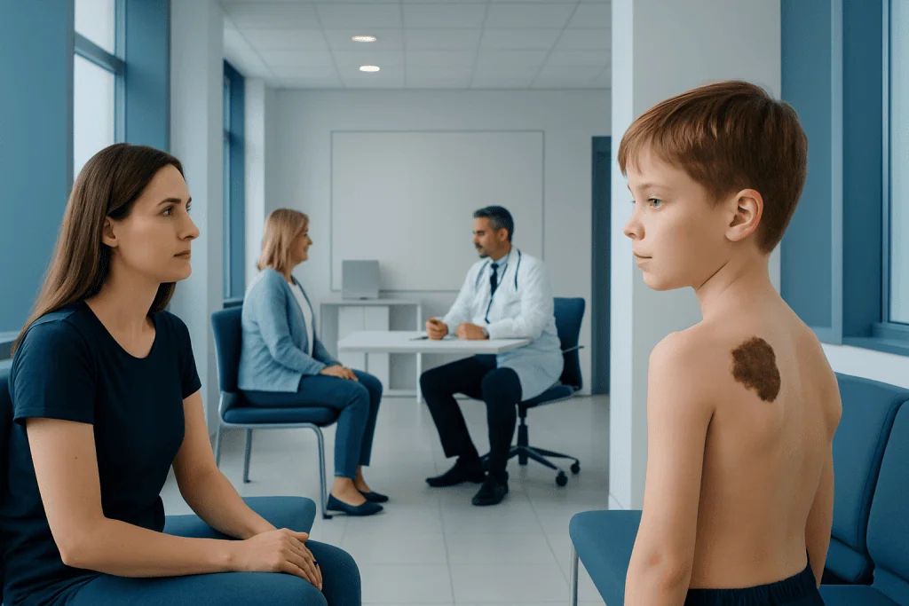
Congenital nevi are moles or pigmented birthmarks that are present at birth or develop very early in life. They are commonly classified by size, and larger lesions may need closer long-term follow-up.
Key Takeaways
- Congenital nevi are moles or pigmented birthmarks that are present at birth or develop very early in life.
- They are commonly classified by size, and larger lesions may need closer long-term follow-up.
- Some congenital nevi change over time in color, thickness, hair growth, or surface texture as a child grows.
- Diagnosis is usually based on a skin examination, with dermoscopy or biopsy used in selected cases.
- Treatment may involve observation, surgical removal, or reconstructive planning depending on medical and cosmetic needs.
- Any rapid or unusual change should be reviewed by a dermatologist or plastic surgeon.
Congenital nevi are pigmented skin lesions present at birth or appearing shortly afterward. They are often harmless, but their size, appearance, and location can affect monitoring, cosmetic concerns, and decisions about treatment.
Overview of congenital nevi
Congenital nevi are moles or pigmented birthmarks that are present at birth or become visible in the first months of life. They develop from melanocytes, the skin cells that produce pigment. These lesions can range from very small spots to larger patches that cover a wider area of skin.
Many congenital nevi are benign and remain stable for years. However, they are important to identify and follow because some may change in appearance as the body grows. Their size, shape, color, location, and surface features help doctors decide whether simple observation is enough or whether treatment should be considered.
From a patient and family perspective, congenital nevi can raise both medical and cosmetic questions. A lesion on the face, scalp, or another visible area may affect confidence or create practical concerns such as irritation from clothing or grooming. In these situations, evaluation may involve both dermatology and plastic surgery specialists, especially when planning treatment for congenital nevi.
Types and how they are classified
Doctors often classify congenital nevi by their expected adult size. Small congenital nevi are the most common. Medium lesions are larger but still limited to a modest area. Large or giant congenital nevi cover a broader region and may involve deeper structures of the skin. This size-based classification helps estimate follow-up needs and shape treatment planning.
Congenital nevi can also look different from one another. Some are light brown, while others are dark brown or nearly black. They may be flat at first and later become thicker, more raised, or more textured. In some cases, they develop coarse hair. The borders may be smooth or somewhat irregular, and small satellite spots can appear near a larger primary lesion.
Location matters too. A nevus on the scalp, eyelid, lip, or another delicate area may need a different approach from one on the trunk or limbs. Lesions near the eye may occasionally require coordinated care involving dermatology, plastic surgery, and reconstructive ophthalmology if function or tissue coverage is affected.
Although congenital nevi are distinct from other skin findings, doctors may sometimes compare them with other congenital or scar-related conditions to guide reconstruction and appearance-related care, such as keloid scars or other visible skin differences.
How congenital nevi can change over time

It is normal for congenital nevi to change as a child grows. Because the skin itself expands, the lesion often grows in proportion with the body. Color may become darker or lighter over time, and the surface can become more pebbled, raised, or uneven. Hair growth within the nevus may also increase with age.
These expected changes do not always mean there is a serious problem. Still, regular observation is important because some changes are more concerning than others. A doctor may want to review a lesion if it develops rapid growth, marked asymmetry, ulceration, bleeding, persistent itching, pain, or a sudden major color change.
Photographs taken at home under similar lighting can help families track gradual changes. This can be especially useful for lesions on the scalp, back, or other areas that are not easy to view often. During follow-up visits, clinicians may compare size and surface details over time to decide whether monitoring remains appropriate or whether a procedure should be discussed.
Causes and possible risk factors
Congenital nevi form during fetal development, when melanocyte cells do not distribute in the usual pattern within the skin. This process is considered a developmental event rather than something caused by anything a parent did or did not do during pregnancy. In most cases, there is no preventable cause.
Researchers believe congenital melanocytic nevi are related to early genetic changes in skin cells during development. These changes are usually sporadic, meaning they happen by chance and are not typically inherited through families. For that reason, having one child with a congenital nevus does not usually mean future children will have the same condition.
Risk considerations are mostly linked to the lesion itself rather than lifestyle factors. Larger nevi, multiple satellite lesions, and certain body locations may need more careful evaluation. In some large or giant nevi, doctors may also assess for associated conditions involving deeper tissues or the nervous system, depending on the pattern and extent of the lesion.
Diagnosis and medical evaluation
Diagnosis usually begins with a clinical skin examination. A dermatologist or plastic surgeon will look at the lesion’s size, color, borders, thickness, hair growth, and location. The medical history may include when the lesion was first noticed, whether it has changed, and whether there are symptoms such as irritation, bleeding, or pain.
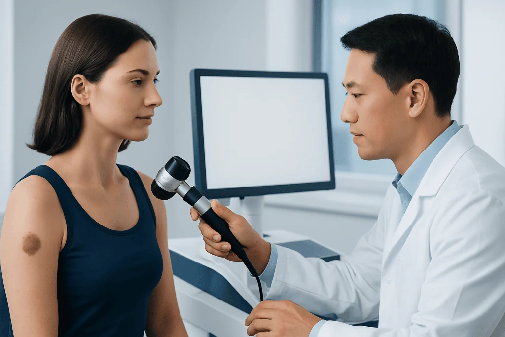
Dermoscopy may be used during the exam. This is a noninvasive tool that magnifies the skin and helps doctors assess pigment patterns and surface structures more closely. In many cases, this is enough to support ongoing observation without immediate intervention.
If a lesion has unusual features, a biopsy may be recommended to examine the tissue under a microscope. For very large congenital nevi or lesions associated with neurological symptoms, additional imaging may occasionally be considered. The goal is to understand not only how the nevus looks on the surface, but also whether there are deeper concerns that need follow-up.
Some congenital conditions are evaluated in a similar multidisciplinary way when appearance and function overlap, such as microtia or syndactyly, where long-term planning balances health, growth, and reconstruction.
Treatment options and when removal is considered
Treatment depends on the size, location, symptoms, appearance, and level of medical concern. Many small and medium congenital nevi can be monitored over time without immediate removal. Observation is often appropriate when the lesion is stable, easy to examine, and not causing discomfort or significant cosmetic concern.
Removal may be considered for several reasons. These include suspicious changes, repeated irritation, difficulty monitoring the area, or a wish to improve appearance and comfort. Surgical excision is the most common treatment when removal is chosen. For larger lesions, treatment may need to be staged over time, sometimes using reconstructive techniques to preserve function and achieve the best possible skin coverage.
In selected cases, plastic and reconstructive specialists may discuss options within broader cosmetic and reconstructive surgery planning, especially for visible lesions or those affecting contour. When a nevus lies on or near the neck, reconstruction may also overlap with techniques used in aesthetic neck contouring if skin quality and appearance are part of the concern.
It is important to understand that not every congenital nevus should or must be removed. Even after treatment, long-term follow-up may still be advised. The best plan is individualized, based on skin examination findings, patient age, personal preferences, and specialist guidance.
Prevention, self-care, and long-term monitoring
Congenital nevi cannot be prevented because they develop before birth. However, good skin care and consistent monitoring can help protect the skin and support early detection of important changes. Sun protection is especially important. Using protective clothing, seeking shade, and applying broad-spectrum sunscreen to exposed skin can reduce additional pigment changes and protect overall skin health.
Families and adults with congenital nevi can benefit from a simple routine of monthly skin observation. It helps to note the lesion’s size, shape, color, thickness, and any symptoms such as tenderness or crusting. Keeping photographs with dates may make subtle changes easier to recognize.
Daily self-care also includes reducing friction and trauma where possible. A nevus located under a bra strap, waistband, or shaving area may become repeatedly irritated. If this happens often, a clinician can advise whether changes in clothing, grooming habits, or treatment would be helpful.
For international patients seeking coordinated assessment, Acibadem International’s multidisciplinary specialists in JCI-accredited hospitals evaluate congenital nevi and plan monitoring, dermatologic care, and reconstructive treatment when needed.
When to see a doctor
A medical review is recommended for any congenital nevus that is new to the doctor, difficult to classify, or causing worry for the patient or family. This is especially important for larger lesions, multiple lesions, or those located on the scalp, face, spine, or genital area, where monitoring and treatment decisions can be more complex.
Prompt assessment is appropriate if the lesion begins to bleed, ulcerate, crust repeatedly, grow rapidly, become painful, or develop a clear change in color or border. These findings do not always mean cancer, but they should not be ignored. A doctor can decide whether closer follow-up, dermoscopy, or biopsy is needed.
Regular specialist follow-up may be advised in childhood and adolescence because the skin and body continue to change during growth. Early consultation can also help families discuss timing, benefits, and limits of treatment in a calm and informed way.
Frequently asked questions
What is the difference between a congenital nevus and a regular mole?
A congenital nevus is present at birth or appears very soon after birth, while many regular moles develop later in childhood or adulthood. Congenital nevi may behave differently over time, especially when they are larger, so they are often followed more closely.
Are congenital nevi dangerous?
Most congenital nevi are benign and do not cause serious health problems. However, some lesions, particularly larger ones, may need regular monitoring because certain changes can require closer evaluation.
Can congenital nevi become cancerous?
There is a recognized but variable risk that some congenital nevi may develop melanoma, and the risk is generally higher in larger lesions. That does not mean most will become cancerous, but it does explain why doctors recommend skin checks and review of any unusual change.
Should a congenital nevus always be removed?
No, removal is not always necessary. Some congenital nevi can be safely observed, while others are removed because of suspicious changes, repeated irritation, location, or cosmetic and reconstructive considerations.
How are congenital nevi treated?
Treatment may include observation, photography-based monitoring, dermoscopic follow-up, or surgical removal. The best approach depends on the nevus size, depth, location, symptoms, and the patient’s age and preferences.
What changes in a congenital nevus should prompt a doctor visit?
A doctor should assess rapid growth, bleeding, ulceration, persistent pain, itching, repeated crusting, or a major change in color or shape. These signs do not confirm a serious problem, but they deserve professional evaluation.
Can children with congenital nevi live normally?
Yes, most children with congenital nevi live full and active lives. Regular skin checks, sun protection, and appropriate specialist follow-up can help families manage the condition confidently and safely.
References
- American Academy of Dermatology
- National Cancer Institute
- DermNet
- British Association of Dermatologists
- American Society of Plastic Surgeons
This article is for general information only and is not a substitute for professional medical advice. Please consult a qualified doctor about your individual situation.
Congenital Nevi in Turkey — costs, top hospitals & a free quote
JCI-accredited · board-certified surgeons · reply within 24h
Add us as a Preferred Source to see more of our trusted health content across Google Search, AI Overviews and Discover.

Related Treatments
Related Conditions
More from the Health Library



Related Specialists

Prof. Dr. Şükrü Yazar
Aesthetic Plastic & Reconstructive Surgery
Assoc. Prof. Dr. Erdem Güven (m)
Aesthetic Plastic & Reconstructive Surgery
Prof. Dr. Hakan Ağır
Aesthetic Plastic & Reconstructive Surgery
Prof. Dr. Bülent Saçak
Aesthetic Plastic & Reconstructive Surgery