

Congenital Nevi: How Dermatologists and Surgeons Assess Size, Location, and Risk
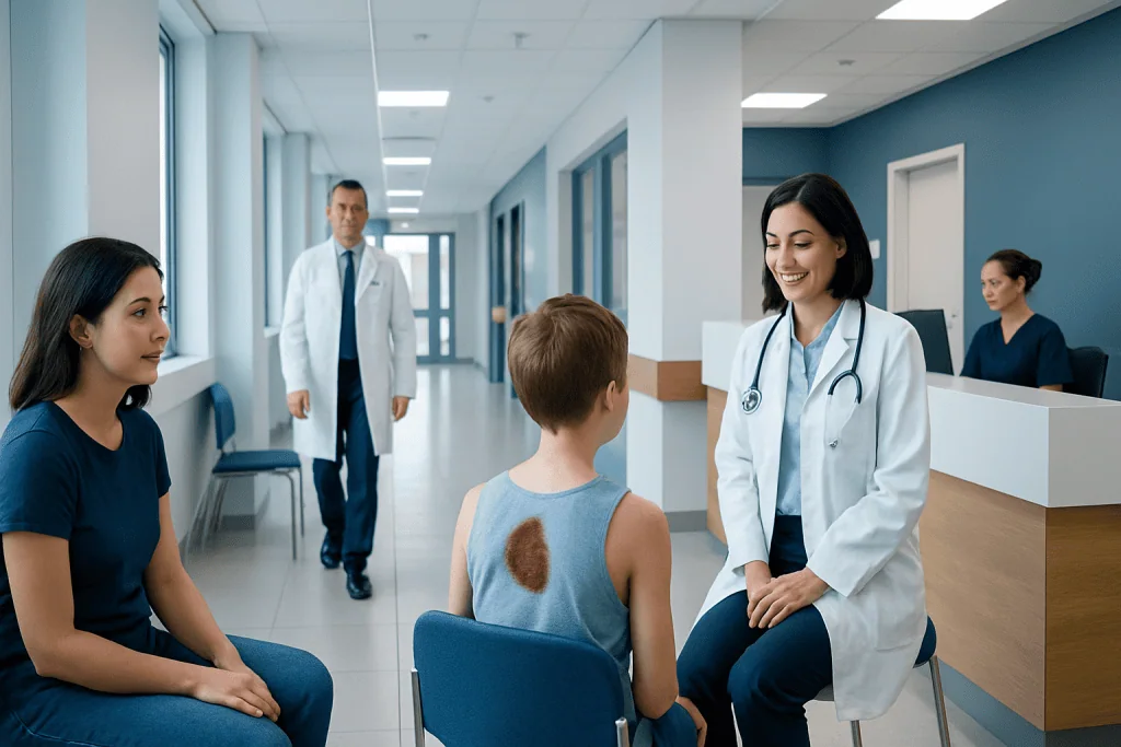
Congenital nevi vary widely in size, color, texture, and body location. Assessment focuses on melanoma risk, symptoms, cosmetic concerns, and practical issues such as friction or difficult monitoring.
Key Takeaways
- Congenital nevi vary widely in size, color, texture, and body location.
- Assessment focuses on melanoma risk, symptoms, cosmetic concerns, and practical issues such as friction or difficult monitoring.
- Not every congenital nevus needs removal; many can be followed safely with regular skin checks and photographs.
- Treatment planning may involve dermatology, plastic surgery, pediatrics, and sometimes imaging specialists.
- Large or complex lesions may need staged treatment and long-term follow-up.
Congenital nevi are moles or pigmented birthmarks present at birth or appearing shortly afterward. Dermatologists and surgeons assess their size, location, appearance, and possible future changes to decide whether observation, imaging, or treatment is the best approach.
Overview of Congenital Nevi
Congenital nevi, also called congenital melanocytic nevi, are pigmented skin lesions that are present at birth or become noticeable in the first weeks or months of life. They develop from pigment-producing cells called melanocytes. Some are small and flat, while others are thicker, darker, hairy, or raised. Their appearance can change gradually as a child grows.
Doctors usually describe congenital nevi by their projected adult size, because a lesion seen in infancy may enlarge as the body grows. In broad terms, they may be classified as small, medium, large, or giant. This size estimate matters because it can influence follow-up, treatment planning, and discussion of possible health risks.
For many families, the first concern is whether the lesion is dangerous. Most congenital nevi are benign. However, specialists evaluate them carefully because some carry a higher lifetime risk of melanoma than ordinary moles, especially when they are large, numerous, or associated with other findings. Cosmetic and emotional concerns are also important, particularly when the nevus is on the face, scalp, hands, or other highly visible areas.
How Size, Location, and Appearance Are Assessed
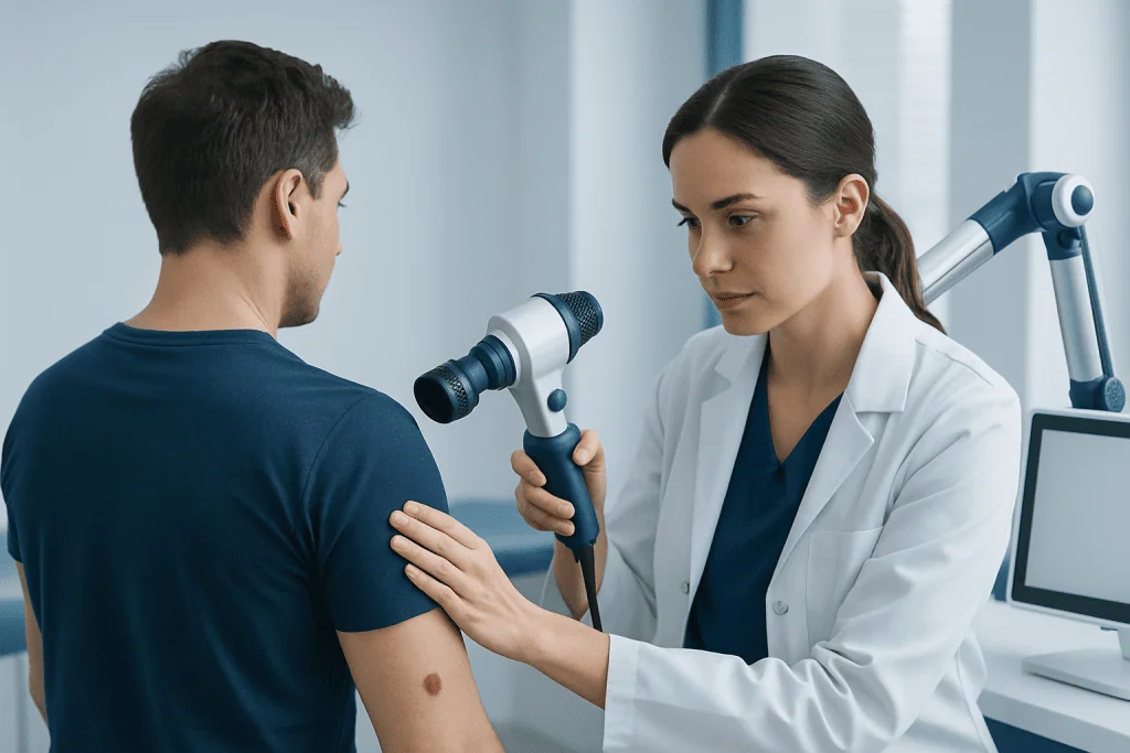
Assessment begins with a detailed skin examination. The doctor looks at the lesion’s diameter, borders, color pattern, thickness, texture, hair growth, and whether there are any nodules or ulcerated areas. Measurements and clinical photographs are often taken to help monitor change over time. In children, the doctor may estimate how large the nevus is likely to become in adulthood based on its location and current size.
Location is especially important. A congenital nevus on the scalp may be harder to watch because hair can hide subtle changes. Lesions on the eyelids, lips, nose, ears, hands, feet, or genital area may affect function, hygiene, or comfort. A nevus in an area subject to repeated friction, such as under clothing straps or around joints, may become irritated more easily.
Doctors also consider whether satellite nevi are present. These are smaller pigmented spots around the main lesion or elsewhere on the body. Having multiple lesions does not automatically mean a serious problem, but it may prompt more detailed follow-up. If the nevus is near the eye or delicate facial structures, planning may involve specialists in reconstructive ophthalmology or plastic surgery to balance safety, function, and appearance.
In practice, assessment is not only about cancer risk. It also includes how easy the lesion will be to monitor, whether it affects daily life, and how the child or adult feels about it. This broader view helps families and clinicians choose between observation and treatment in a thoughtful, individualized way.
Symptoms and Changes That Need Attention

Many congenital nevi cause no symptoms at all. They may simply be darker or more textured than the surrounding skin. Some become more raised, rough, or hairy over time, and these changes can be part of their natural evolution. As children grow, the nevus usually grows in proportion to the body rather than suddenly expanding on its own.
Even so, doctors advise watching for specific changes. Features that deserve medical review include rapid enlargement that seems out of proportion to growth, new firm lumps, repeated bleeding, ulceration, persistent itching, pain, or a clear change in color pattern. These signs do not always mean cancer, but they should be assessed promptly.
The skin surface can also become dry or irritated, especially in areas exposed to rubbing. In some cases, a nevus may be confused with or coexist with other skin conditions. Scar-related changes after prior treatment may also need careful review, particularly in people prone to raised scars such as keloid scars. A specialist can help distinguish routine evolution from changes that need biopsy or treatment.
Causes, Risk Factors, and Related Conditions
Congenital nevi arise during fetal development, when melanocytes develop or migrate differently in the skin. They are not caused by anything a parent did during pregnancy, and in most cases they are not inherited in a simple way. They usually occur sporadically. This can be reassuring for families who worry they may have prevented it.
The main risk factors that guide medical follow-up are lesion size, number of satellite lesions, body location, and certain imaging or neurologic findings when present. Large and giant congenital nevi are generally monitored more closely because they carry a greater risk of melanoma than small lesions. Risk may also be influenced by lesions in the central body axis, such as the back or scalp, where associated findings can occasionally be more complex.
Some people with large or multiple congenital nevi may be evaluated for neurocutaneous melanocytosis, a rare condition involving melanocyte deposits in the central nervous system. This is not present in most children with congenital nevi, but specialists may consider brain or spine imaging when a lesion is very large, located over the spine, or accompanied by neurologic symptoms. The goal is careful screening where appropriate, not routine alarm.
Congenital nevi may also be seen alongside other congenital differences, though they are usually isolated. In multidisciplinary pediatric and plastic surgery settings, specialists may also care for children with conditions such as microtia or syndactyly, which highlights how treatment planning often involves both medical and reconstructive considerations tailored to growth and development.
Diagnosis and Monitoring
Diagnosis is usually clinical, meaning it is based on the lesion’s history and examination by a dermatologist or surgeon experienced in pigmented lesions. Dermoscopy, a noninvasive magnified skin examination, can reveal patterns that help distinguish congenital nevi from other moles or skin growths. In many cases, this is enough to plan safe follow-up.
Biopsy is not required for every congenital nevus. It may be recommended if a particular area looks atypical, develops a new nodule, ulcerates, bleeds repeatedly, or shows changes that are difficult to explain. Sampling can help clarify whether the tissue is benign, irritated, or suspicious for melanoma. The decision is individualized because biopsy in children and in large lesions must balance diagnostic value with scarring and comfort.
For selected infants and children, imaging such as MRI may be advised, especially when the nevus is large, numerous, or associated with neurologic concerns. This is done to look for rare internal melanocytic involvement. Follow-up often includes serial photographs, skin maps, and scheduled re-examinations, which help doctors compare the lesion over time rather than relying on memory alone.
Families are often taught what normal change may look like and which warning signs should prompt an earlier visit. This shared monitoring approach can make long-term care more manageable, especially for lesions in hard-to-see places like the scalp, back, or behind the ears.
Treatment Options and Surgical Planning
Treatment is individualized. Some congenital nevi are best managed with observation alone, especially if they are small, stable, and easy to monitor. Others may be treated because of cancer-related concern, recurrent irritation, functional effects, or the emotional impact of a highly visible lesion. There is no single approach that fits every patient.
When removal is considered, surgery is usually the main option. Small lesions may sometimes be removed in one procedure with direct closure. Larger lesions may require staged excision, tissue expansion, local flaps, or skin grafting so that the wound can be closed with the best possible functional and cosmetic result. In selected cases, families may be referred for cosmetic (aesthetic) surgery or reconstructive planning, particularly when the nevus affects the face or other visible areas.
It is important to understand that removing a nevus may reduce visible pigment and improve monitoring or appearance, but it does not always eliminate every melanocyte, especially in deeper tissues or very large lesions. For this reason, long-term follow-up may still be recommended after treatment. Surgeons also discuss likely scars, timing in relation to growth, and whether treatment should be done in stages.
Laser and other pigment-reducing methods are sometimes discussed, but they do not replace full surgical assessment because they may lighten the lesion without removing all nevus cells. For patients exploring options, information from a dedicated congenital nevi treatment page can help frame questions before a specialist visit. In complex cases, plastic surgeons may also consider broader body aesthetics and reconstruction strategies when planning closure and contour after excision.
Prevention, Self-care, and When to See a Doctor
Congenital nevi cannot be prevented, because they form before birth. What families can do is support healthy skin care and consistent monitoring. Sun protection is important for all skin types and especially helpful for pigmented lesions. Practical steps include shade, protective clothing, and regular use of broad-spectrum sunscreen on exposed skin, according to age-appropriate guidance from a clinician.
At home, it helps to take clear photographs every few months using similar lighting and angles, especially for lesions that are difficult to see. Parents and adults should watch for bleeding, crusting, persistent itching, pain, ulceration, or a new bump within the nevus. If the lesion is on the scalp, parting the hair regularly can make monitoring easier.
A doctor should evaluate any congenital nevus that is large, changing noticeably, repeatedly irritated, or causing emotional distress. Early consultation is also wise when a lesion affects the eyelid, lips, nose, ear, hands, feet, or genital area, where growth may affect function or make future surgery more complex. In rare cases, headaches, vomiting, developmental changes, seizures, or other neurologic symptoms alongside large or multiple nevi may prompt urgent review.
Specialist care may involve dermatology, pediatrics, radiology, and plastic surgery working together. Near the end of the care pathway, some international patients choose centers such as Acibadem International, where multidisciplinary specialists in JCI-accredited hospitals assess and treat congenital nevi with both medical safety and reconstructive planning in mind.
Frequently asked questions
Are congenital nevi the same as ordinary moles?
No. Congenital nevi are present at birth or appear very soon afterward, while ordinary acquired moles usually develop later. They can look similar, but congenital nevi may behave differently and are assessed with special attention to size and long-term change.
Do all congenital nevi need to be removed?
No, many do not need removal. Doctors may recommend observation when a lesion is small, stable, and easy to monitor. Removal is considered when there are concerning changes, functional issues, repeated irritation, or important cosmetic and emotional concerns.
Does removing a congenital nevus completely prevent melanoma?
Not always. Surgery may reduce visible nevus tissue, but deeper or surrounding melanocytes can sometimes remain, especially in large lesions. That is why follow-up may still be needed even after treatment.
How do doctors estimate risk in a congenital nevus?
They look at several factors together, including projected adult size, location, surface changes, the presence of satellite lesions, and any symptoms. Large or giant lesions usually need closer follow-up than small lesions, but the plan is always individualized.
Can a congenital nevus change as a child grows?
Yes. Many congenital nevi naturally become darker, more raised, more textured, or hairier over time. Growth in proportion to the child's body is expected, but sudden or unusual change should be reviewed by a doctor.
When is imaging such as MRI considered?
MRI is not needed for every child with a congenital nevus. It may be considered for very large lesions, multiple lesions, or when there are neurologic symptoms or patterns that raise concern for rare associated conditions. The doctor decides this based on the full clinical picture.
References
- American Academy of Dermatology
- British Association of Dermatologists
- DermNet
- National Organization for Rare Disorders
- American Cancer Society
This article is for general information only and is not a substitute for professional medical advice. Please consult a qualified doctor about your individual situation.
Congenital Nevi in Turkey — costs, top hospitals & a free quote
JCI-accredited · board-certified surgeons · reply within 24h
Add us as a Preferred Source to see more of our trusted health content across Google Search, AI Overviews and Discover.

Related Treatments
Related Conditions
More from the Health Library



Related Specialists

Prof. Dr. Şükrü Yazar
Aesthetic Plastic & Reconstructive Surgery
Assoc. Prof. Dr. Erdem Güven (m)
Aesthetic Plastic & Reconstructive Surgery
Prof. Dr. Hakan Ağır
Aesthetic Plastic & Reconstructive Surgery
Prof. Dr. Bülent Saçak
Aesthetic Plastic & Reconstructive Surgery