Echocardiogram: Heart Ultrasound Findings Explained

An echocardiogram uses sound waves to create moving images of the heart and does not use radiation. Common findings include measurements of heart size, pumping strength, valve function, wall thickness, and pressures inside the heart.
Key Takeaways
- An echocardiogram uses sound waves to create moving images of the heart and does not use radiation.
- Common findings include measurements of heart size, pumping strength, valve function, wall thickness, and pressures inside the heart.
- Ejection fraction is one important number, but it is only one part of the overall interpretation.
- Mild findings are common and may only require monitoring, while more significant abnormalities may need additional testing or treatment.
- Results should always be interpreted together with symptoms, medical history, physical examination, and other tests.
An echocardiogram is a safe, noninvasive ultrasound test that helps doctors see how the heart muscle, valves, and blood flow are working. Understanding common findings can make the report less confusing and help patients discuss next steps with their cardiologist.
Overview: What Is an Echocardiogram?
An echocardiogram, often called an echo or heart ultrasound, is a test that uses high-frequency sound waves to create images of the heart in real time. It allows a cardiologist to see the heart chambers, valves, major blood vessels, and the motion of the heart muscle. Because it uses ultrasound rather than X-rays, it does not expose the patient to radiation.
The most common type is a transthoracic echocardiogram, where a small handheld probe is moved across the chest with ultrasound gel. In some cases, a transesophageal echocardiogram may be recommended, where a specialized probe is passed into the esophagus to obtain clearer images from behind the heart. Stress echocardiography combines ultrasound images with exercise or medication to assess how the heart performs under strain.
Doctors may request an echocardiogram for many reasons, including shortness of breath, chest discomfort, palpitations, fainting, a heart murmur, high blood pressure, suspected valve disease, previous heart attack, heart failure evaluation, or follow-up of known heart conditions. The test is also used before certain surgeries or cancer treatments when heart function needs to be assessed.
What an Echocardiogram Report Usually Includes
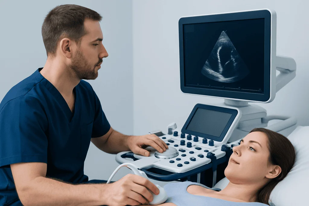
An echocardiogram report can look technical because it contains measurements, descriptions, and a final interpretation. The most helpful way to read it is to focus on the main categories: heart chamber size, pumping function, valve structure and leakage, valve narrowing, heart wall thickness, pressure estimates, and the appearance of the sac around the heart, called the pericardium.
The report may describe the left ventricle, which is the main pumping chamber that sends oxygen-rich blood to the body. It may also describe the right ventricle, which pumps blood to the lungs. The left and right atria, the upper chambers of the heart, are assessed for enlargement, which can be associated with conditions such as high blood pressure, valve disease, atrial fibrillation, or long-standing pressure changes.
Many reports include terms such as normal, mild, moderate, or severe. These words help classify how significant a finding appears on the ultrasound. However, the meaning depends on the patient’s age, body size, symptoms, blood pressure at the time of the test, and overall health, so the final explanation should come from a qualified doctor.
Ejection Fraction and Pumping Strength Explained

Ejection fraction, often abbreviated as EF, is one of the most commonly discussed echocardiogram findings. It estimates the percentage of blood pumped out of the left ventricle with each heartbeat. A normal EF generally means the main pumping chamber is squeezing well, although exact reference ranges may vary slightly by laboratory and clinical context.
A reduced ejection fraction can be seen in some forms of heart failure, previous heart attack, cardiomyopathy, certain valve conditions, or rhythm problems. A preserved ejection fraction means the heart’s squeezing ability is normal, but a person may still have symptoms if the heart is stiff, the valves are abnormal, or pressures are elevated. This is why EF should not be viewed as the only measure of heart health.
The report may also mention regional wall motion. This describes whether all parts of the heart muscle move normally. If one area moves less strongly than expected, it may suggest prior injury, reduced blood supply, or another heart muscle problem. Sometimes further testing, such as a stress test, coronary imaging, or cardiac MRI, is needed to clarify the cause.
Heart Valves: Leakage, Narrowing, and Murmurs
The heart has four valves: the mitral, aortic, tricuspid, and pulmonary valves. They open and close to keep blood moving in the correct direction. An echocardiogram is one of the most important tests for evaluating valve structure and function, especially when a doctor hears a murmur with a stethoscope.
Valve regurgitation means a valve is leaking backward. Mild leakage, especially of the mitral or tricuspid valve, is a common ultrasound finding and often does not require treatment if the heart size and function are normal. Moderate or severe leakage may cause the heart chambers to enlarge over time and may require closer follow-up, medication to manage contributing conditions, or in selected cases a valve procedure.
Valve stenosis means a valve is narrowed and does not open fully. This can make the heart work harder to push blood through the valve. The report may include measurements such as valve area, pressure gradients, and blood flow velocity to estimate severity. Symptoms such as breathlessness, chest discomfort, dizziness, or reduced exercise tolerance are especially important when valve narrowing is found.
Other Common Findings: Wall Thickness, Pressures, and Fluid
An echocardiogram may measure the thickness of the heart muscle, especially the left ventricular wall. Thickening can occur with long-standing high blood pressure, certain valve conditions, athletic conditioning, or inherited heart muscle diseases. Mild thickening is often managed by treating underlying causes such as blood pressure, while more unusual patterns may lead to additional evaluation.
The report may estimate pressures in the right side of the heart and lungs. Elevated pulmonary pressure estimates can occur for many reasons, including lung disease, sleep apnea, left-sided heart disease, blood clots in the lungs, or valve problems. Because ultrasound pressure estimates are indirect, doctors interpret them carefully and may recommend further testing if the finding is significant or symptoms are present.
The pericardium, the thin sac around the heart, is also assessed. A small amount of fluid may be described as a pericardial effusion. Many small effusions are monitored, especially if the patient feels well, but larger effusions or those associated with chest pain, infection, inflammatory disease, kidney disease, or cancer history may need closer medical assessment.
How Doctors Interpret Results and Decide Next Steps
An echocardiogram is most useful when combined with the patient’s story. The same finding may have different meanings in different people. For example, mild valve leakage in a healthy person with no symptoms may simply be observed, while a similar finding in someone with worsening shortness of breath may prompt more detailed evaluation.
Doctors may compare the current echocardiogram with previous studies to see whether a condition is stable or changing. They also consider blood pressure, heart rhythm, blood tests, electrocardiogram findings, medications, and other imaging tests. A clear trend over time can be more meaningful than a single measurement.
Possible next steps may include no immediate action, lifestyle and risk factor management, medication adjustment, repeat echocardiography after a defined interval, referral to a valve or heart failure specialist, or additional tests. These may include stress testing, Holter monitoring, CT coronary angiography, cardiac MRI, or invasive cardiac catheterization when clinically appropriate.
Preparing for the Test, Safety, and When to See a Doctor
A standard transthoracic echocardiogram usually requires little preparation. The patient may be asked to remove clothing from the upper body and wear a gown. Small stickers called electrodes are placed on the chest to monitor heart rhythm, and the ultrasound probe is moved over different areas to capture images. Some pressure from the probe may be felt, but the test is generally painless.
Patients should tell their doctor or sonographer if they are pregnant, have had previous heart surgery, have implanted devices, or have difficulty lying flat. For transesophageal or stress echocardiography, special instructions may apply, such as fasting or arranging transportation afterward if sedation is used. The medical team provides individualized guidance before these tests.
Medical advice should be sought promptly if symptoms such as new or worsening chest pain, fainting, severe shortness of breath, sudden swelling, rapid irregular heartbeat, or blue lips occur. For nonurgent symptoms or questions about an echocardiogram report, a cardiology appointment can help explain the findings and plan follow-up. Acibadem International’s multidisciplinary specialists and JCI-accredited hospitals diagnose and treat heart conditions for international patients, including evaluation with echocardiography when appropriate.
Frequently asked questions
Is an echocardiogram the same as an ECG?
No. An echocardiogram is an ultrasound scan that shows the heart’s structure, pumping function, valves, and blood flow. An ECG, or electrocardiogram, records the electrical activity of the heart and is useful for rhythm problems, signs of strain, or evidence of past or current heart injury.
Does a normal echocardiogram mean the heart is completely healthy?
A normal echocardiogram is reassuring because it suggests normal heart structure and function in the areas assessed. However, it does not rule out every heart condition, such as early coronary artery disease or intermittent rhythm problems. Doctors interpret it alongside symptoms, examination findings, ECG results, blood tests, and other investigations if needed.
What does mild valve regurgitation mean?
Mild valve regurgitation means a small amount of blood leaks backward through a valve. This is a common echocardiogram finding and often does not cause symptoms or require treatment. Follow-up depends on which valve is involved, whether the heart chambers are normal in size, and whether the patient has symptoms.
What is a good ejection fraction?
A normal ejection fraction generally indicates that the left ventricle is pumping an expected proportion of blood with each beat, although exact ranges can vary. A reduced ejection fraction may suggest weakened pumping function, while symptoms can still occur even when EF is preserved. The cardiologist will explain what the number means in the context of the full report.
Can an echocardiogram detect blocked arteries?
A resting echocardiogram does not directly show most coronary artery blockages. It can sometimes show indirect signs, such as an area of heart muscle that does not move normally. If blocked arteries are suspected, a doctor may recommend a stress test, coronary CT angiography, or cardiac catheterization.
How often should an echocardiogram be repeated?
The timing depends on the reason for the test and the findings. Some people with normal results may not need another echo unless symptoms develop, while those with valve disease, heart failure, or cardiomyopathy may need scheduled monitoring. The follow-up interval should be set by a cardiologist or treating physician.
This article is for general information only and is not a substitute for professional medical advice. Please consult a qualified doctor about your individual situation.
Ultrasound in Turkey — costs, top hospitals & a free quote
JCI-accredited · board-certified surgeons · reply within 24h
Add us as a Preferred Source to see more of our trusted health content across Google Search, AI Overviews and Discover.
Check your numbers in seconds
BMI, calories, due date, blood pressure and 30+ more clinical calculators — free, instant, doctor-reviewed ranges.

Check-up & Preventive Medicine
Comprehensive health screenings designed for early detection and prevention.
1 specialists in this unitRelated Treatments
Related Conditions
More from the Health Library

Pulmonary Hypertension: What Happens If It Goes Untreated?

High Cholesterol: Heart Risk, Blood Tests, and Treatment Goals

Deep Vein Thrombosis: Warning Signs and Urgent Treatment

Heart Bypass Surgery for Multivessel Coronary Disease: Why CABG May Be Preferred

Cardiology Checkup: Tests You May Need and What They Mean