Hysteroscopy Before IVF: When the Uterus Needs a Closer Look

Hysteroscopy uses a thin camera inserted through the cervix to examine the inside of the uterus. It may be recommended before IVF if there are abnormal ultrasound findings, heavy or irregular bleeding, recurrent miscarriage, or repeated unsuccessful embryo transfers.
Key Takeaways
- Hysteroscopy uses a thin camera inserted through the cervix to examine the inside of the uterus.
- It may be recommended before IVF if there are abnormal ultrasound findings, heavy or irregular bleeding, recurrent miscarriage, or repeated unsuccessful embryo transfers.
- Common findings include polyps, submucosal fibroids, adhesions, a uterine septum, or inflammation that may need further evaluation.
- Some conditions can be treated during the same procedure, but timing depends on the finding, the patient’s IVF plan, and recovery needs.
- Hysteroscopy is generally well tolerated, but it is an invasive procedure and should be used when the expected benefit is clear.
Hysteroscopy before IVF is a procedure that allows a doctor to look directly inside the uterus and, when needed, treat problems that may interfere with embryo implantation. It is not necessary for every patient, but it can be helpful when ultrasound findings, symptoms, or previous IVF outcomes suggest the uterine cavity needs closer assessment.
Overview
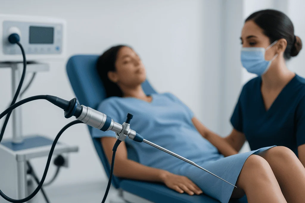
Hysteroscopy before IVF is a targeted way to examine the uterine cavity, the space where an embryo is expected to implant and develop. During the procedure, a doctor passes a very thin telescope-like instrument, called a hysteroscope, through the vagina and cervix into the uterus. The camera provides a direct view of the uterine lining and the shape of the cavity.
In fertility care, hysteroscopy is most often considered when there is a reason to suspect that something inside the uterus may reduce the chance of implantation or increase the risk of miscarriage. It can be purely diagnostic, meaning the doctor only looks and assesses, or operative, meaning small instruments are used to treat a problem during the same procedure.
Not every person preparing for IVF treatment needs hysteroscopy. Many patients can be evaluated with ultrasound, saline infusion sonography, or other imaging. Hysteroscopy becomes especially useful when these tests suggest an abnormality, when symptoms are present, or when previous embryo transfers have not resulted in pregnancy despite good-quality embryos and appropriate treatment planning.
Why the Uterus Matters in IVF
IVF focuses on several important steps: ovarian stimulation, egg retrieval, fertilization, embryo development, and embryo transfer. Even when the embryo is healthy, implantation also depends on a receptive uterine lining and a cavity that can support early pregnancy. This is why uterine evaluation is an important part of a complete fertility assessment.
Problems within the uterine cavity may act like a physical or inflammatory barrier. For example, a polyp or submucosal fibroid may change the surface of the lining, while scar tissue can distort the cavity. A uterine septum or adhesions may reduce the space available for implantation. In some cases, subtle inflammation of the lining may be considered if symptoms or history point in that direction.
It is important to keep expectations realistic. Hysteroscopy can find and treat certain uterine causes of reduced fertility, but it does not address all reasons IVF may be unsuccessful. Egg quality, sperm factors, embryo genetics, hormone balance, endometrial receptivity, and medical conditions all play a role. A balanced infertility evaluation looks at both uterine and non-uterine factors.
When Hysteroscopy May Be Recommended Before IVF
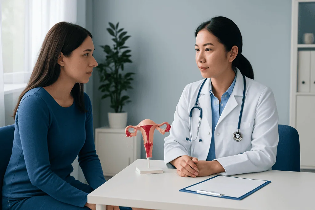
A fertility specialist may recommend hysteroscopy before IVF when imaging shows a possible uterine cavity abnormality. This may include suspected endometrial polyps, fibroids that project into the uterine cavity, scar tissue, retained tissue after pregnancy loss, or an unusual uterine shape. Hysteroscopy can confirm what is present and help guide treatment.
It may also be considered when a patient has symptoms such as heavy menstrual bleeding, bleeding between periods, pelvic discomfort, or a history of infection or uterine surgery. Previous procedures such as dilation and curettage, cesarean section, myomectomy, or treatment for miscarriage can sometimes be associated with changes inside the uterus, although many patients heal without problems.
Another common reason is repeated implantation failure or recurrent pregnancy loss, especially when other causes have already been reviewed. In these situations, hysteroscopy may reveal small abnormalities that were not clear on ultrasound. However, routine hysteroscopy for every IVF patient remains debated, and many guidelines support a selective, individualized approach rather than automatic use.
What Hysteroscopy Can Detect and Treat
Hysteroscopy gives the doctor a direct view of the uterine lining, the openings of the fallopian tubes into the uterus, and the contour of the cavity. This makes it especially useful for detecting focal problems that may be missed or uncertain on routine ultrasound. The findings can vary from a completely normal cavity to abnormalities that require treatment before embryo transfer.
Common findings may include:
- Endometrial polyps: Soft overgrowths of the uterine lining that may be associated with irregular bleeding or implantation concerns.
- Submucosal fibroids: Fibroids that bulge into the cavity and can alter its shape.
- Intrauterine adhesions: Bands of scar tissue that may narrow or distort the cavity.
- Uterine septum: A congenital band of tissue that divides part of the uterine cavity.
- Retained tissue or inflammation: Findings that may need removal, biopsy, or additional testing.
If a treatable abnormality is found, operative hysteroscopy may be performed either at the same appointment or in a planned second procedure. Small polyps can often be removed, adhesions can be divided, and selected septa or fibroids can be treated. The decision depends on the size and type of the finding, available equipment, anesthesia plan, and patient safety.
What to Expect During the Procedure
Hysteroscopy is usually scheduled after menstrual bleeding has ended and before ovulation, when the uterine lining is thinner and the view is clearer. Patients may be asked to avoid pregnancy in that cycle before the procedure. The care team will provide instructions about eating, drinking, medications, and whether someone should accompany the patient home.
The procedure may be done in an outpatient clinic or an operating room. Some diagnostic hysteroscopies use local pain relief or mild sedation, while operative hysteroscopy may require deeper sedation or anesthesia. A sterile fluid is gently used to expand the uterine cavity so the doctor can see the lining clearly. The hysteroscope passes through the cervix, so no abdominal incision is needed.
The duration varies. A simple diagnostic hysteroscopy may be brief, while removal of a polyp, fibroid, or scar tissue can take longer. Afterward, patients are monitored for a short time. Mild cramping, light bleeding, or watery discharge can occur for a few days. The clinic will explain when normal activities, sexual intercourse, and fertility treatment steps can safely resume.
Benefits, Limitations, and Possible Risks
The main benefit of hysteroscopy is accuracy. Unlike imaging tests that infer what is happening inside the uterus, hysteroscopy allows direct visualization. It can also combine diagnosis and treatment, which may shorten the pathway for patients who need correction of a clear cavity problem before embryo transfer.
At the same time, hysteroscopy has limitations. A normal hysteroscopy does not guarantee implantation, and an abnormal finding does not always mean it is the only cause of infertility. Some uterine factors are microscopic or functional and may not be visible. Also, the benefit of treating very small or uncertain findings should be discussed carefully, because unnecessary procedures can delay IVF and add cost or discomfort.
Hysteroscopy is generally considered safe when performed by trained clinicians, but it is still a medical procedure. Possible risks include bleeding, infection, cervical injury, uterine perforation, fluid-related complications, or anesthesia-related reactions. These complications are uncommon, and the care team reduces risk through appropriate patient selection, sterile technique, careful monitoring, and clear aftercare instructions.
Timing Hysteroscopy With IVF Treatment
The best timing depends on why hysteroscopy is being done and whether treatment is needed. If the hysteroscopy is diagnostic and the cavity is normal, the IVF plan may continue without major delay. If a polyp, fibroid, adhesion, or septum is treated, the doctor may recommend waiting for the uterus to heal before embryo transfer. This is particularly relevant when a fresh transfer was being considered.
In many IVF programs, embryos can be frozen and transferred in a later cycle if uterine treatment is needed. This allows time for healing and for the lining to return to an appropriate pattern. The waiting period varies according to the procedure performed, the amount of tissue treated, and the patient’s overall fertility plan.
Hysteroscopy should be integrated into a broader fertility strategy rather than viewed as a stand-alone solution. Patients being treated for female infertility may also need evaluation of ovulation, ovarian reserve, tubes, sperm parameters, endocrine conditions, and embryo development. The fertility team will weigh whether hysteroscopy is likely to change management before recommending it.
Recovery, Self-Care, and When to See a Doctor
Most patients recover quickly after hysteroscopy. Rest on the day of the procedure is usually advised, especially if sedation or anesthesia was used. Light cramping can often be managed with the pain relief recommended by the doctor. Patients may be advised to avoid tampons, swimming, and sexual intercourse for a short period to reduce infection risk, particularly after operative treatment.
Patients should follow the specific instructions given by their clinic, because aftercare differs depending on whether tissue was removed, a biopsy was taken, or adhesions were treated. Any prescribed medication should be taken exactly as directed. If pathology results are expected, a follow-up visit is important to review findings and decide when IVF or embryo transfer can proceed.
A doctor should be contacted promptly if there is heavy bleeding, fever, worsening pelvic pain, foul-smelling discharge, dizziness, or symptoms that feel unusual for the patient. For international patients, Acibadem International’s multidisciplinary fertility specialists and JCI-accredited hospitals can diagnose and treat uterine cavity conditions within individualized IVF care plans. As with any fertility decision, patients should discuss benefits, alternatives, and timing with a qualified reproductive medicine specialist.
Frequently asked questions
Is hysteroscopy required before every IVF cycle?
No. Hysteroscopy is not required for every IVF patient. It is usually considered when ultrasound or other tests suggest a uterine cavity problem, when symptoms are present, or after repeated unsuccessful embryo transfers or pregnancy losses.
Can hysteroscopy improve IVF success?
Hysteroscopy may help when it finds and treats a problem that could interfere with implantation, such as a polyp, submucosal fibroid, adhesion, or septum. However, it does not guarantee pregnancy, because IVF outcomes depend on many factors including embryo quality, age, sperm factors, and overall health.
Is hysteroscopy painful?
Discomfort varies by patient and by the type of procedure. Some diagnostic hysteroscopies cause mild cramping, while operative procedures may be done with sedation or anesthesia. The care team will discuss pain control options before the appointment.
How soon can IVF continue after hysteroscopy?
If hysteroscopy is only diagnostic and results are normal, treatment may continue soon afterward, depending on the clinic’s plan. If tissue is removed or the uterine cavity is repaired, the doctor may recommend waiting for healing before embryo transfer.
What is the difference between diagnostic and operative hysteroscopy?
Diagnostic hysteroscopy is used to look inside the uterus and identify any abnormalities. Operative hysteroscopy uses small instruments through the hysteroscope to treat certain findings, such as removing a polyp or dividing scar tissue.
Can hysteroscopy detect endometriosis?
Hysteroscopy examines the inside of the uterus, so it does not diagnose endometriosis outside the uterine cavity. Endometriosis is usually evaluated through symptoms, imaging in selected cases, and sometimes laparoscopy. A fertility specialist can decide which tests are appropriate.
References
- American Society for Reproductive Medicine
- European Society of Human Reproduction and Embryology
- American College of Obstetricians and Gynecologists
- National Institute for Health and Care Excellence
- Royal College of Obstetricians and Gynaecologists
This article is for general information only and is not a substitute for professional medical advice. Please consult a qualified doctor about your individual situation.
IVF & fertility treatment in Turkey — success rates and costs
JCI-accredited · board-certified surgeons · reply within 24h
Add us as a Preferred Source to see more of our trusted health content across Google Search, AI Overviews and Discover.

Related Treatments
Related Conditions
More from the Health Library



Related Specialists

Prof. Dr. Bülent Tıraş
Gynecology & Obstetrics
Prof. Dr. Mete Güngör
Gynecology & Obstetrics
Prof. Dr. Faruk Suat Dede
Gynecology & Obstetrics
Prof. Dr. Tansu Küçük
Gynecology & Obstetrics