Hysteroscopy Before IVF: When the Uterus Needs Checking

Hysteroscopy allows direct inspection of the uterine cavity and can often treat small abnormalities during the same procedure. It may be recommended before IVF if scans suggest polyps, fibroids, adhesions, a septum, abnormal bleeding or repeated implantation failure.
Key Takeaways
- Hysteroscopy allows direct inspection of the uterine cavity and can often treat small abnormalities during the same procedure.
- It may be recommended before IVF if scans suggest polyps, fibroids, adhesions, a septum, abnormal bleeding or repeated implantation failure.
- Routine hysteroscopy for every IVF patient is not universally recommended; the decision should be individualized.
- Most diagnostic hysteroscopies are short procedures, and recovery is usually quick, but mild cramping or spotting can occur.
- The timing of IVF after hysteroscopy depends on whether the procedure was diagnostic only or included surgical treatment.
Hysteroscopy before IVF is a camera-based procedure used to look inside the uterus when doctors suspect a uterine cavity problem that may affect embryo implantation or pregnancy. It is not necessary for every IVF patient, but it can be helpful when ultrasound findings, symptoms, miscarriage history or previous unsuccessful embryo transfers suggest that the uterus needs closer evaluation.
Overview
Hysteroscopy before IVF is a procedure in which a fertility specialist or gynecologist uses a thin telescope-like instrument, called a hysteroscope, to look directly inside the uterine cavity. The uterus is where an embryo must implant and grow, so its shape and lining are important parts of fertility assessment. During hysteroscopy, the doctor can identify conditions such as endometrial polyps, certain fibroids, scar tissue, or a uterine septum that may not always be fully clear on standard imaging.
In fertility care, hysteroscopy is usually considered after an initial evaluation with pelvic ultrasound, saline infusion sonography, or other tests. It is not automatically needed for everyone planning IVF treatment. Many people proceed to IVF without hysteroscopy when scans are normal and there are no concerning symptoms. However, when the uterine cavity needs a closer look, hysteroscopy can provide direct visual information and, in some cases, allow treatment at the same time.
The main goal is to make sure the inside of the uterus is as favorable as possible for embryo transfer. This does not mean hysteroscopy guarantees pregnancy, because IVF success depends on many factors, including age, egg and sperm quality, embryo development, genetics, hormonal health and overall medical history. Instead, hysteroscopy is one tool that may help identify and correct specific uterine factors before an embryo is placed.
Why the Uterus May Need Checking Before IVF
The uterine cavity is lined by the endometrium, a specialized tissue that changes during the menstrual cycle and supports embryo implantation. Even small changes in the cavity, depending on their size and location, may interfere with embryo placement or the early stages of pregnancy. For this reason, fertility teams often assess the uterus as part of a broader evaluation for female infertility.
Hysteroscopy may be recommended if there are symptoms such as abnormal uterine bleeding, spotting between periods, heavy periods, pelvic discomfort, or bleeding after fertility medications. It may also be advised if ultrasound suggests a polyp, a submucosal fibroid, fluid in the cavity, scarring, or an unusually shaped uterus. Some patients are offered hysteroscopy after recurrent miscarriage or repeated unsuccessful embryo transfers, especially when other explanations have not been found.
Common uterine findings that hysteroscopy can help diagnose include:
- Endometrial polyps: soft tissue overgrowths from the uterine lining.
- Submucosal fibroids: benign muscle growths that bulge into the uterine cavity.
- Intrauterine adhesions: bands of scar tissue, sometimes called Asherman syndrome.
- Uterine septum: a congenital band of tissue dividing part of the uterine cavity.
- Chronic inflammation or irregular lining: findings that may require additional testing or treatment.
Who May Benefit and Who May Not Need It

The decision to perform hysteroscopy before IVF should be individualized. It is more likely to be useful when there is a specific reason to suspect a uterine cavity problem. Examples include an abnormal ultrasound, a history of intrauterine procedures, previous uterine infection, difficult embryo transfer, recurrent implantation failure, recurrent pregnancy loss, or unexplained bleeding. Patients with prior dilation and curettage, cesarean-related uterine concerns, myomectomy, or surgery for uterine anomalies may also need closer assessment.
On the other hand, many patients with a normal uterine cavity on high-quality imaging and no symptoms may not gain additional benefit from routine hysteroscopy. Medical societies and fertility guidelines generally encourage careful selection rather than performing invasive procedures for everyone. A fertility specialist weighs the possible benefit of identifying a treatable abnormality against the cost, discomfort, timing and small procedural risks.
It is also important to remember that not all IVF challenges are caused by the uterus. Some unsuccessful cycles relate to embryo chromosome status, ovarian response, sperm factors, endometrial timing, immune or endocrine conditions, or chance. A balanced infertility evaluation looks at both partners and the full reproductive picture before deciding whether hysteroscopy is the right next step.
How Hysteroscopy Is Performed
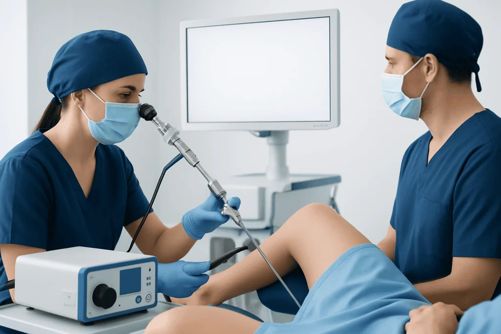
Hysteroscopy is usually performed through the vagina and cervix, without abdominal incisions. The doctor gently passes a narrow hysteroscope into the uterus and uses sterile fluid to expand the cavity so the lining can be seen clearly. A camera transmits images to a screen, allowing the doctor to examine the shape of the cavity, the openings of the fallopian tubes, and the appearance of the endometrium.
There are two broad types: diagnostic hysteroscopy and operative hysteroscopy. Diagnostic hysteroscopy is mainly for looking and confirming whether the cavity is normal. Operative hysteroscopy uses small instruments through the hysteroscope to remove or correct selected abnormalities, such as polyps, small fibroids, adhesions or a septum. Sometimes a planned diagnostic procedure becomes operative if a treatable finding is identified and consent has been obtained.
The procedure may be done in an office setting, day surgery unit or operating room, depending on the patient’s needs, the expected complexity and local practice. Pain control also varies. Some patients need only simple pain relief, while others may receive local anesthesia, sedation or general anesthesia, especially if operative treatment is planned. The healthcare team explains preparation, fasting instructions if needed, and what to expect before the appointment.
Timing Before IVF and What Results Mean
Hysteroscopy is often scheduled in the first half of the menstrual cycle, after bleeding has stopped and before ovulation. At this time, the lining is usually thinner, visibility is better and the chance of an undetected pregnancy is low. Patients may be asked to avoid unprotected intercourse in the cycle of the procedure or to take a pregnancy test beforehand, depending on clinic protocol.
If hysteroscopy is purely diagnostic and the uterus looks normal, IVF planning may often continue without much delay. If a polyp or other abnormality is removed, the doctor may recommend waiting until the lining has healed before embryo transfer. The waiting period can vary from one cycle to several weeks or longer, depending on the extent of treatment, whether a biopsy was taken, whether infection treatment is needed, and whether the transfer will be fresh or frozen.
Results should be interpreted in context. A normal hysteroscopy can be reassuring because it reduces concern about major cavity abnormalities. An abnormal result can be useful when it identifies a treatable factor, but it does not prove that the finding was the only cause of infertility or IVF failure. The fertility plan may also include embryo assessment, hormonal optimization, sperm evaluation, or procedures such as intracytoplasmic sperm injection when appropriate for male-factor or fertilization concerns.
Benefits, Limitations and Possible Risks
The main advantage of hysteroscopy is direct visualization. Unlike imaging tests that infer the shape of the cavity, hysteroscopy allows the doctor to see the inside of the uterus in real time. It can also be both diagnostic and therapeutic, meaning that some problems can be treated during the same procedure. For selected patients, this may reduce uncertainty before embryo transfer and help the fertility team plan care more precisely.
However, hysteroscopy has limitations. It does not assess egg quality, sperm function, embryo genetics, tubal function outside the uterine openings, or all causes of miscarriage. Very small or microscopic problems may require biopsy or laboratory testing, and not every visual finding requires treatment. For this reason, the procedure should be part of a complete fertility strategy rather than a stand-alone solution.
Hysteroscopy is generally considered a safe and commonly performed gynecologic procedure, but no procedure is risk-free. Possible side effects include mild cramping, light bleeding, temporary watery discharge, nausea after anesthesia, or shoulder discomfort from positioning. Less common complications include infection, heavier bleeding, cervical injury, uterine perforation, fluid overload, or complications related to anesthesia. Patients should receive clear instructions about warning signs and follow-up.
Recovery, Self-Care and Preparing for the Next Step
Recovery after diagnostic hysteroscopy is often quick, and many patients return to usual light activities within a short time, depending on how they feel and what type of anesthesia was used. Mild cramps and spotting can occur for a few days. After operative hysteroscopy, recovery may take longer, and the doctor may advise avoiding tampons, swimming, sexual intercourse or strenuous exercise for a specified period to reduce infection risk and support healing.
Patients should follow their clinic’s instructions about medications, antibiotics if prescribed, pain relief, and when to restart fertility medications. It is important not to begin an embryo transfer cycle until the fertility specialist confirms that the uterine lining has healed and the timing is appropriate. If tissue was removed or a biopsy was taken, pathology results may guide further management.
Practical preparation can make the experience easier. Patients may want to ask whether the procedure is diagnostic or operative, what anesthesia is planned, whether someone should accompany them home, how soon results will be discussed, and how the findings could change the IVF timeline. Keeping a written list of questions helps ensure that decisions are understood, especially during emotionally demanding fertility treatment.
When to See a Doctor
Anyone planning IVF should discuss uterine cavity assessment with a qualified fertility specialist, particularly if there has been abnormal bleeding, recurrent miscarriage, previous uterine surgery, suspected polyps or fibroids, pelvic infection, or repeated unsuccessful embryo transfers. A doctor can decide whether ultrasound, saline sonography, hysterosalpingography, MRI, hysteroscopy or a combination of tests is most appropriate.
After hysteroscopy, patients should contact their healthcare team promptly if they develop fever, worsening pelvic pain, heavy bleeding, foul-smelling discharge, dizziness, fainting, or symptoms that feel unusual for their recovery. These signs do not necessarily mean a serious problem, but they should be assessed so that treatment can be given if needed.
For international patients seeking fertility evaluation, Acibadem International provides access to multidisciplinary specialists and JCI-accredited hospitals that diagnose and treat reproductive conditions, including uterine cavity problems before IVF. The most suitable approach depends on the patient’s medical history, test results and personal fertility goals, and should be planned through an individualized consultation.
Frequently asked questions
Is hysteroscopy required before every IVF cycle?
No. Hysteroscopy is not required for every person before IVF. It is usually considered when symptoms, previous history or imaging suggest a uterine cavity abnormality, or after repeated implantation failure or miscarriage.
Can hysteroscopy improve IVF success?
Hysteroscopy may help selected patients if it identifies and treats a problem such as a polyp, adhesion, fibroid protruding into the cavity or uterine septum. However, it does not guarantee pregnancy, and IVF success depends on many factors including embryo quality, age, sperm factors and overall reproductive health.
Is hysteroscopy painful?
Some patients feel mild to moderate cramping, similar to menstrual cramps, especially during office hysteroscopy. Pain control options vary and may include simple pain medicine, local anesthesia, sedation or general anesthesia depending on the procedure and the patient’s needs.
How soon can IVF start after hysteroscopy?
If hysteroscopy is diagnostic only and the uterus is normal, IVF may often continue with little delay. If tissue is removed or surgery is performed, the doctor may recommend waiting until the uterine lining has healed, which can vary by case.
What is the difference between hysteroscopy and ultrasound?
Ultrasound uses sound waves to create images of the uterus and ovaries from outside the cavity. Hysteroscopy places a small camera inside the uterus, allowing direct visualization and, in some cases, treatment of abnormalities during the same procedure.
Can polyps or fibroids be removed during hysteroscopy?
Many endometrial polyps and some fibroids that extend into the uterine cavity can be removed during operative hysteroscopy. Whether this is possible depends on the size, location and type of the growth, as well as the patient’s medical situation.
This article is for general information only and is not a substitute for professional medical advice. Please consult a qualified doctor about your individual situation.
IVF & fertility treatment in Turkey — success rates and costs
JCI-accredited · board-certified surgeons · reply within 24h
Add us as a Preferred Source to see more of our trusted health content across Google Search, AI Overviews and Discover.

Related Treatments
Related Conditions
More from the Health Library

IVF After Tubal Ligation: Options, Testing, and Success Factors

Male Age and IVF: Sperm Quality, Embryo Development, and Risks

IVF for Unexplained Infertility: Tests, Treatment Steps, and Expectations

IVF in Turkey: How to Choose the Right Fertility Clinic

Embryo Quality in IVF: Grading, Transfer Decisions, and What It Means

When Is a Lady Fertile? How the Fertile Window Really Works
Gynecology & Obstetrics Specialists at Acibadem

Dr. Cemile Alçay
Gynecology & Obstetrics
Assoc. Prof. Dr. Ceren Yıldız Eren
Gynecology & Obstetrics
Prof. Dr. Çetin Kılıççı
Gynecology & Obstetrics
Assoc. Prof. Dr. Cevat Rıfat Cündübey
Gynecology & Obstetrics