Keratoconus: Blurred Vision from Corneal Thinning

Keratoconus affects the cornea, the clear front surface of the eye, and can cause increasing astigmatism and visual distortion. Symptoms often begin in adolescence or early adulthood and may progress gradually over several years.
Key Takeaways
- Keratoconus affects the cornea, the clear front surface of the eye, and can cause increasing astigmatism and visual distortion.
- Symptoms often begin in adolescence or early adulthood and may progress gradually over several years.
- Diagnosis is usually made with a detailed eye examination and corneal mapping tests such as corneal topography or tomography.
- Treatment may include glasses, specialty contact lenses, corneal cross-linking, intracorneal ring segments, or corneal transplant in advanced cases.
- Avoiding eye rubbing and managing allergies may help reduce irritation and mechanical stress on the cornea.
Keratoconus is an eye condition in which the normally round cornea gradually thins and bulges into a cone-like shape, causing blurred or distorted vision. Early diagnosis and appropriate treatment can help protect vision and reduce the need for more invasive procedures.
Overview
Keratoconus is a progressive condition that affects the cornea, the clear dome-shaped window at the front of the eye. In a healthy eye, the cornea has a smooth, rounded shape that helps focus light sharply onto the retina. In keratoconus, the corneal tissue becomes thinner and weaker, allowing it to bulge forward into an irregular cone-like shape.
This change in shape can bend incoming light unevenly, leading to blurred vision, distorted vision, glare, and frequent changes in glasses prescription. The condition usually affects both eyes, although one eye may be more affected than the other. It often starts during the teenage years or early adulthood and may progress slowly over time.
Keratoconus is not usually painful, and many people continue daily activities with appropriate treatment and follow-up. The main goal of care is to improve vision and, when possible, slow or stop progression before the cornea becomes severely distorted. Modern diagnostic imaging and treatments such as corneal cross-linking have improved the ability to detect and manage keratoconus earlier.
Symptoms of Keratoconus
The symptoms of keratoconus can be subtle at first. A person may notice that vision becomes blurry despite wearing glasses, or that a new prescription improves vision only for a short time. As the cornea becomes more irregular, standard glasses may no longer provide clear, stable vision.
Common symptoms include:
- Blurred, distorted, or double vision in one eye
- Increasing nearsightedness or astigmatism
- Frequent changes in eyeglass or contact lens prescription
- Glare, halos, or difficulty seeing at night
- Light sensitivity
- Eye strain, headaches, or squinting to see clearly
- Contact lenses that become uncomfortable or fit poorly
In more advanced keratoconus, the cornea can become very irregular or develop scarring, which further reduces visual clarity. Rarely, a sudden swelling of the cornea may occur due to a small break in the inner corneal layer; this is called acute corneal hydrops and needs prompt ophthalmic care.
Causes and Risk Factors

The exact cause of keratoconus is not fully understood. It is believed to involve a combination of genetic, biomechanical, environmental, and inflammatory factors that weaken the corneal structure. In many patients, the corneal collagen fibers that normally help maintain shape and strength appear less able to resist normal pressure inside the eye.
Several factors may increase the likelihood of developing or worsening keratoconus. A family history of keratoconus can raise risk, although many people with the condition have no known affected relatives. Allergic eye disease, chronic eye irritation, and repeated eye rubbing are also commonly associated with keratoconus and may contribute to progression.
Risk factors and associated conditions can include:
- Family history of keratoconus
- Frequent or forceful eye rubbing
- Allergic conjunctivitis or atopic conditions such as eczema or asthma
- Certain connective tissue or genetic conditions
- Poorly fitting contact lenses that irritate the eye
Having a risk factor does not mean a person will definitely develop keratoconus. However, people with persistent eye allergies, rapidly changing astigmatism, or a family history should have regular eye examinations, especially during the years when keratoconus most often begins.
How Keratoconus Is Diagnosed
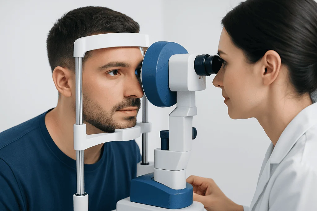
Keratoconus is diagnosed by an eye care specialist through a combination of symptoms, vision testing, slit-lamp examination, and specialized corneal imaging. A standard eye exam may show increasing astigmatism or reduced best-corrected vision, but early keratoconus can be difficult to identify without corneal mapping.
Corneal topography and corneal tomography are important tests used to evaluate the shape, curvature, and thickness of the cornea. These scans create detailed maps that can detect subtle steepening, thinning, or asymmetry before major symptoms appear. Pachymetry may also be used to measure corneal thickness.
Diagnosis may also involve checking how well glasses or contact lenses correct vision and looking for clinical signs such as corneal thinning, fine stress lines, or scarring. Because keratoconus can progress, repeat testing over time is often recommended to determine whether the cornea is changing and whether treatments to stabilize it should be considered.
Treatment Options
Treatment depends on the stage of keratoconus, the degree of visual distortion, corneal thickness, and whether the condition is progressing. In early cases, glasses or soft contact lenses may provide adequate vision. As the cornea becomes more irregular, specialty contact lenses are often needed to create a smoother focusing surface.
Specialty lenses may include rigid gas permeable lenses, hybrid lenses, scleral lenses, or custom soft lenses designed for irregular corneas. Scleral lenses rest on the white part of the eye and vault over the cornea, which can improve comfort and visual quality for many people with more advanced irregularity. Proper fitting by an experienced contact lens specialist is important to protect the cornea and maintain comfort.
Corneal cross-linking is a treatment used to strengthen the cornea and slow or stop progression. It typically involves applying riboflavin eye drops to the cornea and activating them with controlled ultraviolet light. Cross-linking is not primarily intended to eliminate the need for glasses or contact lenses; its main purpose is to stabilize the cornea and reduce the risk of further worsening.
In selected patients, intracorneal ring segments may be placed within the cornea to help flatten and regularize its shape. In advanced cases with significant scarring, severe thinning, or vision that cannot be improved with lenses, corneal transplantation may be considered. The choice of treatment is individualized, and long-term follow-up remains important even after successful treatment.
Prevention and Self-care
Keratoconus cannot always be prevented, especially when genetic or structural factors are involved. However, some steps may help reduce irritation and avoid additional mechanical stress on the cornea. One of the most important recommendations is to avoid rubbing the eyes, particularly with force or frequency.
People with itchy eyes should speak with an eye doctor about safe ways to manage allergies, dryness, or inflammation. Treatment may include lubricating eye drops, allergy eye drops, cold compresses, or other therapies depending on the cause of irritation. Managing allergies can make it easier to avoid rubbing and improve day-to-day comfort.
Helpful self-care habits include:
- Attend scheduled eye exams and repeat corneal imaging when advised
- Use contact lenses only as prescribed and maintain proper hygiene
- Report sudden changes in vision or lens comfort promptly
- Wear protective eyewear during activities that may risk eye injury
- Avoid sleeping in contact lenses unless specifically instructed
Self-care does not replace medical treatment, especially if keratoconus is progressing. Early monitoring is valuable because treatments such as corneal cross-linking are generally most useful before severe corneal distortion or scarring develops.
When to See a Doctor
A person should schedule an eye examination if vision becomes increasingly blurred, distorted, or difficult to correct with glasses. Frequent prescription changes, worsening astigmatism, night glare, or contact lenses that no longer fit comfortably are also reasons to seek evaluation. Teenagers and young adults with a family history of keratoconus should consider regular eye checks even if symptoms are mild.
Prompt medical attention is recommended for sudden vision loss, new corneal cloudiness, significant eye pain, marked redness, or sudden sensitivity to light. These symptoms may have causes other than keratoconus and should be assessed by a qualified eye care professional.
International patients who need evaluation or treatment for keratoconus can access multidisciplinary eye care services at Acibadem International, where specialists in JCI-accredited hospitals diagnose and manage corneal conditions. As with any medical decision, the best approach should be discussed with an ophthalmologist who can assess the individual eye findings and treatment goals.
Frequently asked questions
Is keratoconus the same as astigmatism?
Keratoconus can cause astigmatism, but they are not the same condition. Astigmatism means the eye has an uneven focusing shape, while keratoconus is a progressive thinning and bulging of the cornea. Some people with astigmatism do not have keratoconus.
Can keratoconus make a person go blind?
Keratoconus can significantly reduce vision if it becomes advanced, but complete blindness is uncommon. Many patients achieve useful vision with glasses, specialty contact lenses, cross-linking, or surgery when needed. Regular monitoring helps identify progression early.
Does corneal cross-linking cure keratoconus?
Corneal cross-linking does not usually restore the cornea to a normal shape or remove the need for vision correction. Its main purpose is to strengthen the cornea and reduce the chance of further progression. Some patients still need glasses or specialty contact lenses after treatment.
Are contact lenses safe for keratoconus?
Contact lenses are commonly used to improve vision in keratoconus and can be safe when properly fitted and cared for. Specialty lenses, such as rigid gas permeable or scleral lenses, may provide better vision than standard soft lenses. Regular follow-up is important to check comfort, fit, and corneal health.
Can children or teenagers have keratoconus?
Yes, keratoconus often begins in adolescence and may progress more actively in younger patients. Early symptoms can include changing glasses prescriptions, eye rubbing, and reduced visual clarity. Prompt evaluation is important because early treatment may help preserve vision.
What should people with keratoconus avoid?
People with keratoconus should avoid frequent or forceful eye rubbing, as it may contribute to corneal irritation and progression. They should also avoid wearing poorly fitting contact lenses or using eye drops without medical guidance. Any sudden vision change should be checked by an eye care professional.
This article is for general information only and is not a substitute for professional medical advice. Please consult a qualified doctor about your individual situation.
Explore treatments in Turkey — costs, top hospitals & a free quote
JCI-accredited · board-certified surgeons · reply within 24h
Add us as a Preferred Source to see more of our trusted health content across Google Search, AI Overviews and Discover.

Related Treatments
More from the Health Library

Can You Get LASIK With Astigmatism? Eligibility Explained

PRK vs LASEK: Key Differences in Recovery and Results

Orbital Tumors: Symptoms, Imaging, and When Orbit Surgery Is Needed

When to Go to Urgent Care for Eye Problems

How Old Do You Have to Be to Get LASIK?