

Nuclear Stress Test vs Stress Echocardiogram: Which Heart Test May Be Better?
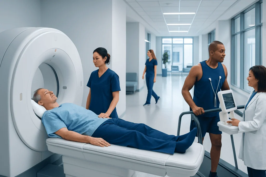
A nuclear stress test focuses on blood flow to the heart muscle, while a stress echocardiogram focuses on heart movement and valve function. Neither test is universally better; the most suitable choice depends on the clinical situation.
Key Takeaways
- A nuclear stress test focuses on blood flow to the heart muscle, while a stress echocardiogram focuses on heart movement and valve function.
- Neither test is universally better; the most suitable choice depends on the clinical situation.
- Stress echocardiography avoids radiation, but image quality can be limited in some people.
- Nuclear stress testing can be especially helpful when doctors need more detail about reduced blood flow or prior heart damage.
- Preparation, medications, and the ability to exercise may affect which test is recommended.
- A cardiologist uses test results together with symptoms, examination findings, and other heart tests to guide next steps.
A nuclear stress test and a stress echocardiogram are both common ways to evaluate how the heart responds to exercise or medication-induced stress. The better test depends on the question being asked, the person’s symptoms, body type, medical history, and how clearly images can be obtained.
Overview: how these two heart tests differ
When a doctor needs to learn how the heart performs under stress, two widely used options are the nuclear stress test and the stress echocardiogram. Both are designed to detect signs that the heart may not be getting enough blood during activity, which can happen with coronary artery disease. They can also provide clues about heart function, exercise capacity, and whether symptoms such as chest discomfort or shortness of breath may be related to the heart.
A stress echocardiogram uses ultrasound to create moving pictures of the heart before and after exercise, or after a medicine that makes the heart work harder. The main goal is to see how well the heart muscle contracts and whether the valves open and close normally. A nuclear stress test uses a small amount of radioactive tracer and a special camera to show blood flow to the heart muscle at rest and during stress.
Because these tests look at slightly different aspects of heart health, one is not automatically superior to the other. In many cases, the choice depends on what the doctor most needs to know. For example, if structural heart problems or valve disease are suspected, stress echocardiography may be especially useful. If blood flow patterns, prior scarring, or the extent of ischemia need closer assessment, nuclear imaging may add valuable information.
What a stress echocardiogram shows
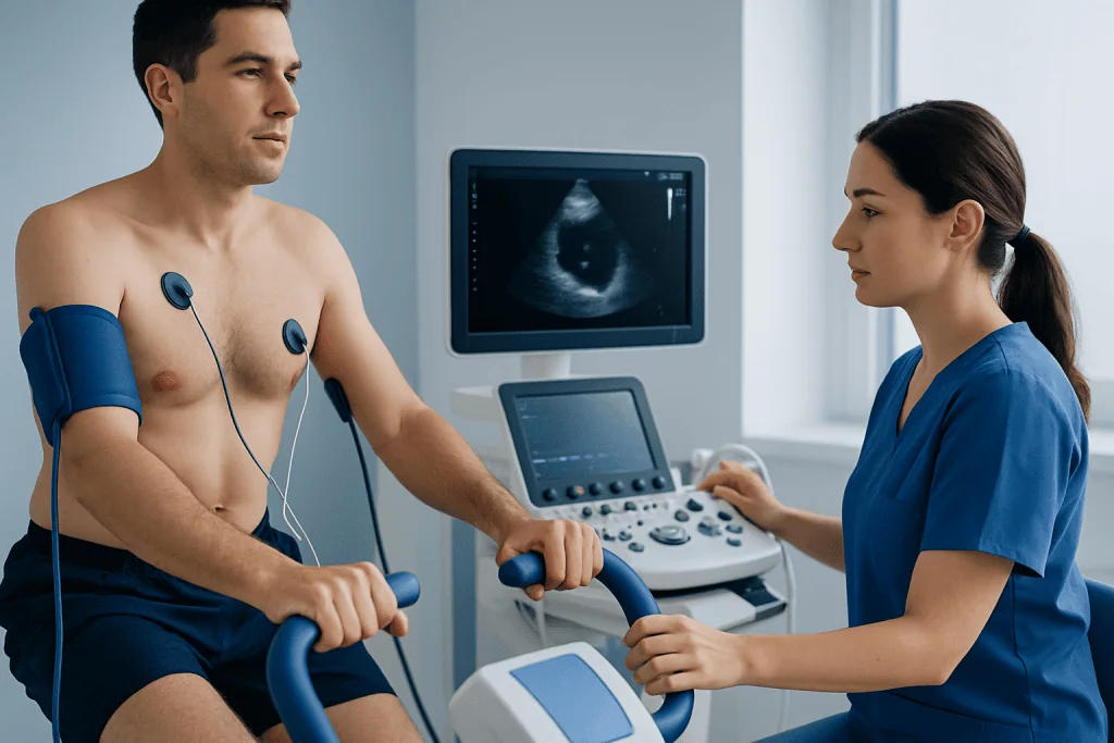
A stress echocardiogram combines a standard echocardiogram with stress, either from exercise on a treadmill or bicycle or from medication when exercise is not possible. Ultrasound images are taken before stress and again when the heart rate is elevated. The doctor compares these images to see whether any part of the heart muscle moves less effectively during stress, which can suggest a reduced blood supply.
This test can also reveal changes in pumping function and help assess valve problems such as aortic regurgitation or other forms of valve disease. In some people, it may help evaluate symptoms linked to heart failure or cardiomyopathy. Because ultrasound shows heart motion in real time, it can be especially helpful when the clinical question involves both blood supply and the mechanics of the heart.
One important advantage is that stress echocardiography does not use ionizing radiation. It is usually completed relatively quickly and may be preferred in younger patients or in those who should minimize radiation exposure. However, the quality of the images depends on how well the ultrasound can “see” the heart through the chest wall, which may be more difficult in some individuals.
What a nuclear stress test shows
A nuclear stress test evaluates how blood flows through the heart muscle at rest and during stress. After a small amount of tracer is injected into a vein, a camera detects the tracer and creates images that show where blood flow is normal and where it may be reduced. Areas that receive less tracer during stress may suggest narrowed coronary arteries, while areas with reduced uptake both at rest and stress can point to old injury or scar tissue.
This type of test is often used when a doctor wants a clearer picture of perfusion, meaning the blood supply to the heart itself. It can be useful if a person has symptoms that suggest angina, a history of myocardial infarction, or an abnormal resting ECG that makes other stress tests harder to interpret. Nuclear imaging may also help estimate how extensive a blood flow problem is, which can guide decisions about medication, further imaging, or procedures.
The main limitation is that it involves radiation exposure, even though the dose is generally kept as low as reasonably possible. It also takes longer than a stress echo because images are usually obtained both before and after stress. Depending on the tracer and protocol, the test may require waiting periods between the injection and imaging portions.
When one test may be better than the other
A stress echocardiogram may be a better choice when the doctor needs information about heart wall motion, valve function, or exercise-related symptoms without radiation. It may also be favored when a patient is able to exercise and has a body type that allows clear ultrasound images. In experienced hands, it is a very effective test for many people with suspected ischemia or unexplained shortness of breath.
A nuclear stress test may be preferred when there is a strong need to evaluate blood flow patterns in more detail, when prior ECG changes make interpretation difficult, or when stress echo images are likely to be suboptimal. It may also be helpful in people with previous heart damage, prior revascularization, or more complex risk assessment questions. If the concern is how much of the heart muscle is affected by reduced blood flow, nuclear imaging can be particularly informative.
Doctors also consider practical factors. These include the patient’s age, ability to exercise, weight and body habitus, lung disease, heart rhythm issues, pregnancy status, and whether they have had previous heart surgery or implanted devices. In some circumstances, a cardiologist may instead recommend another test altogether, such as coronary CT angiography, cardiac MRI, or invasive angiography with possible stent procedures if symptoms or risk are high.
In short, the better test is the one that best answers the clinical question with the fewest limitations for that individual patient. The decision is personalized rather than one-size-fits-all.
How doctors choose the right test
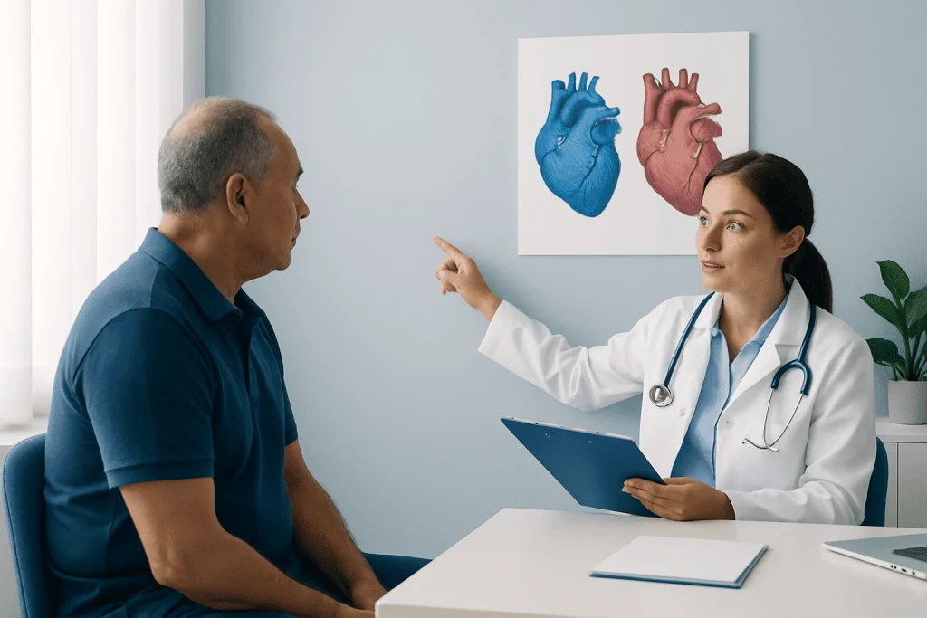
Choosing between these tests starts with a careful review of symptoms, medical history, physical examination, and baseline studies such as an ECG. Symptoms like chest pressure with exertion, unusual breathlessness, reduced exercise tolerance, or a pattern suggestive of stable angina may lead to stress testing. The doctor then estimates how likely it is that the symptoms are due to reduced blood flow in the coronary arteries.
Risk factors also influence the decision. High blood pressure, diabetes, smoking, abnormal cholesterol levels, kidney disease, family history of early heart disease, and older age all matter. Management of high blood pressure and abnormal cholesterol levels is an important part of the larger picture, whether or not a stress test shows ischemia.
The doctor also considers whether the patient can exercise adequately. Exercise-based testing provides helpful information about symptoms, blood pressure response, and fitness level. If exercise is not possible because of joint pain, neurologic problems, lung disease, or frailty, medicines can be used to simulate the effect of exercise for either a nuclear stress test or, in some cases, a stress echocardiogram.
Results are interpreted in context. A normal result is reassuring, but it does not completely rule out every heart condition. Likewise, an abnormal result does not always mean a blocked artery will require a procedure. Some patients improve with medication, lifestyle changes, and follow-up, while others may need more testing or treatment such as coronary artery bypass surgery in selected cases.
What to expect before, during, and after testing
Preparation instructions vary slightly by center, so patients should follow the guidance given by their care team. In general, they may be asked to avoid caffeine for a period before the test, especially for certain nuclear stress protocols, and to avoid heavy meals shortly before testing. Some heart medications may need temporary adjustment, but this should only be done if the doctor specifically advises it.
During a stress echocardiogram, electrodes are placed on the chest to monitor the heart rhythm, and blood pressure is checked throughout the test. Ultrasound images are taken, then the patient exercises or receives medication to increase heart workload. Additional images are obtained quickly at peak stress or immediately afterward. The test team watches closely for symptoms, ECG changes, or blood pressure responses.
During a nuclear stress test, a tracer is injected into a vein, and images are taken with a special camera. Stress is created with exercise or medication, followed by another tracer injection and another set of images. The test usually takes longer than stress echocardiography because image acquisition may occur after a waiting period. Patients can typically return to usual activities afterward unless their doctor advises otherwise.
Mild fatigue is common after either test, especially if exercise was involved. Serious complications are uncommon because testing is supervised by trained staff with equipment to respond if needed. Patients should ask when results will be available and what the next step will be if the test is normal, unclear, or abnormal.
Benefits, limitations, and safety considerations
Both tests are considered generally safe when performed for the right reasons and under medical supervision. Their main benefit is that they help identify whether symptoms may be related to reduced blood flow to the heart, while also helping estimate risk and guide treatment decisions. They can reduce uncertainty and help doctors avoid either under-treating important disease or over-testing low-risk symptoms.
Stress echocardiography offers the benefit of no radiation exposure and direct visualization of heart motion and valves. Its limitations include dependence on image quality and the expertise needed to capture and interpret images at the right moment. In some patients, especially those with obesity, certain lung conditions, or unusual chest anatomy, the pictures may be less clear.
Nuclear stress testing provides strong information about myocardial perfusion and can identify areas of reversible ischemia or previous damage. Its limitations include radiation, longer test time, and the need for tracer injection. Some medications or foods may interfere with the stress portion, so careful preparation matters.
Neither test should be viewed in isolation. If symptoms are severe, worsening, or highly suggestive of significant heart disease, doctors may move more quickly to advanced evaluation. Near the end of the care pathway, multidisciplinary specialists at Acibadem International and its JCI-accredited hospitals diagnose and treat heart conditions for international patients when coordinated specialist care is needed.
When to speak with a doctor urgently
A planned stress test is usually arranged to evaluate stable symptoms, but some warning signs need immediate medical attention rather than routine testing. New or severe chest pain, chest pressure that lasts more than a few minutes, fainting, sudden shortness of breath, or symptoms that occur at rest can signal an emergency. These situations should be assessed urgently.
Patients should also seek prompt advice if they develop palpitations with dizziness, unusual weakness, or swelling that is getting worse. People who have known heart disease, diabetes, or multiple risk factors should not ignore subtle but persistent changes in exercise tolerance. Early assessment often helps clarify whether symptoms are related to the heart or another cause.
If a stress test has already been performed, patients should discuss the results with their doctor and ask what they mean for daily life, medicines, and future follow-up. The best next step may involve lifestyle changes, risk-factor treatment, additional imaging, or referral to a heart specialist. A personalized plan is the safest approach.
Frequently asked questions
Is a nuclear stress test more accurate than a stress echocardiogram?
Not in every situation. A nuclear stress test may give more detailed information about blood flow to the heart muscle, while a stress echocardiogram may be better for evaluating heart motion and valve function. The most appropriate test depends on the patient’s symptoms, history, and the clinical question.
Does a stress echocardiogram involve radiation?
No. A stress echocardiogram uses ultrasound, so it does not expose the patient to ionizing radiation. This can make it an attractive option for some people, although image quality may vary from person to person.
Why would a doctor order a nuclear stress test instead of a stress echo?
A doctor may prefer a nuclear stress test when blood flow to the heart needs closer assessment, when previous ECG findings make interpretation harder, or when ultrasound images are likely to be limited. It can also help evaluate prior heart damage or the extent of ischemia.
Can both tests be done without exercise?
Yes. If a person cannot exercise enough for accurate testing, medications can be used to place the heart under stress. The choice of medicine and protocol depends on the type of test and the patient’s health status.
How should someone prepare for these tests?
Preparation usually includes avoiding certain foods, drinks, or medications for a period before the test, especially caffeine before many nuclear stress studies. Instructions vary, so patients should carefully follow the testing center’s guidance and ask before stopping any prescription medication.
What happens if the test result is abnormal?
An abnormal result does not automatically mean surgery or a procedure is needed. The doctor will consider the severity of the findings, symptoms, risk factors, and other test results before recommending medication changes, further imaging, or interventions.
This article is for general information only and is not a substitute for professional medical advice. Please consult a qualified doctor about your individual situation.
Explore treatments in Turkey — costs, top hospitals & a free quote
JCI-accredited · board-certified surgeons · reply within 24h
Add us as a Preferred Source to see more of our trusted health content across Google Search, AI Overviews and Discover.
Check your numbers in seconds
BMI, calories, due date, blood pressure and 30+ more clinical calculators — free, instant, doctor-reviewed ranges.

Cardiology Department
Diagnosis and treatment of heart and vascular conditions, from prevention to advanced interventional procedures.
93 specialists in this unitRelated Treatments
Related Conditions
More from the Health Library
Peripheral Artery Disease vs Deep Vein Thrombosis: How to Tell the Difference

Pulmonary Hypertension: What It Means and How Doctors Confirm It

Aortic Aneurysm: How It Is Found and When Surgery Is Needed
Coronary Angiography: Procedure, Risks, and Results
Heart Failure: Shortness of Breath, Swelling, and Modern Treatment
Cardiac Rehabilitation After Bypass Surgery: When to Start and How It Helps
Cardiology Specialists at Acibadem

Prof. Dr. Ahmet Oytun Baykan
Cardiology
Prof. Dr. Ahmet Tulga Ulus
Cardiovascular Surgery
Prof. Dr. Ahmet Ümit Güllü
Cardiovascular Surgery
Prof. Dr. Aleks Değirmencioğlu
Cardiology