What Is a Diagnostic Mammogram After an Abnormal Screening?

A diagnostic mammogram is a follow-up test used to examine a specific breast finding in more detail. An abnormal screening result does not automatically mean breast cancer.
Key Takeaways
- A diagnostic mammogram is a follow-up test used to examine a specific breast finding in more detail.
- An abnormal screening result does not automatically mean breast cancer.
- Extra mammogram views and sometimes breast ultrasound are commonly used together.
- Results may show a benign change, the need for short-term follow-up, or the need for biopsy.
- Prompt follow-up helps clarify findings and supports early diagnosis when treatment is needed.
A diagnostic mammogram after an abnormal screening mammogram is a more detailed breast imaging test used to look closely at a specific area. In many cases, it helps show that a finding is benign, but it can also guide the next steps if more evaluation is needed.
Overview
A screening mammogram is designed to look for early breast changes before symptoms appear. If the screening exam shows an area that is unclear or looks different from expected, the next step is often a diagnostic mammogram. This is a more focused imaging test that takes additional pictures of one or both breasts to study a specific finding more carefully.
A diagnostic mammogram after abnormal screening does not mean that cancer has been found. Many callback findings turn out to be normal overlapping tissue, cysts, benign calcifications, or other non-cancerous breast changes. The purpose of the test is to clarify what was seen on screening and decide whether it is harmless, needs monitoring, or needs further testing.
Unlike routine screening, a diagnostic mammogram is tailored to the patient. The radiologist may request special angles, magnified images, or compression views during the visit. In many breast centers, the images are reviewed while the patient is still there so that additional views or a breast ultrasound can be performed right away if needed.
Why a Screening Mammogram May Be Called Abnormal
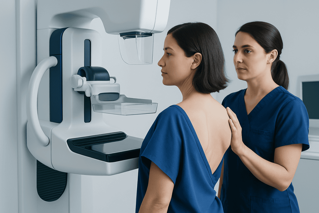
An abnormal screening mammogram means that the radiologist saw something that needs a closer look. It does not describe a diagnosis by itself. Screening images are taken in standard views, and sometimes these views are not enough to fully define an area.
Common reasons for recall include an area of asymmetry, a possible mass, calcifications, architectural distortion, or a region that may simply represent overlapping breast tissue. Dense breast tissue can also make interpretation more difficult and may increase the chance that extra imaging is recommended.
Prior mammograms are very helpful for comparison. A finding that seems new may need diagnostic evaluation, while a stable area that has not changed over time may be reassuring. If older images are available from another center, sharing them can sometimes reduce unnecessary follow-up.
- Overlapping normal tissue that mimics a lump
- Small calcium deposits called calcifications
- A possible cyst or solid mass
- Areas that look distorted or less clearly defined
- Technical factors such as motion or incomplete visualization
What Happens During a Diagnostic Mammogram
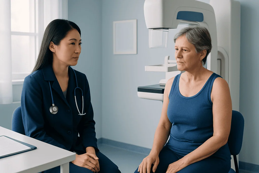
A diagnostic mammogram is similar to a screening mammogram, but it is more targeted. The breast is positioned on the mammography unit, and gentle compression is used to spread the tissue so the images are clearer. The technologist takes additional views that focus on the area of concern.
Depending on the finding, the radiologist may request spot compression views, magnification views, or images from different angles. Spot compression presses on a small part of the breast to separate tissue and improve detail. Magnification views enlarge a small area, which is especially useful for evaluating calcifications.
Many patients also have a breast ultrasound during the same visit, particularly if the mammogram suggests a lump or if the breast tissue is dense. Ultrasound can help distinguish a fluid-filled cyst from a solid lesion and may offer more information about the shape and margins of a finding. In some cases, advanced imaging such as breast MRI may be recommended if the mammogram and ultrasound do not fully answer the question.
The examination usually takes longer than a routine screening visit because the images are customized and may be reviewed while the patient waits. Although compression can be uncomfortable for a short time, it should not last long, and the staff can often adjust positioning to make the experience easier.
How Results Are Interpreted
After the images are reviewed, the radiologist assigns an assessment category to guide follow-up. Some findings are clearly benign and require no extra testing beyond routine screening. Others are probably benign and may need a short-interval follow-up mammogram, often in several months, to confirm stability.
If a finding remains suspicious after diagnostic imaging, the radiologist may recommend a biopsy. This does not mean cancer is certain; it means that imaging alone cannot confidently show that the area is benign. Tissue sampling is the most reliable way to make a diagnosis in those cases.
Patients may hear terms such as mass, asymmetry, distortion, or calcifications. These are imaging descriptions, not final diagnoses. For example, calcifications are common and often harmless, but some patterns need closer review. A solid mass may still be benign, such as a fibroadenoma, while a cyst is usually a non-cancerous fluid-filled sac.
If biopsy is needed, options may include image-guided procedures such as breast biopsy performed with mammographic, ultrasound, or MRI guidance. The best method depends on which imaging test shows the finding most clearly.
Possible Next Steps After the Exam
There are several possible outcomes after a diagnostic mammogram. The most reassuring result is that the area is normal or clearly benign, allowing the patient to return to routine screening. Another common outcome is a recommendation for short-term follow-up imaging to make sure a probably benign finding does not change over time.
When the images suggest a specific benign condition, the report may name it. Examples include a simple breast cyst, benign calcifications, fat necrosis, or a likely fibroadenoma. In other situations, further evaluation may be needed for a suspicious area or when symptoms such as a new lump, nipple discharge, or skin changes are present.
If additional testing is advised, this may include breast ultrasound or a biopsy. Some patients are also referred to a breast specialist to review the imaging findings and discuss options. If a diagnosis such as breast cancer is eventually confirmed, early evaluation helps treatment planning begin as soon as possible.
For international patients who need coordinated evaluation, Acibadem International’s multidisciplinary specialists and JCI-accredited hospitals provide diagnostic and treatment services for breast conditions.
How to Prepare and What to Bring
Preparation for a diagnostic mammogram is usually simple. It is often helpful to schedule the exam for a time when the breasts are less tender, if possible. On the day of the test, patients are generally advised not to use deodorant, powders, creams, or lotions under the arms or on the breasts because these products can appear on images.
Bringing prior mammogram images and reports can be very important, especially if the screening was done at another facility. Comparison with earlier studies may help the radiologist decide whether a finding is stable and benign or whether it is truly new. Patients should also bring a list of symptoms, prior breast procedures, hormone use, and family history if relevant.
Comfortable two-piece clothing can make the visit easier because only the upper garments need to be removed. If the patient has breast implants, is pregnant, or could be pregnant, the imaging team should be informed before the exam. Questions are welcome, and understanding the reason for the test often helps reduce anxiety.
When to See a Doctor Promptly
Patients should follow up promptly after being called back for diagnostic imaging, even though many findings are benign. Delays can prolong uncertainty and may postpone diagnosis if a problem does need attention. Timely evaluation is especially important if there are new breast symptoms in addition to an abnormal screening result.
A doctor should be contacted sooner if there is a new breast lump, persistent focal pain, nipple discharge that is spontaneous or bloody, skin dimpling, nipple inversion that is new, or redness and swelling that do not improve. These symptoms do not always mean cancer, but they should be assessed by a qualified clinician.
If the results are unclear, patients can ask for an explanation in plain language, whether comparison images are needed, and what the next recommended step is. Shared decision-making is important, and a second radiology or breast specialist opinion may be reasonable in selected cases. The goal is not to create alarm, but to make sure the finding is understood and managed appropriately.
Frequently asked questions
Does an abnormal screening mammogram mean cancer?
No. An abnormal screening mammogram means that an area needs a closer look, not that cancer has been diagnosed. Many callback findings are caused by overlapping tissue, cysts, or other benign breast changes.
What is the difference between a screening mammogram and a diagnostic mammogram?
A screening mammogram is a routine test done when there are no breast symptoms. A diagnostic mammogram is a follow-up exam that uses additional, targeted images to evaluate a specific area or symptom in more detail.
Will a diagnostic mammogram hurt?
It may feel uncomfortable because breast compression is needed to obtain clear images, but the pressure is usually brief. Patients can tell the technologist if they are in pain so positioning can be adjusted when possible.
Why might an ultrasound be done after a diagnostic mammogram?
Ultrasound is often used to add more information, especially when a lump is suspected or the breasts are dense. It can help show whether a finding is a fluid-filled cyst or a solid area that needs closer evaluation.
How soon are results available?
This depends on the imaging center, but many facilities review diagnostic images during the visit and may discuss initial findings the same day. A formal report is then sent to the referring doctor, who explains the next steps if needed.
If the radiologist recommends a biopsy, does that mean the finding is cancer?
Not necessarily. A biopsy is recommended when imaging cannot confirm that a finding is benign with enough confidence. Many biopsies show non-cancerous results, but tissue testing is the safest way to make a clear diagnosis.
This article is for general information only and is not a substitute for professional medical advice. Please consult a qualified doctor about your individual situation.
Explore treatments in Turkey — costs, top hospitals & a free quote
JCI-accredited · board-certified surgeons · reply within 24h
Add us as a Preferred Source to see more of our trusted health content across Google Search, AI Overviews and Discover.

More from the Health Library

Neurophysiology for Seizures and Fainting: Which Tests Help Find the Cause?

What Is a Diagnostic Mammogram Result Called Back For?

What Happens During a Blood Test Visit?
When to Get Imaging for Breast Symptoms: Lumps, Pain, and Nipple Changes

Does Quest Accept Walk-Ins? How Lab Check-In Works and When You May Need an Appointment