Congenital Nevi Treatment
Congenital nevi are birthmarks present at birth that may require monitoring or removal for cosmetic reasons, irritation, or melanoma risk. Treatment is individualized with dermatologic assessment and surgical planning.
Quick answer
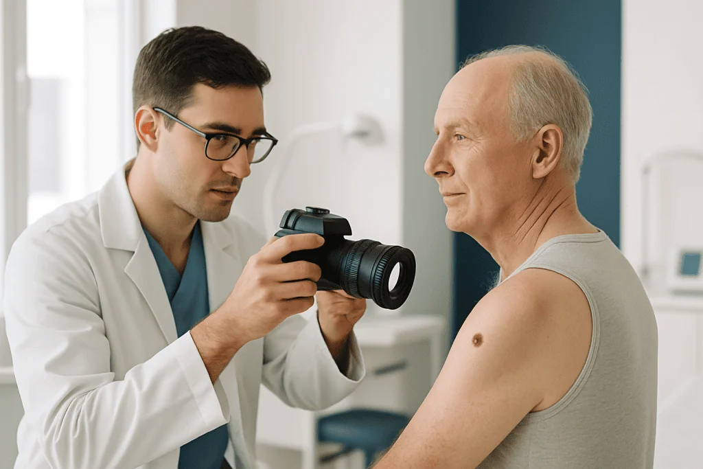
A congenital nevus is a mole present at birth or appearing shortly afterwards, formed by clusters of pigment cells in the skin. Treatment ranges from specialist monitoring with dermoscopy and photography to surgical excision, which may be done in one procedure or in stages using tissue expansion or grafts for larger nevi. Removed tissue is examined by pathology to confirm the diagnosis.
Congenital Nevi: Deciding Whether to Monitor or Treat a Nevus
A congenital nevus is a mole that is present at birth or becomes visible during the first weeks to months of life. It forms when pigment-producing cells, called melanocytes, cluster in the skin during development in the womb, creating a patch that may be flat or raised, smooth or textured, and anywhere from light brown to nearly black. Treatment for congenital nevi covers the full range of care these birthmarks may need: careful specialist assessment, structured long-term monitoring, and, when there is a sound medical or personal reason, surgical removal, staged excision or reconstruction.
You may also see the same word spelt naevus in British English texts; the meaning is identical, and the plural in either spelling is nevi or naevi. Whichever spelling your records use, the clinical questions are the same. Is this nevus one that can safely be watched? Is it one that should be examined more closely, photographed, biopsied or removed? And if removal is chosen, what will the skin look like afterwards? This page walks through each of those questions in turn.
For many families, a congenital nevus is first noticed in the delivery room or at an early paediatric visit. Some are small, flat and easy to keep an eye on. Others are larger, darker, raised, hairy, or sit on the face, scalp, trunk, hands, feet or genital area, where they may affect appearance, clothing comfort, hygiene or self-confidence as a child grows. Parents usually have several questions at once: is this birthmark dangerous, will it grow, should it be removed now or watched over time, and if surgery is needed, will it leave a scar? Adults with congenital nevi often have their own concerns, particularly if the lesion has changed, becomes irritated, or affects personal comfort in social, professional or intimate settings.
Treatment matters because congenital nevi are not all the same. Size, depth, location, colour pattern, surface texture and behaviour over time all influence both medical risk and treatment planning. Most congenital nevi do not become melanoma, but some types, particularly very large or giant congenital melanocytic nevi, are associated with a higher lifetime melanoma risk and may need closer surveillance. In other cases, treatment is considered for repeated irritation, bleeding, difficulty monitoring, or cosmetic and psychological reasons. A plan that fits one patient may be entirely wrong for another.
A congenital nevus belongs to the broad family of conditions present from birth, but it behaves very differently from most of them. Unlike structural problems such as congenital heart diseases or congenital hydrocephalus, a congenital nevus is confined to the skin in the great majority of cases, and for many people it never needs anything beyond periodic checks. You can read more about how conditions present from birth are assessed on our congenital disease page.
At Acibadem, care for congenital nevi is individualised. Dermatology, plastic and reconstructive surgery, paediatric specialists, pathology, radiology and oncology expertise may be involved depending on the patient’s age, nevus characteristics and risk profile. The goal is not simply to remove a visible mark, but to make a careful medical decision: when observation is appropriate, when biopsy or imaging is needed, and when removal can be performed with the best possible functional and aesthetic planning.
Are a nevus and mole the same thing?
Yes, nevus is simply the medical term for what most people call a mole. Doctors prefer the medical word because it allows precise classification: a melanocytic nevus is a benign collection of pigment cells, and further terms describe when it appeared, how deep the cells sit, and how it looks under magnification. When you read “congenital melanocytic nevus” in a clinic letter, it means a mole made of pigment cells that was present at or around birth. Nothing about the word itself implies danger; the descriptive details that follow it, size, pattern, stability, carry the clinically useful information.
What does a skin nevus look like?
A skin nevus typically looks like a round or oval patch or bump that is darker than the surrounding skin, with a reasonably even colour and a defined border. Congenital nevi in particular may be larger than ordinary moles, oval or geographic in shape, and light brown to dark brown or black. The surface can be smooth, pebbled, thickened or velvety, and coarse hair often grows within the lesion, especially as a child gets older. Some congenital nevi darken, lighten or become more raised during childhood and adolescence, often in proportion to normal growth. Some larger lesions are surrounded by smaller “satellite” nevi scattered across nearby skin. Because appearance alone does not settle every question, dermatologists use magnified examination and photographic comparison rather than relying on a single glance.
What Congenital Nevi Treatment Is
Congenital nevi treatment refers to the medical assessment, monitoring and, when appropriate, removal or reduction of a congenital melanocytic nevus. A melanocytic nevus is a benign proliferation of melanocytes, the cells that give skin its pigment, and the congenital form arises before birth rather than later in life. These cells create a visible lesion that may range from light brown to dark brown or black, with a surface that can be smooth, raised, thickened or hairy.
“Treatment” can mean several quite different things. For a small, stable nevus in a low-risk area, treatment may consist of dermatological examination, dermoscopic imaging, photographic monitoring and education about the changes worth noting. For a nevus that is changing, difficult to assess, symptomatic or cosmetically distressing, treatment may involve surgical excision. In larger nevi, complete removal may require staged procedures, tissue expansion, skin grafting or reconstructive techniques carried out over months. In selected cases, laser-based treatment may be discussed for surface pigmentation or hair reduction, but a laser does not remove all nevus cells and is not a substitute for medical surveillance where melanoma risk is a concern.
Because congenital nevi can extend into deeper layers of the skin, the choice of treatment rests on clinical examination and realistic goals. Surgical removal can take away visible pigment and remove the area that would otherwise need monitoring, but scars are expected after any excision, no honest surgeon will tell you otherwise. A well-planned procedure aims to place scars in natural skin lines or less conspicuous areas where possible, to protect nearby structures, and to preserve function.
In children, timing is especially important. Some procedures are easier when a child is older and there is more skin to work with; others may be considered earlier if the nevus sits where staged reconstruction can produce a better result over time, or if there are specific medical concerns. A sensible plan weighs the child’s growth, anaesthesia safety, family preferences and psychosocial development together, rather than treating the nevus in isolation.
What is the difference between congenital nevi and acquired nevi?
The difference between congenital nevi and acquired nevi is timing: congenital nevi are present at birth or emerge shortly afterwards, while acquired nevi develop later in childhood, adolescence or adulthood. The distinction matters clinically. Congenital nevi tend to be larger than acquired moles, their pigment cells often reach deeper into the dermis and sometimes around hair follicles, and they grow in proportion to the child rather than appearing on previously clear skin. Congenital nevi are also classified by their projected adult size, small, medium, large or giant, because size correlates with how they are monitored and how removal, if chosen, would be planned. Acquired nevi are usually smaller, more superficial and far more numerous over a lifetime. Both types are benign in the great majority of cases, but the surveillance approach for a giant congenital lesion differs considerably from that for an ordinary acquired mole.
What is a compound nevus?
A compound nevus is a mole whose pigment cells sit in both the upper layer of the skin (the epidermis) and the deeper layer (the dermis). Dermatologists classify melanocytic nevi by depth: junctional nevi lie at the boundary between epidermis and dermis and tend to be flat; intradermal nevi sit entirely within the dermis and are often raised and pale; compound nevi combine both levels. This classification is more than academic. Congenital nevi frequently show deep, compound-type or deeper involvement, which is one reason surface treatments such as laser cannot reach every nevus cell, and why pathology examination of removed tissue gives information that visual inspection cannot.
Who May Need Evaluation or Treatment for a Congenital Nevus
Anyone with a congenital nevus can benefit from at least one specialist evaluation, particularly if the lesion is large, unusual in appearance, changing, or located somewhere hard to examine, such as the scalp or back. Many patients do not need removal at all, but an expert assessment clarifies whether monitoring is sufficient or whether additional testing or treatment should be considered. If your child was born with a nevus, that first structured assessment also gives you a documented baseline against which every future observation can be compared.
Typical features of congenital nevi include a mole or pigmented patch present from birth, gradual enlargement in proportion to body growth, darker or somewhat uneven colouration, hair growth within the lesion, and a surface that may become raised or textured over the years. Some congenital nevi are surrounded by smaller satellite nevi. Very large lesions may cover part of the scalp, face, back, buttocks or limbs, sometimes crossing joints or the midline.
Diagnosis usually begins with a clinical skin examination. A dermatologist evaluates the size, shape, border, colour, texture and distribution of the nevus, as well as the total number and pattern of other pigmented lesions on the body. Dermoscopy, a non-invasive, magnified examination of the skin, helps identify pigment patterns that are typical or atypical. Standardised photography allows objective comparison over time. If a suspicious area is found, a biopsy may be recommended. In patients with large or giant congenital nevi, particularly when many satellite lesions are present or the nevus involves the head, neck or spinal region, additional evaluation may be discussed to look for rare associated conditions such as neurocutaneous melanosis, in which pigment cells are also present in the membranes around the brain and spinal cord.
How rare are giant congenital melanocytic nevi?
Giant congenital melanocytic nevi are rare. Small congenital nevi are relatively common findings at newborn examinations, medium lesions are considerably less frequent, and the giant form, usually defined by its projected size in adulthood, is seen only occasionally even in large dermatology centres. Published estimates vary depending on how “giant” is defined and which populations were studied, so precise figures should be treated cautiously. What matters practically is that rarity concentrates experience: giant lesions are best assessed in centres where dermatology, plastic surgery, paediatrics and pathology routinely work together, because both the surveillance strategy and any reconstructive plan are more complex than for a small nevus.
Referral for treatment is commonly considered when a nevus bleeds, ulcerates, becomes painful, changes rapidly, develops a new firm nodule, or is repeatedly irritated by clothing, shaving, sports equipment or hygiene routines. Treatment may also be considered when a lesion causes significant emotional distress or social discomfort, particularly on the face, neck, hands or other exposed areas. Emotional burden is a legitimate indication, not a lesser one; for a school-age child, a highly visible facial nevus can shape daily experience in ways adults underestimate.
Whoever performs the assessment, earlier photographs, previous biopsy reports and notes about prior laser or surgical treatments make any consultation far more informative, because they show how the nevus has behaved over years rather than how it looks on a single day. Baby photographs are often surprisingly useful for establishing whether growth has been proportional.
Melanoma Risk: What a Nevus Can and Cannot Tell You
Can a nevus be cancerous?
Most nevi are benign and stay benign, but melanoma, a skin cancer arising from pigment cells, can develop within or near a nevus, which is why change over time is the single most useful observation a patient or parent can make. A nevus itself is not cancer; the concern is the small possibility of malignant transformation, and that possibility is not evenly distributed across all moles. Stable size, colour and texture over years are reassuring features. New firmness, persistent ulceration, rapid asymmetric growth or a distinctly different-looking area within a lesion are the kinds of findings that lead a dermatologist to recommend biopsy, because microscopy answers questions that the naked eye cannot.
Are congenital nevi associated with malignancy?
Most congenital nevi never become malignant, but the association with melanoma is real and depends heavily on the size and type of the lesion. Small and medium congenital melanocytic nevi carry a low lifetime risk, and melanoma arising in them, when it occurs at all, tends to appear after childhood. Very large and giant congenital nevi carry a higher lifetime risk, and in these lesions melanoma can occasionally arise earlier and deeper in the skin, where it is harder to see. This is why large lesions are followed more closely, why a new nodule within one is taken seriously, and why removed tissue is always examined by pathology. Risk assessment is individual: it considers the nevus itself, satellite lesions, family history and examination findings together, not size alone.
Can congenital nevi fade, or do they go away?
Congenital nevi do not usually go away on their own. They typically persist for life and grow in proportion to the body, and while their colour can shift, some lighten gradually, others darken or become more raised during adolescence, genuine spontaneous disappearance is uncommon. Occasionally a nevus develops a pale halo or patchy lightening; this is usually an immune phenomenon rather than a sign of trouble, but any abrupt or uneven change in an established lesion is worth documenting with photographs and discussing at a routine dermatology review, simply because “different” is the finding that examination and dermoscopy are designed to interpret. Planning around the assumption that a congenital nevus will fade is not realistic; planning around monitoring or removal is.
Conditions and Indications Addressed by Congenital Nevi Treatment
Congenital nevi treatment addresses both medical and quality-of-life indications. The right approach depends on the nevus type, size category, location, symptoms and the patient’s own priorities.
- Small congenital melanocytic nevi: often monitored unless they change, become irritated or are cosmetically concerning. Excision may be considered when the lesion sits in a favourable location and removal can be achieved with a simple closure and a single, well-placed scar.
- Medium congenital melanocytic nevi: monitored or removed depending on appearance, location, patient age and preference. Surgical planning weighs scar placement against the amount of surrounding skin available for closure.
- Large or giant congenital melanocytic nevi: these need the most detailed assessment. Management may include long-term surveillance, staged excision, tissue expansion, grafting or combined reconstructive techniques, with melanoma risk and psychosocial impact considered side by side.
- Symptomatic nevi: lesions that itch, bleed, crack, ulcerate, catch on clothing or become repeatedly inflamed may warrant removal to improve comfort and stop the cycle of trauma and healing.
- Changing or clinically atypical areas: a new nodule, rapid colour change, persistent ulceration, asymmetry or a different-looking region within the nevus may require biopsy or excision so that pathology can examine the tissue.
- Cosmetic and psychosocial concerns: visible congenital nevi can affect self-image, school experience, social confidence and personal comfort. A treatment decision can legitimately include emotional wellbeing alongside medical factors.
- Lesions in difficult-to-monitor areas: nevi on the scalp, back, buttocks or genital area are hard for patients to observe themselves. Closer dermatological follow-up, or removal of selected areas, may be advised.
The indication for treatment is never determined by size alone. A small lesion in a sensitive location may cause significant distress, while a larger lesion may be best managed with careful surveillance if removal would create a greater functional or aesthetic burden than the nevus itself. The most appropriate plan balances risk reduction, appearance, safety and the patient’s stage of life.
Should a nevus be removed?
Not every nevus should be removed, and for many congenital nevi the honest recommendation after assessment is monitoring rather than surgery. Removal makes sense when a lesion shows suspicious features, when it is repeatedly traumatised or symptomatic, when it sits somewhere that cannot realistically be monitored, or when its appearance causes distress that outweighs the certainty of a scar. Removal makes less sense when a lesion is stable and reassuring on dermoscopy, when excision would create a functional problem near an eyelid, lip or joint, or when the scar burden of a very large removal would exceed the benefit. The question is not “can it be removed”, most can, but “does removal serve this patient better than surveillance does”. That is a judgement made with a specialist, case by case.
How Congenital Nevi Treatment Is Performed
Initial Assessment and Planning
Treatment begins with a detailed consultation. The physician reviews your medical history, any family history of melanoma, previous changes in the nevus, symptoms, prior procedures and current concerns. In children, the consultation also covers growth patterns, developmental stage and anaesthesia considerations. The skin examination looks not only at the main nevus but at the total number and distribution of other pigmented lesions across the body, because context changes interpretation.
Dermoscopic assessment is used to examine pigment structures invisible to the naked eye. Clinical photography documents the baseline appearance. If any area looks suspicious, a biopsy may be performed before definitive planning; the sample goes to pathology to establish whether it contains benign nevus cells, atypical changes or malignancy. Definitive surgical decisions are better made with that answer in hand than without it.
For large or complex lesions, especially on the face, scalp, trunk or over joints, planning may bring in plastic and reconstructive surgery and, where useful, imaging. The team considers how much tissue can be removed safely, whether the wound can be closed directly, and how to protect structures such as eyelids, lips, the nose, ears, nerves and joints. If staged reconstruction is needed, the plan may span several months or longer, and you should expect that timeline to be discussed openly at the outset rather than discovered along the way.
Preparation Before Treatment
Preparation depends on the chosen approach. For a simple excision under local anaesthesia, you may need only basic pre-procedure instructions. For larger operations, particularly in children, or whenever general anaesthesia is planned, preoperative evaluation may include blood tests, an anaesthesia assessment, a review of your regular medications by the treating team, and guidance about fasting before surgery.
It helps the team to know about allergies, bleeding tendencies, previous reactions to anaesthesia, regular medications and supplements, and any personal or family history of abnormal scarring or keloids. Photographs taken at different ages are genuinely valuable, because they show whether the nevus has grown in proportion to the body or changed in an unexpected way: a distinction that shapes both urgency and technique.
For patients travelling from abroad, an initial review of records and photographs can sometimes take place before the journey, which helps establish whether a visit is likely to involve observation, biopsy, surgery or staged planning. Final decisions, however, almost always require an in-person examination; skin is assessed by touch and magnification as much as by sight, and no photograph fully replaces that.
Observation and Dermatologic Monitoring
Not every congenital nevus needs removing, and structured observation is itself a form of treatment. Monitoring is typically recommended when a lesion looks clinically reassuring, has remained stable, and causes neither symptoms nor distress. It usually includes regular dermatological examinations at an interval suited to the individual lesion, dermoscopic documentation, and clear education for the patient or parents about which changes are worth recording.
The changes dermatologists ask you to note include new or persistent pain, bleeding, ulceration, growth out of proportion to body growth, a new firm lump within the lesion, marked colour change, or a wound over the nevus that does not heal. Photographing the lesion at home under consistent lighting, next to a ruler or coin for scale, makes those observations far more useful at the next review. Sun protection is also part of sensible care: sunscreen does not eliminate melanoma risk, but limiting ultraviolet exposure is a reasonable component of long-term skin health and may reduce additional pigmentation change in and around the nevus.
Surgical Excision
Surgical excision is the most direct method of removing a congenital nevus. The surgeon removes the visible lesion together with a margin of surrounding skin appropriate to the clinical situation, sends the tissue to pathology for microscopic examination, and closes the wound, often in layers, to support healing and reduce tension on the skin. Small congenital nevi can frequently be removed in a single outpatient procedure under local anaesthesia, sometimes with sedation depending on age, anxiety and location. Duration varies from a brief procedure to a longer operation for larger or anatomically delicate lesions.
A typical single-stage excision follows a predictable sequence:
- Marking and planning: the excision lines are drawn, oriented where possible along natural skin creases to make the eventual scar less conspicuous.
- Anaesthesia: local anaesthetic is injected, or general anaesthesia is administered for children and larger procedures.
- Excision: the nevus is removed with its planned margin, down to a depth judged appropriate for the lesion.
- Layered closure: deeper stitches take tension off the skin edge, and the surface is closed with fine sutures.
- Pathology: the removed tissue is examined under the microscope to confirm the diagnosis and check the margins.
- Dressing and instructions: the wound is dressed and you receive written guidance on care, activity and follow-up.
For medium or large nevi, direct closure may not be possible without excessive tension on the skin. In those cases, the surgeon may recommend staged excision, removing portions of the nevus over several operations so the surrounding skin can stretch gradually. Staged excision trades a longer overall timeline for a more controlled scar, and it is commonly chosen when preserving function and appearance matters most.
Tissue Expansion and Reconstructive Techniques
For selected large congenital nevi, tissue expansion may be used. A temporary expander, essentially a deflated silicone balloon, is placed under healthy skin near the nevus and gradually filled over a series of visits, stretching the skin above it. Once enough new skin has been generated, the nevus is excised and the expanded skin is moved to cover the defect. The technique’s great advantage is that the reconstruction uses skin with the same colour, texture and hair pattern as the surrounding area, which grafts from distant sites cannot match.
Tissue expansion demands planning and patience. The expander is visible as a swelling during the filling phase, expansion sessions can be uncomfortable, there is a risk of infection or expander problems, and the total treatment takes months. It is not suitable for every patient or every body area, and the team should talk you through the practicalities, appearance during expansion, the visit schedule, and what happens if a stage does not go to plan, before you commit.
When neither direct closure nor expansion is suitable, skin grafting or local flap reconstruction may be considered. A graft moves a layer of skin from another part of the body to cover the excised site; a flap moves nearby tissue while keeping its blood supply attached. Both are effective, established techniques, but both create additional scars and require attentive postoperative care, and grafted skin may differ in colour and texture from its surroundings. These trade-offs are discussed honestly in advance, because a technically successful operation that surprises the patient is not a success.
Laser and Adjunctive Treatments
Laser treatment is sometimes discussed for congenital nevi, and its limits deserve plain statement. A laser can lighten surface pigment, improve texture or reduce hair, and in carefully selected cases that may be worthwhile. But congenital nevus cells frequently extend deep into the dermis, beyond the reach of surface treatment, so laser does not remove all nevus tissue. It can also alter the pigment pattern in ways that make future dermoscopic monitoring harder to interpret. For these reasons, laser should be considered only after dermatological evaluation, with clearly agreed goals, and never as a substitute for surveillance where melanoma risk is a consideration.
Hair reduction may be considered separately when coarse hair within a nevus causes irritation or cosmetic concern. It changes appearance and comfort, not medical risk, and decisions about hair should be kept distinct from decisions about surveillance or excision.
Technology Used in Diagnosis and Treatment
Modern congenital nevi care uses technology to sharpen judgement rather than replace it. Dermoscopy reveals pigment networks, globules, vessels and structural patterns in magnified detail. Digital photography supports objective comparison across visits and years. In complex cases, imaging can assist with surgical mapping or with evaluating deeper structures near the lesion.
In the operating theatre, magnification, fine instruments, modern anaesthesia monitoring and reconstructive planning tools support precision and safety. Pathology examination remains the essential final step whenever tissue is removed: it provides microscopic confirmation of the diagnosis and identifies any atypical or malignant change that examination from the surface could not detect.
Recovery After Treatment
Recovery depends on the procedure. After a small excision, most patients return to light activities within days, with instructions to keep the area clean, protect the incision and avoid stretching the wound. Sutures may be absorbable or may need removal at a follow-up visit. Bruising, swelling, tightness and mild discomfort are common early on and usually settle gradually.
After larger excisions, grafts, flaps or tissue expansion, recovery is more involved: dressing changes, activity restrictions, wound checks and staged follow-up visits. Children may need practical guidance about school, sport, bathing and scar care. Scar maturation continues for many months, scars typically pass through a pink, firm phase before softening and lightening, and patience during this period is part of a realistic plan.
Why Acting Early Matters
Early specialist evaluation does not mean early surgery. It means early clarity. A congenital nevus that has been examined, photographed and documented can be followed with confidence, and you can make decisions from information rather than worry. For large or complex nevi, early consultation may also widen the options, because reconstructive planning can be aligned with a child’s growth and development instead of working against it.
Delaying assessment makes it harder to know whether any change is new or long-standing. If a nevus develops bleeding, ulceration, a firm lump, persistent pain or rapid change, waiting delays the pathology answer that settles what is happening. Melanoma arising in congenital nevi is uncommon overall, but the consequences are significant when it does occur, particularly in higher-risk lesions, which is precisely why the higher-risk lesions are the ones followed most closely.
Delay can also narrow reconstructive choices. A lesion that is repeatedly traumatised or inflamed may heal with thicker scarring, making later surgery more complicated. For children, postponed evaluation can prolong distress when a nevus is highly visible and affecting confidence at school. Equally, early assessment often delivers the opposite of surgery: reassurance that observation is appropriate, and the avoidance of a procedure that was never needed.
The right timing is personal. It depends on medical risk, appearance, the child’s age, surgical feasibility, anaesthesia considerations and family values. A careful plan protects you from both overtreatment and undertreatment, and a plan made early is easier to keep careful.
Benefits of Congenital Nevi Treatment
The potential benefits depend on whether treatment means monitoring, excision, reconstruction or a combination. Framed honestly, they look like this:
| Benefit | What It Means for You |
|---|---|
| Clear diagnosis and risk assessment | A specialist evaluation establishes whether the nevus appears benign, needs monitoring, or requires biopsy or removal, replacing uncertainty with a plan. |
| Improved ability to monitor the skin | Dermoscopic imaging and standardised photography create a baseline, so future changes are easier to identify and interpret. |
| Removal of symptomatic tissue | Excision can end the cycle of irritation, bleeding and catching on clothing caused by a raised or exposed lesion. |
| Cosmetic and psychosocial improvement | When appropriate, treatment can reduce the visibility of a nevus and support confidence in social, school or professional settings. |
| Pathology confirmation | Removed tissue is examined microscopically, providing more definitive information than any visual assessment alone. |
| Individualised reconstructive planning | For larger lesions, staged procedures or tissue expansion can balance removal goals against function and appearance. |
Recovery Timeline After Congenital Nevi Treatment
Recovery varies with the size of the procedure, its location, the patient’s age and the reconstruction method, but most patients follow a broadly similar pattern of wound healing followed by slow scar maturation. Use the timeline below as orientation, not as a schedule; your own surgeon’s instructions take precedence.
| Time Period | What Patients Can Expect |
|---|---|
| Day 1 | Mild discomfort, swelling, tightness and a dressing over the treated area are common. You receive instructions for wound care, bathing, medications and activity limits. |
| First week | The incision or graft is monitored for healing. Bruising and swelling usually begin to settle. Physical activity may be restricted to protect the wound. |
| First month | Most simple excisions are well healed at the surface, though the scar may remain pink or firm. Larger reconstructions may still need dressing care and follow-up visits. |
| Several months | Scars gradually soften and mature. Scar care may include sun protection, silicone therapy, massage or other measures your surgeon recommends. |
| Longer term | Dermatological monitoring continues, especially if part of the nevus remains or you have multiple congenital nevi. Any planned staged procedures are scheduled as agreed. |
Factors That Influence Outcomes
A good result in congenital nevi treatment is shaped by several factors: the nevus’s size, depth, location and surface characteristics, and whether it can be removed in one stage. Lesions on the face, scalp, hands, feet, joints and genital area demand especially careful planning, because scars and tissue movement in these areas affect function as well as appearance.
Age matters. In infants and young children, skin elasticity and ongoing growth can work in the surgeon’s favour, but anaesthesia and postoperative care require genuine paediatric expertise. In adolescents and adults, the patient can take a direct part in decision-making, but the lesion may be larger and the surrounding skin less adaptable than it was in early childhood.
Your skin type and scarring tendency also shape the result. Some people form thicker, darker, wider or raised scars regardless of surgical technique. Previous surgery, infection, tension on the wound, smoking, certain medical conditions and inconsistent wound care can all impair healing. Sun exposure during scar maturation may darken the scar or the surrounding skin, which is why protection during those months is repeatedly emphasised.
For large or giant congenital nevi, expectations must be realistic and should be set before the first operation, not after it. Complete removal may not be possible, or may require several stages over a long period. Even when most visible pigment is gone, scars, graft colour differences or texture changes can remain. In some cases the safest, most sensible approach is partial removal of the most concerning or most visible areas, combined with continued monitoring of the rest.
Pathology findings can change the plan. If atypical changes or melanoma are identified in removed tissue, further excision, imaging, sentinel lymph node evaluation or oncology consultation may follow, depending on the diagnosis. These situations are uncommon, but they are exactly why expert assessment and microscopic tissue examination matter whenever suspicious features are present.
Above all, strong outcomes rest on a shared understanding between you, your family, the dermatologist and the surgeon. The best plan is one in which medical safety, reconstructive feasibility, cosmetic priorities and long-term surveillance have all been discussed openly before anything is done.
How Congenital Nevi Care Is Organised at Acibadem
Congenital nevi care at Acibadem is built around coordinated specialties rather than a single department. A small, stable lesion may need nothing more than a dermatology assessment with a surgical opinion. A large congenital nevus in a child may draw on paediatric anaesthesia, plastic and reconstructive surgery, dermatology and pathology together, with radiology or neurology involved in selected cases. When examination or pathology raises concern, the case can be reviewed jointly so that the plan reflects more than one specialist’s view.
The diagnostic pathway follows the same logic described throughout this page: dermoscopic evaluation, standardised photography, pathology review of any removed tissue, and reconstructive planning that respects both safety and appearance. Technology supports clinical judgement; it does not replace an experienced examination or an individualised plan. Where a nevus forms part of a wider picture of conditions present from birth, care can be aligned with the teams described on our congenital correction page.
The recommendation that comes out of an assessment is not automatic. Sometimes it is observation rather than surgery; sometimes biopsy, excision, staged removal, tissue expansion or simply a defined schedule of dermatological monitoring. The emphasis is on matching the approach to the patient’s medical risk, anatomy, age and priorities, including the honest possibility that the best treatment is none at all.
For patients who travel for care, follow-up is planned before the journey home: wound care instructions, pathology results when available, and clear recommendations for future monitoring. If staged procedures are planned, the expected schedule and the practical implications are set out in advance, and where ongoing dermatology follow-up can happen locally, the relevant findings and images are prepared so that the physician at home can continue surveillance without gaps.
Making a Considered Decision
A congenital nevus can raise understandable concern, especially when it is large, visible, changing or present in a child. The essential first step is a careful assessment by specialists who understand both the medical and the reconstructive sides of these lesions. With a proper evaluation, many patients can be monitored safely and never need surgery; when treatment is warranted, careful planning can reduce symptoms, improve appearance and support long-term skin surveillance.
Whatever path you take, the decision to monitor or remove a congenital nevus works best when it rests on documented history, photographs across the years, any biopsy reports, a record of changes, combined with clear medical guidance, realistic expectations about scars and staging, and respect for the patient’s own comfort and goals. A nevus is, in the end, a known quantity once it has been properly assessed. That knowledge, more than any single procedure, is what turns worry into a manageable plan.
Preparation
- A dermatologist or plastic surgeon evaluates the nevus size, location, symptoms, and melanoma risk, often using dermoscopy and clinical photography. Blood tests or anesthesia assessment may be needed for larger excisions. Patients may be asked to stop certain blood-thinning medicines and follow fasting instructions if sedation or general anesthesia is planned.
Aftercare
- Keep the wound clean and dry as instructed, and attend scheduled dressing and suture-removal appointments. The removed tissue is usually sent for pathology review. Protect the area from sun exposure and follow long-term skin checks, especially for large or changing congenital nevi.
Frequently Asked Questions
What affects the cost of congenital nevus treatment?
Cost depends on the nevus size, location, depth, symptoms, cosmetic goals, need for biopsy or pathology, anesthesia type, surgical technique, reconstruction needs, hospital stay, and follow-up plan. A specialist review is needed to estimate the most appropriate pathway.
How can I get a personalised quote from Acibadem?
You can request a free consultation and share clear photos, previous dermatology reports, biopsy results if available, and a short medical history. The international patient team can coordinate specialist review and provide a personalised treatment plan and quote.
Is removal always required for congenital nevi?
No. Some congenital nevi are monitored, while others may be removed because of irritation, cosmetic concerns, difficult location, or clinical concern about change. The decision should be made with a dermatologist or relevant surgical specialist.
Does the quoted package usually include pathology?
Packages may include pathology when tissue is removed, but inclusions vary by case. It is important to confirm whether consultation, dermoscopy, surgery, anesthesia, pathology, dressings, follow-up, translation, and transfers are included.
Will there be a scar after congenital nevus removal?
Any surgical removal can leave a scar. Scar planning depends on the nevus location, skin tension, closure method, patient age, healing tendency, and whether staged excision or reconstruction is needed.
Is this information medical or financial advice?
No. This is general educational information. Treatment suitability and final cost can only be confirmed after specialist assessment, and a free consultation can help clarify the expected approach and package details.
Medically reviewed by the Acıbadem International Medical Board September 13, 2026
See our medical review board →
Update history
- PublishedJune 8, 2026
- Medical review approvedSeptember 13, 2026
- Last content updateSeptember 12, 2026
Trusted care for international patients
From the Acibadem Blog

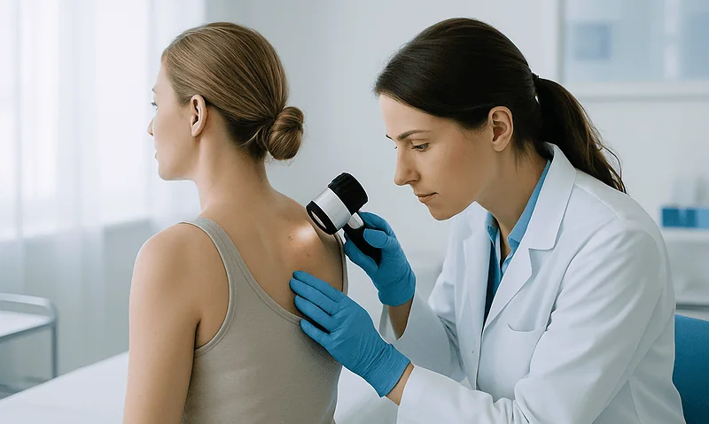
What a Congenital Nevus Check Involves: Dermoscopy, Photo Mapping and Tracking Change
A congenital nevus check is a skin examination of a birthmark mole. A clinician inspects the whole nevus and the surrounding skin, uses a…
When Does a Congenital Nevus Need Removal? How Dermatologists Weigh Monitoring Against Surgery
A congenital nevus needs removal only when a specific reason outweighs the scar and anesthesia involved: a suspicious change on examination, a lesion too…
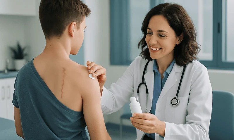
Scar and Sun Care After Congenital Nevus Removal: Dressings, Sutures and the Months After
After congenital nevus removal, scar care usually means keeping the incision covered and dry for the first day or two, following your surgeon's plan…
Doctors Performing This Treatment



Aesthetic Plastic & Reconstructive Surgery
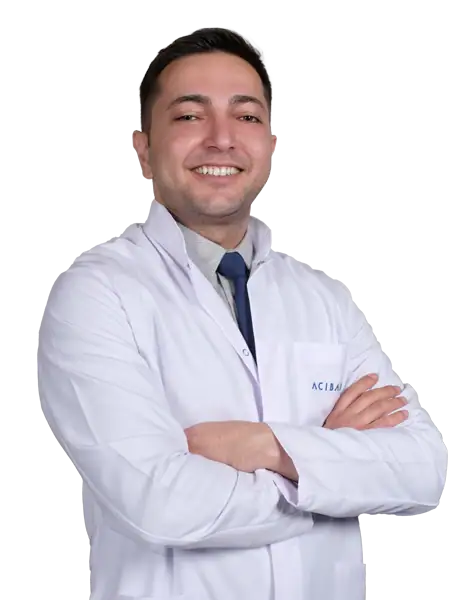
Prof. Mehmet Veli Karaaltın, MD

Aesthetic Plastic & Reconstructive Surgery
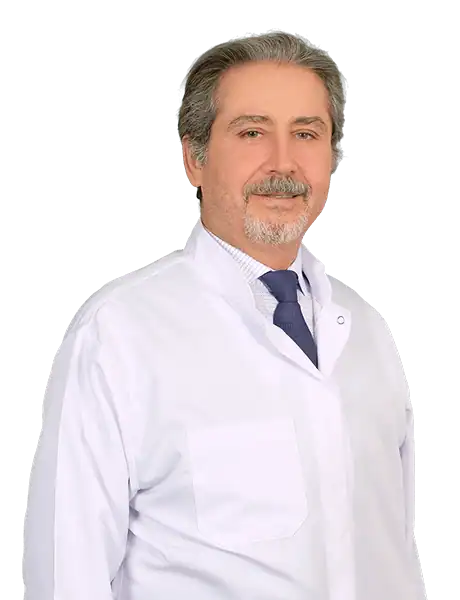
Assoc. Prof. Erdem Güven, MD

Aesthetic Plastic & Reconstructive Surgery
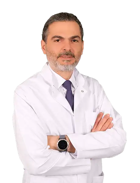
Assoc. Prof. Ahmet Küçükçelebi, MD
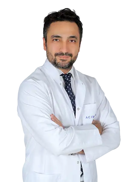
Aesthetic Plastic & Reconstructive Surgery
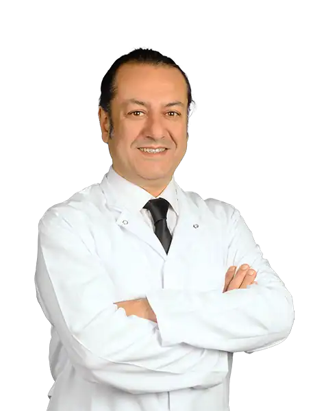
Assoc. Prof. Mehmet Altıparmak, MD

Aesthetic Plastic & Reconstructive Surgery