Hyperparathyroidism Treatment
Hyperparathyroidism treatment manages overactive parathyroid glands that raise calcium levels and may affect bones, kidneys, and overall health. Care may include monitoring, medication, or parathyroid surgery.
Quick answer
Hyperparathyroidism is a hormonal condition in which one or more parathyroid glands produce too much parathyroid hormone (PTH), raising blood calcium and drawing calcium from the bones. Treatment depends on the type and severity: some patients are safely monitored with regular blood tests, some take medication, and many with primary disease have a parathyroidectomy, surgery to remove the overactive gland tissue.
Hyperparathyroidism: When the Body Makes Too Much Parathyroid Hormone
Hyperparathyroidism is a hormonal condition in which one or more of the parathyroid glands, four small glands that sit behind the thyroid in the neck, produce too much parathyroid hormone, known as PTH. Excess PTH pushes blood calcium upwards and pulls calcium out of the bones. Treatment ranges from structured monitoring and medication through to surgery that removes the overactive gland tissue.
Many people first hear the word hyperparathyroidism after a routine blood test, at a point when they feel mostly well. Being told your calcium level is high can be confusing when nothing obvious seems wrong. Others reach the diagnosis after months or years of fatigue, bone or joint discomfort, kidney stones, stomach upset, changes in mood, or a vague sense that something is not right. Because these symptoms are subtle and overlap with many other conditions, hyperparathyroidism is sometimes missed, or monitored for a long time before a clear treatment plan takes shape.
Left unaddressed, the condition can quietly affect bone strength, kidney function, cardiovascular health, digestion and quality of life. That does not mean every patient needs an operation, some can be safely observed for years, but it does mean the diagnosis deserves a precise answer rather than a shrug. This page explains what hyperparathyroidism is, what causes it, how it is diagnosed, and how monitoring, medication and surgery each fit into treatment.
At Acibadem, care for hyperparathyroidism is planned around the full medical detail of each case. The aim is to identify the cause of the high PTH and calcium, measure the strain the disease has placed on bones and kidneys, and select the safest appropriate pathway: observation, medication, minimally invasive parathyroid surgery, or a more extensive neck exploration where the disease pattern demands it.
The parathyroid glands: the ‘para glands’ behind your thyroid
The parathyroid glands, often shortened to ‘para glands’ in everyday searches, are usually four glands, each roughly the size of a grain of rice, sitting behind the thyroid gland at the front of the neck. Despite the shared name and the shared address, they have nothing to do with the thyroid’s job of regulating metabolism. Their single task is to keep the calcium level in your blood inside a narrow healthy range. Their position varies more than most organs: some glands sit tucked against the back of the thyroid, others lie lower in the neck or even behind the breastbone. This anatomical variability, together with their tiny size, is a large part of why parathyroid surgery is treated as a specialist field rather than routine neck surgery.
What does parathyroid hormone (PTH) do?
Parathyroid hormone, or PTH, is the chemical signal the parathyroid glands release when blood calcium falls. It raises calcium in three ways: it releases calcium stored in bone, it tells the kidneys to retain calcium rather than excrete it, and it activates vitamin D so the gut absorbs more calcium from food. When calcium returns to normal, PTH production should fall: a feedback loop that works much like a thermostat. In hyperparathyroidism this loop is broken. The overactive gland keeps releasing PTH regardless of the calcium level, so calcium keeps rising while the skeleton steadily loses mineral. One detail matters more than most patients realise: a PTH value inside the laboratory’s ‘normal’ range can still be abnormal if calcium is high, because in a healthy person a high calcium should switch PTH production almost off. Calcium and PTH must always be interpreted together.
What is hypercalcemia?
Hypercalcemia is the medical term for a blood calcium level above the normal range, and it is the laboratory signature of most primary hyperparathyroidism. Calcium is essential for nerves, muscles, heart rhythm and bone, but too much of it circulating in the blood causes trouble of its own: thirst, frequent urination, constipation, nausea, poor concentration and fatigue. Hyperparathyroidism is one of the most common explanations for hypercalcemia found on routine testing, but it is not the only one, certain cancers, some medications, vitamin D excess and inherited calcium disorders can raise calcium as well. Separating these causes is the first task of any proper evaluation, and it rests on measuring PTH alongside calcium rather than reacting to either value alone.
Types and Causes of Hyperparathyroidism
‘Hyperparathyroidism’ is a description, not a single disease. Treatment depends heavily on which type you have, because the underlying mechanism, and therefore the right response, differs in each case.
What causes hyperparathyroidism?
The most common cause of hyperparathyroidism is a benign growth, called an adenoma, in a single parathyroid gland. The adenoma produces PTH continuously and ignores the body’s feedback signals. Less often, several glands enlarge together, a pattern known as hyperplasia. Parathyroid cancer exists but is rare; it is considered when calcium and PTH are extremely high or when imaging and examination findings raise specific concern. Several factors make the condition more likely: previous radiation treatment to the neck, long-term treatment with lithium, chronic kidney disease, persistent vitamin D deficiency, and certain inherited syndromes in which parathyroid overactivity runs in families. In many patients, though, no specific trigger is ever identified: an adenoma simply develops, and the search for a reason matters less than the plan for dealing with it.
What are the three main causes of hyperparathyroidism?
Doctors classify hyperparathyroidism into three forms, primary, secondary and tertiary, according to why the glands have become overactive. The distinction shapes everything that follows, from which specialist leads your care to whether surgery is the right answer.
Primary hyperparathyroidism means the problem lies in the gland itself. Usually one gland carries an adenoma; sometimes several glands are enlarged. Calcium is typically high, PTH is high or inappropriately normal, and the rest of the body is reacting to a gland that has stopped listening. This is the form most often discovered on routine blood tests, and the form for which surgery is most often the definitive treatment.
Secondary hyperparathyroidism means the glands are responding, appropriately, at first, to a problem elsewhere. Chronic kidney disease and vitamin D deficiency are the usual culprits: both make it harder for the body to maintain calcium, so the parathyroid glands work overtime to compensate. Calcium may be low, normal or high depending on the stage of disease and treatment history. Here the first response is usually medical, aimed at the underlying condition rather than the glands.
Tertiary hyperparathyroidism can develop after long-standing secondary disease, most often in people with kidney failure. After years of overwork, the glands become autonomous: they carry on overproducing PTH even after the original trigger has improved, for example, after a successful kidney transplant. At this stage the gland behaviour resembles primary disease, and surgery frequently returns to the discussion.
How rare is hyperparathyroidism?
Hyperparathyroidism is not rare. It sits among the more common hormone disorders, diagnosed most often in women and in adults over fifty, and secondary forms are frequent in people with chronic kidney disease. Because modern laboratory panels routinely include calcium, most cases in countries with regular blood testing are now discovered incidentally, before symptoms become obvious. What is genuinely uncommon is parathyroid cancer, which accounts for a very small fraction of cases. The practical point is simple: if your calcium has come back high more than once, this diagnosis is common enough to be worth investigating properly rather than dismissing as a laboratory quirk.
Symptoms of Hyperparathyroidism and Who May Need Treatment
Some patients have no obvious symptoms at all; others live for years with complaints that quietly erode daily function. Treatment decisions rest not only on how you feel, but on whether the disease is placing measurable stress on your bones, kidneys or other systems, which is why the evaluation always pairs your story with laboratory and imaging evidence.
What are the symptoms of hyperparathyroidism?
The classic teaching summarises hyperparathyroidism as ‘bones, stones, groans and moans’, bone pain, kidney stones, digestive upset, and mood or cognitive changes. In practice, patients describe a broader and vaguer mix, and any combination of the following can appear:
- Fatigue, weakness and reduced stamina that has no other clear explanation
- Increased thirst and frequent urination, driven by high calcium
- Constipation, nausea, loss of appetite or general abdominal discomfort
- Bone pain, joint discomfort and muscle aches
- Poor concentration, memory complaints, low mood or irritability
- Disturbed, unrefreshing sleep
- Kidney stones or repeated urinary symptoms
- Low bone density, or fractures after minor injuries
Because none of these is specific, people are sometimes treated for depression, irritable bowel, fibromyalgia or ‘just ageing’ before the endocrine cause is found. A careful review of past laboratory results is often the most revealing step of all: mild calcium elevation that has appeared repeatedly over several years deserves attention, especially when PTH was never checked alongside it, or was checked and found not appropriately suppressed. Kidney stones, recurring urinary problems, unexplained osteoporosis or a fragility fracture are sometimes the first sign that the condition has been present for a long time.
Does hyperparathyroidism cause weight gain?
Weight gain is not a classic or diagnostic feature of hyperparathyroidism, and the condition does not directly alter how the body stores fat. That said, some patients do gain weight while living with it, and the connection is usually indirect: persistent fatigue, low mood, disturbed sleep and muscle weakness all tend to reduce physical activity, and reduced activity over months or years shifts weight upwards. Other patients lose weight because of nausea and poor appetite. The honest answer is that weight change in either direction neither confirms nor excludes the diagnosis. If your weight has changed and your calcium is high, the two facts belong in the same conversation with your doctor, but the calcium and PTH values, not the scales, are what settle the question.
Patient situations that often lead to treatment
Treatment is commonly recommended when hyperparathyroidism causes symptoms, kidney stones, reduced kidney function, significant calcium elevation, osteoporosis, fragility fractures or increased urinary calcium. Younger patients are often advised to consider surgery even with mild disease, because they face a longer lifetime of exposure to excess PTH and its cumulative effects. Another common path to treatment runs through years of monitoring: calcium levels drift upwards, bone density scans show steady decline, or symptoms that were once easy to ignore become harder to live with, and the balance of the decision shifts.
Patients with kidney-disease-related hyperparathyroidism follow a different logic. Treatment escalates when PTH stays high despite medical therapy, when bone disease progresses, when calcium and phosphorus become difficult to control together, or when the enlarged glands no longer respond adequately to medication. In these situations, care usually involves nephrology, endocrinology and endocrine surgery working from the same plan rather than a single specialist deciding alone.
Diagnosis: How Hyperparathyroidism Is Confirmed
The diagnosis of hyperparathyroidism is biochemical. Scans and imaging come later, and they answer a different question. Understanding that order protects you from two common mistakes: dismissing the diagnosis because a scan was negative, and rushing towards surgery because a scan showed something.
How can you test for hyperparathyroidism?
Hyperparathyroidism is tested for with blood tests that measure calcium and PTH together, high calcium with a high or inappropriately normal PTH is the defining pattern. Depending on the picture, physicians also check ionised calcium, phosphorus, vitamin D, kidney function and sometimes magnesium, because each of these can shift the interpretation. A 24-hour urine calcium collection helps separate true primary hyperparathyroidism from rarer inherited conditions that mimic it but should not be operated on. Beyond the biochemistry, two further tests measure what the disease has already done: a bone density scan shows whether the skeleton has weakened, and kidney imaging checks for stones or calcium deposits. None of this requires guesswork, but it does require the right tests in the right order, and it often requires repeating values to confirm a trend rather than reacting to a single result.
Localisation imaging before surgery
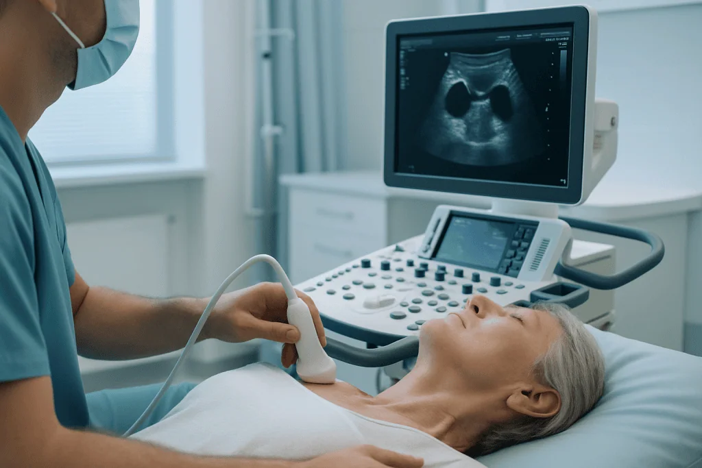
Imaging is used to locate the overactive gland or glands once surgery is on the table, not to prove the diagnosis itself. High-resolution neck ultrasound is usually the first step, and it examines the thyroid at the same time; if an ultrasound is part of your work-up, the practical points in this ultrasound preparation and exam-day checklist apply. Nuclear medicine scans show parathyroid tissue by its metabolic activity, and cross-sectional imaging adds detailed anatomy when earlier studies disagree, when a gland may sit in an unusual position, or when the neck has been operated on before. One point deserves emphasis because it causes real confusion: negative imaging does not mean you do not have hyperparathyroidism, and it does not always mean surgery is off the table. It usually means the gland is small or awkwardly placed, and that the operation calls for greater surgical experience, not abandonment of the plan.
Conditions and Indications Hyperparathyroidism Treatment Addresses
Treatment may be considered for several related conditions, each requiring its own balance of monitoring, medication and surgical planning:
- Primary hyperparathyroidism: overproduction of PTH from one or more parathyroid glands, usually with high calcium. Surgery is often considered when guideline criteria are met or symptoms are present.
- Parathyroid adenoma: a benign overactive gland, and the most common cause of primary disease. When localised accurately, it can often be treated with a focused operation through a small incision.
- Multigland parathyroid hyperplasia: enlargement of several glands at once. This may require bilateral neck exploration and removal of more than one gland, with careful preservation of enough functioning tissue.
- Secondary hyperparathyroidism: usually driven by chronic kidney disease or vitamin D deficiency. Treatment typically begins with medical management and correction of the underlying metabolic problem.
- Tertiary hyperparathyroidism: persistent gland overactivity after long-standing secondary disease, often following kidney transplantation or prolonged kidney failure. Surgery enters the discussion when biochemical control remains difficult.
- Recurrent or persistent hyperparathyroidism: disease that returns or continues after previous treatment. Evaluation requires a detailed review of the earlier operation, pathology reports, imaging and laboratory trends before anything else is planned.
- Hyperparathyroidism affecting bones or kidneys: treatment may be recommended when osteoporosis, fractures, kidney stones, nephrocalcinosis or declining kidney function are traced back to abnormal PTH and calcium metabolism.
The right pathway depends on whether the disease is primary, secondary or tertiary; whether one gland or several are involved; whether the neck has been operated on before; and how the condition is affecting your long-term health. Two patients with identical calcium values can reasonably receive very different plans.
How Hyperparathyroidism Treatment Is Performed
Care follows a structured pathway: confirm the diagnosis, understand the anatomy, weigh the options, and then act, or deliberately wait. Some patients do not need immediate surgery and can be followed carefully. Others benefit from a planned operation. Each stage exists to reduce risk and remove guesswork.
Preparation and medical evaluation
Before any treatment decision, the medical team reviews prior blood tests, imaging, medications, supplements, kidney history, bone density results and any previous neck operations. Calcium and PTH values are always interpreted as a pair, because a normal-range PTH can still be inappropriate when calcium is high. Vitamin D status matters twice over: deficiency can push PTH upwards and muddy the diagnosis, and it also influences how calcium behaves after surgery.
You will be asked about kidney stones, fractures, digestive symptoms, concentration and mood, voice changes, swallowing, family history, and the use of medicines such as lithium or thiazide diuretics that can affect calcium, decisions about any of these medicines rest with your treating doctor, but the team needs the full list to interpret your results correctly. If surgery is planned, vocal cord function may be assessed in selected patients, particularly those with previous neck or thyroid surgery, existing voice symptoms, or professional voice demands.
Laboratory tests are usually repeated close to treatment to confirm current values, and imaging is scheduled so the surgical team has everything needed for safe planning. Where other conditions coexist, thyroid nodules found on the same ultrasound, or established kidney disease, consultations with the relevant specialists are built into the same evaluation rather than left for later.
Monitoring and medication-based treatment
Not every patient with hyperparathyroidism needs an operation now, or ever. In selected patients with mild primary disease who do not meet surgical criteria and prefer observation, physicians recommend structured monitoring: periodic calcium and kidney function tests, PTH and vitamin D assessment where appropriate, bone density scans on a set schedule, and evaluation for kidney stones if symptoms develop. The word ‘structured’ is doing real work in that sentence, monitoring is a plan with dates attached, not an open-ended decision to do nothing.
Medication has its place when surgery is not appropriate, when a patient is not ready for it, or when the condition is secondary to kidney disease. Depending on the situation, physicians may prescribe vitamin D replacement, medicines that lower calcium by changing how the parathyroid gland senses calcium, or bone-protective therapies for patients with osteoporosis. In chronic kidney disease, the medical toolkit widens further: dietary phosphorus management, phosphate binders, active vitamin D compounds and other agents selected and adjusted by nephrology specialists.
One honest limitation belongs in this section. Medication can control certain biochemical values and protect bone health, but in primary hyperparathyroidism it does not correct the underlying gland abnormality. The adenoma remains, and so does the need for ongoing treatment and testing. That distinction, managing the disease versus removing its source, is the heart of the conversation when monitoring and surgery are compared.
Parathyroid surgery: step by step
When surgery is indicated, the operation is a parathyroidectomyremoval of the overactive parathyroid tissue. It is usually performed under general anaesthesia, though selected cases may use other anaesthesia approaches depending on patient and surgeon preference. The surgical plan hinges on whether imaging points to a single abnormal gland or to possible multigland disease. A focused, minimally invasive operation typically proceeds like this:
- Anaesthesia and positioning. You are placed under anaesthesia with the neck gently extended, and monitoring is established.
- A small incision. The surgeon makes a short cut low in the neck, planned along natural skin lines so the eventual scar sits as discreetly as possible.
- Locating the gland. Guided by the preoperative imaging, the surgeon identifies the abnormal parathyroid gland while protecting the thyroid, blood vessels and nerves around it.
- Removing the overactive tissue. The enlarged gland is carefully freed and removed.
- Intraoperative PTH confirmation. Because PTH has a short half-life, blood taken during the operation can show whether the hormone level falls appropriately once the gland is out, real-time biochemical evidence that the source of overproduction has been dealt with.
- Closure and pathology. The incision is closed, and the removed tissue goes to the pathology laboratory for formal examination.
If imaging is unclear, if multigland disease is suspected, or if the diagnosis is secondary or tertiary hyperparathyroidism, the surgeon performs a bilateral neck exploration instead: all parathyroid glands are examined, and abnormal tissue is removed according to the disease pattern. In some of these operations, a portion of parathyroid tissue is deliberately left in place, or implanted at another site in the body, to preserve some natural hormone function and reduce the risk of permanently low calcium afterwards.
Throughout either approach, the team works with particular care around the recurrent laryngeal nerves, which control the vocal cords, and around the thyroid gland and nearby vessels. Nerve monitoring may be used in selected operations to help identify and protect nerve function. This attention is not ceremony; it is the difference between a small, well-tolerated neck operation and a complicated one.
Technology used in diagnosis and surgery
Modern parathyroid care draws on several technologies. High-resolution ultrasound identifies enlarged glands and surveys the thyroid at the same sitting. Nuclear medicine imaging highlights overactive parathyroid tissue by its metabolic behaviour. Cross-sectional imaging supplies detailed anatomy for reoperations, ectopic glands and cases where earlier studies conflict. In theatre, intraoperative PTH testing provides biochemical feedback within the operation itself, while magnification, refined instruments, careful anaesthesia monitoring and selective nerve monitoring keep the surgery controlled in a delicate part of the neck. For complex disease, though, the most important ‘technology’ is not a device at all: it is the integration of laboratory interpretation, imaging review, surgical judgement, pathology and postoperative calcium management into one coherent plan.
How long does treatment take?
Duration varies with complexity. A focused parathyroid operation can be relatively short; multigland or reoperative surgery takes longer. Afterwards, patients are observed for calcium levels, voice quality, swallowing comfort and general recovery from anaesthesia. Some go home the same day or after a short hospital stay; those with kidney disease, severe bone disease, multigland operations or a higher risk of calcium shifts stay under monitoring for longer. It helps to plan around the whole pathway, preoperative testing, the operation itself and the early follow-up blood tests, rather than the operation alone. The exact schedule depends on your case and your physician’s recommendations.
Recovery after treatment
After parathyroid surgery, most patients notice mild neck discomfort, throat irritation or temporary swallowing discomfort, all of which usually settle gradually. Calcium levels can fall as the body adjusts to normal PTH signalling, which is why blood tests continue after the operation. Some patients feel tingling around the lips or in the fingertips, or muscle cramps, signs of low calcium that the team asks you to mention so levels can be checked and corrected promptly. Calcium and vitamin D supplementation is often prescribed for a short period, and sometimes longer, depending on your situation.
Patients who arrived at surgery with significant bone disease can experience a more pronounced shift of calcium back into the skeleton, known as hungry bone syndrome. It is more common after severe or long-standing hyperparathyroidism, in secondary disease, and in patients with kidney failure, and it calls for close laboratory monitoring and careful calcium replacement rather than alarm. Follow-up covers calcium and PTH testing, wound review, discussion of the pathology findings, and long-term planning for bone and kidney health. Bone density needs time to rebuild, and stone prevention may remain part of your care depending on your history.
Why Acting Early Matters
Hyperparathyroidism usually progresses slowly, which can make it feel less than urgent. But the arithmetic of the disease is cumulative: every additional year of elevated PTH and calcium is a year of gradual bone mineral loss, ongoing stone risk in susceptible kidneys, and, for some patients, persistent fatigue, cognitive fog, constipation or muscle weakness that quietly narrows daily life. The bones may drift towards osteoporosis; the kidneys may develop stones or calcium deposits; kidney function can decline in vulnerable patients.
Delay can also make eventual treatment more complex. Long-standing disease contributes to more significant bone remodelling, a higher chance of pronounced calcium shifts after surgery, and greater overall medical complexity. In kidney-related hyperparathyroidism, prolonged uncontrolled PTH brings bone pain, vascular calcification risk and a mineral balance that becomes progressively harder to manage.
Early evaluation does not mean early surgery. It means understanding the disease accurately, measuring what it has already done, and choosing the timing deliberately. For some patients, careful monitoring is safe and entirely appropriate. For others, postponement is simply a way of letting preventable complications develop, and the evaluation exists to tell those two groups apart.
How long can you live with hyperparathyroidism?
Many people live with mild hyperparathyroidism for years, and mild primary disease under proper monitoring is often compatible with a normal life span. For most patients the question is not survival but accumulation: what the excess PTH and calcium are doing, year on year, to bones, kidneys, heart and everyday wellbeing. Severe, untreated hypercalcemia is a different and more serious matter, which is one reason significant calcium elevation weighs towards treatment rather than watching. The realistic framing is this: the condition is rarely an emergency, but it is also rarely harmless over decades, and the decision between monitoring and treatment should be an informed one revisited at intervals, not a decision made once and forgotten.
Benefits of Hyperparathyroidism Treatment
What treatment can realistically achieve depends on the type of hyperparathyroidism, the option chosen, and your baseline bone, kidney and metabolic health.
| Benefit | What It Means for You |
|---|---|
| Improved calcium control | Effective treatment can bring calcium metabolism closer to a healthy range and reduce the risks associated with persistently high calcium. |
| Protection of bone health | Treating excess PTH may help slow ongoing bone loss and support long-term fracture prevention, especially alongside appropriate bone care. |
| Reduced kidney stone risk | For patients whose stones are driven by hyperparathyroidism, treatment may lower the chance of future stone formation and support kidney preservation. |
| Relief of certain symptoms | Some patients notice improvement in fatigue, muscle weakness, concentration, mood, digestion or general wellbeing once calcium and PTH are corrected. |
| A clear long-term plan | A structured evaluation settles whether monitoring, medication or surgery is the right route, and how follow-up will be organised afterwards. |
Recovery Timeline After Parathyroid Surgery
Recovery varies with the type of operation, calcium behaviour and overall health, but the following reflects what many patients experience after uncomplicated parathyroid surgery.
| Time Period | What Patients Can Expect |
|---|---|
| Day 1 | Mild neck discomfort, throat irritation and tiredness are common. Calcium is checked, and supplements may be started if needed. |
| First week | Most patients resume light daily activities. The incision is monitored, and the care team reviews any tingling, cramps, voice change, fever or increasing swelling. |
| First month | Energy and swallowing comfort usually continue to improve. Follow-up blood tests confirm that calcium and PTH are stable. |
| Longer term | Bone density, kidney stone risk and vitamin D status are followed over time. Patients with complex disease may need ongoing endocrinology or nephrology care. |
Factors That Influence Outcomes
A good result begins with an accurate diagnosis. Calcium and PTH patterns must be read correctly, and other causes of abnormal calcium metabolism ruled in or out. Vitamin D deficiency, kidney disease, medications, familial calcium disorders and previous neck operations can each reshape both the diagnosis and the plan, which is why the evaluation is broader than a single blood test.
The type of hyperparathyroidism is among the most decisive factors. A single-gland adenoma in primary disease is usually more straightforward than multigland hyperplasia, secondary or tertiary disease, or a recurrence after earlier surgery. Imaging quality and interpretation matter too, but always in combination with the biochemistry: an experienced team knows that a scan can mislead, and that success depends on matching imaging findings to the laboratory diagnosis rather than trusting either alone.
Surgical experience carries particular weight because the glands are small and inconsistently located. Some sit against the thyroid; others lie lower in the neck or behind the breastbone. Prior thyroid or parathyroid surgery leaves scar tissue and altered anatomy, and in those cases careful preoperative planning and specialist review are not optional extras: they are the plan.
Patient factors shape recovery as well. Severe bone disease, very high PTH, chronic kidney disease, vitamin D deficiency and long-standing disease all call for closer calcium monitoring after surgery. Patients with osteoporosis may need continuing bone-specific therapy even after successful parathyroid treatment, and those with a stone history may need metabolic stone evaluation, hydration guidance and follow-up to reduce recurrence.
Finally, communication belongs on this list. Outcomes are better when you understand why a treatment is recommended, what the alternatives are, what to expect in the weeks after surgery, and how long-term monitoring will be handled. Coordination between the treating physicians and your local doctor, particularly for laboratory follow-up and medication adjustments made by your own treating doctor, is part of the outcome, not an afterthought.
How Acibadem Organises Multidisciplinary Hyperparathyroidism Care
Hyperparathyroidism rarely belongs to a single specialty, and care at Acibadem is built around that fact. Evaluation and treatment bring together physicians experienced in endocrine disorders, neck surgery, imaging, anaesthesia, pathology and nephrology, so that the biochemical diagnosis, the anatomy and the long-term plan are considered by the right people at the same time rather than in sequence.
In practice, that means endocrinologists assess the biochemical diagnosis and the case for medical treatment or surgery; endocrine surgeons set the operative strategy and discuss the risks openly, including low calcium, bleeding, infection, scarring and voice changes; radiologists and nuclear medicine specialists work on localising abnormal glands when surgery is planned; and nephrologists join for patients with chronic kidney disease, a dialysis history, kidney transplantation or complex mineral metabolism. In selected complex cases, multidisciplinary case discussions align the plan before treatment begins rather than after questions arise.
Treatment plans are individual because the disease is. A patient with mild primary hyperparathyroidism and stable bone density may need careful monitoring and clear education. A patient with recurrent kidney stones and a well-localised adenoma may benefit from a focused operation. A patient with kidney failure and severe secondary disease needs a different operation and closer calcium management afterwards. A patient with persistent disease after prior surgery needs painstaking imaging review and a more cautious operative strategy. These distinctions shape both safety and recovery, and they are why the pathway starts with review rather than with a scheduled theatre slot. The team explains expected recovery time, wound care and follow-up testing at each step, and written medical summaries support continuity with your own physicians afterwards.
Living With and After Hyperparathyroidism
Hyperparathyroidism is a treatable condition, and for many patients the right plan meaningfully reduces long-term risks to the bones and kidneys while restoring a more normal metabolic balance. The most important single step is a careful evaluation that confirms the diagnosis, identifies the type, primary, secondary or tertiary, and sets out honestly whether monitoring, medication or surgery best fits your situation.
After treatment, the work shifts to the long view. Calcium and PTH are rechecked on a schedule; bone density is followed and given the time it needs to recover; vitamin D is kept in range; and patients with a history of stones continue hydration and metabolic follow-up. Patients who choose monitoring rather than treatment carry a parallel responsibility: keeping the agreed testing schedule, so that any change in calcium, kidney function or bone density is caught while the options remain wide open. Either way, the condition rewards the same habit, paying attention to the numbers over time rather than to any single result, and that habit, shared between you, your treating team and your local doctor, is what turns a confusing blood test into a managed, understood part of your health.
Preparation
- Preparation includes blood calcium and parathyroid hormone tests, kidney function assessment, bone density evaluation, and imaging to locate abnormal glands. Patients should share all medications and supplements, especially calcium, vitamin D, blood thinners, and kidney-related medicines. Fasting is usually required before surgery if parathyroidectomy is planned.
Aftercare
- After surgery, calcium levels are monitored closely, and temporary calcium or vitamin D supplements may be prescribed. Most patients return to light daily activities within several days, while strenuous activity should wait until the surgeon approves. Follow-up visits check wound healing, hormone levels, calcium balance, kidney health, and bone recovery.
Frequently Asked Questions
What affects the cost of hyperparathyroidism treatment?
The cost depends on the type of hyperparathyroidism, required blood and urine tests, imaging, specialist consultations, whether surgery is needed, anesthesia, hospital stay, pathology, and follow-up. Travel support, translation, and accommodation coordination may also affect the overall package.
How can I get a personalised quote from Acibadem?
You can request a free consultation and share your medical records, recent calcium and parathyroid hormone results, imaging reports, medication list, and symptom history. The medical team can then review your case and provide a personalised treatment plan and quote.
Is surgery always required for hyperparathyroidism?
No. Some patients may be monitored or treated with medication, while others benefit from parathyroid surgery. The decision depends on symptoms, calcium levels, kidney findings, bone health, gland localization, and overall medical condition.
What is usually included in an international patient package?
Depending on the plan, a package may include specialist consultation, diagnostic tests, imaging review, surgery if indicated, hospital services, translation, care coordination, and follow-up planning. It is important to confirm inclusions before travel.
Will I need tests before receiving a quote?
Existing results can often be reviewed first, but additional tests may be recommended to confirm the diagnosis, assess kidney and bone health, and plan treatment safely. The final quote is usually clearer after the specialist has reviewed the necessary information.
Medically reviewed by the Acıbadem International Medical Board August 31, 2026
See our medical review board →
Update history
- PublishedJune 8, 2026
- Medical review approvedAugust 31, 2026
- Last content updateSeptember 8, 2026
References2
- Hyperparathyroidism nhs.uk
- Hyperparathyroidism my.clevelandclinic.org
Trusted care for international patients
From the Acibadem Blog
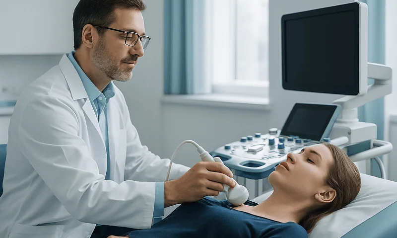
Hyperparathyroidism Work-Up: Calcium and PTH Tests, Bone Density and Kidney Checks Explained
A hyperparathyroidism work-up starts with blood tests for calcium and parathyroid hormone (PTH), usually repeated to confirm the pattern of raised or non-suppressed PTH…
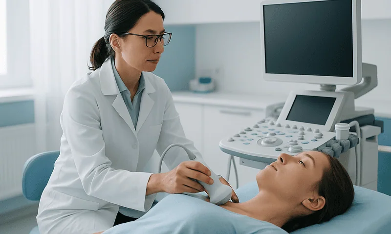
Hyperparathyroidism in Older Adults: How Age and Kidney Function Shape the Treatment Plan
Hyperparathyroidism in older adults is treated according to how high the blood calcium is, how well the kidneys are working, bone strength, symptoms and…

Primary or Secondary Hyperparathyroidism: Why Does the Cause Change the Treatment Path?
Primary hyperparathyroidism begins inside the parathyroid glands themselves, usually a benign growth that oversecretes hormone and pushes blood calcium high, so treatment often targets…