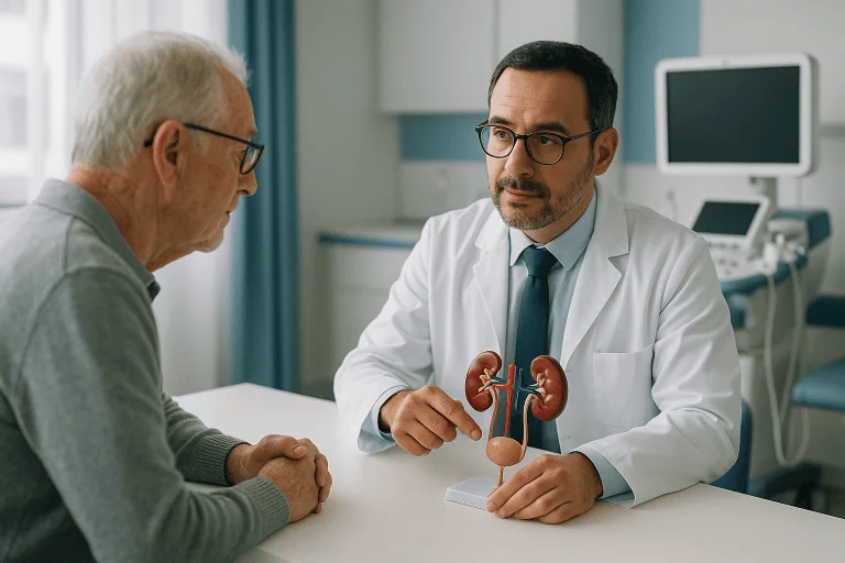
Percutaneous Nephrostomy
Percutaneous nephrostomy is a procedure in which an interventional radiologist places a thin drainage catheter through the skin into a blocked kidney, guided by ultrasound and X-ray. It is used when urine…

Quick answer
Percutaneous nephrostomy is a minimally invasive procedure in which a thin tube is placed through the skin of the back into the kidney to drain urine when the ureter is blocked, often by a stone, tumor, or scarring. It relieves pressure, treats infection, and protects kidney function, usually under local anesthetic with sedation.
What is percutaneous nephrostomy?
A percutaneous nephrostomy is a procedure in which a thin, flexible tube (a catheter) is placed through the skin of the back directly into the kidney. Percutaneous means through the skin, and nephrostomy means an opening into the kidney. The tube allows urine to drain out of the kidney into a bag worn outside the body, bypassing a blocked or damaged section of the urinary tract.
Normally, urine flows from each kidney down a narrow tube called the ureter into the bladder. If the ureter is blocked, urine backs up and the kidney swells. Doctors call this swelling hydronephrosis. A kidney that stays blocked can become infected, and its ability to filter blood can fall over time. Percutaneous nephrostomy relieves this pressure quickly and is usually performed by an interventional radiologist, a doctor who uses imaging such as ultrasound and X-ray to guide procedures through very small openings in the skin.
The procedure is used in several situations, including:
- Blockage of the ureter by a kidney stone, especially when there is also infection or the kidney is not working well.
- Narrowing or compression of the ureter caused by a tumor in the pelvis or abdomen, scar tissue, or pregnancy-related changes.
- Infection in a blocked kidney (pyonephrosis), where infected urine must be drained urgently.
- Leaks or injuries to the ureter, for example after surgery, where diverting urine helps the tissue heal.
- Creating access to the kidney for a later stone-removal procedure or to place an internal drainage tube called a ureteral stent.
In many hospital groups, including Acibadem, care for this condition is shared between the Urology department and interventional radiology, with urologists managing the underlying cause of the blockage.
Who needs percutaneous nephrostomy: candidates and when it is not suitable
Deciding who needs percutaneous nephrostomy depends on how blocked the kidney is, how well it is working, and whether there is infection. Your doctor may recommend it if:
- Imaging shows a swollen, obstructed kidney and your kidney function tests are getting worse.
- You have signs of infection (fever, chills, pain) together with a blocked kidney, which can become life-threatening without drainage.
- An internal ureteral stent could not be placed from below through the bladder, or is not appropriate for your situation.
- You have a single functioning kidney, or both kidneys are blocked, so urgent drainage is needed to keep the body filtering waste.
- Your medical team needs a route into the kidney for a planned stone procedure (percutaneous nephrolithotomy).
There are also situations where the procedure may be delayed or a different approach chosen. It may not be suitable when:
- Blood does not clot normally, for example because of certain medications or a bleeding disorder, until this can be corrected.
- The kidney cannot be reached safely because of its position, bowel loops in the way, or other anatomy seen on imaging.
- The blockage can be relieved just as effectively and more comfortably with a ureteral stent placed from inside the bladder.
- There is a severe, uncontrolled infection of the skin at the planned entry site.
Your doctors will weigh these factors with you. In an emergency, the balance often favors draining the kidney as quickly and safely as possible.
How the percutaneous nephrostomy procedure works
The percutaneous nephrostomy procedure is usually done in an interventional radiology suite rather than a traditional operating room. In plain language, this is what typically happens.
Before the procedure. You will have blood tests to check kidney function and how well your blood clots. Imaging, usually ultrasound or a CT scan (a detailed X-ray scan), confirms which kidney is blocked and where the tube can safely go. If infection is suspected, antibiotics are often started beforehand. You will be asked to sign a consent form after the risks and alternatives have been explained.
During the procedure. You lie on your stomach or slightly turned onto your side. The skin over the affected kidney is cleaned, and a local anesthetic is injected to numb the area. Most patients also receive sedation, medication given through a vein that makes you drowsy and relaxed, though you may remain awake. General anesthesia, in which you are fully asleep, is sometimes used for children or for patients who cannot lie still. Using ultrasound to see the kidney in real time, the doctor passes a fine needle through the skin into the urine-filled part of the kidney. A small amount of urine may be drawn off for testing. A thin guidewire is threaded through the needle, the needle is removed, and the track is gently widened. The drainage catheter is then passed over the wire into the kidney. X-ray contrast dye is often injected to confirm the tube sits correctly. The tip of the catheter usually has a small curl or loop that helps hold it in place. The tube is secured to the skin with stitches, an adhesive device, or both, and connected to a drainage bag. The procedure often takes about 30 to 60 minutes.
After the procedure. You are monitored for several hours. Nurses check your blood pressure, pulse, temperature, and the color and amount of urine draining into the bag. It is common for the urine to be pink or slightly bloody at first, and this usually clears within a day or two. Pain at the tube site is expected and is managed with pain medication. Many patients stay in hospital at least one night, longer if they were admitted with infection or other medical problems.
Preparation for percutaneous nephrostomy
Careful preparation lowers the chance of complications. Your team will give you specific instructions, which commonly include the following:
- Medication review. Tell your doctors about every medication, supplement, and herbal product you take. Blood thinners such as warfarin, apixaban, rivaroxaban, clopidogrel, and in some cases aspirin may need to be paused or adjusted. Never stop a prescribed medication without medical guidance.
- Fasting. If you will have sedation or general anesthesia, you are usually asked not to eat for several hours beforehand. Clear fluids may be allowed up to a set time.
- Allergies. Report any allergy to iodine contrast dye, latex, antibiotics, or anesthetic medications.
- Blood tests. Kidney function, blood count, and clotting tests are checked, and blood may be typed in case a transfusion is ever needed.
- Infection control. Antibiotics are often given before the procedure, particularly when infected urine is suspected.
- Practical arrangements. Because of sedation, you will not be able to drive yourself home. Wear loose clothing and bring a list of your medications.
If the procedure is an emergency, some of these steps are shortened, and your team will focus on the essentials such as clotting tests and antibiotics.
Recovery and aftercare: percutaneous nephrostomy recovery time
Percutaneous nephrostomy recovery time is generally short compared with open surgery, because only a small opening is made in the skin. Many patients are up and walking the same day or the next morning. Discomfort at the tube site typically eases over the first few days, and most people can return to light daily activities within about a week. Heavy lifting, twisting, and contact sports are usually avoided while the tube is in place, since these movements can dislodge it.
Living with the tube requires some routine care, which nurses will teach you or a family member before discharge:
- Keep the dressing clean and dry. The dressing is usually changed every few days or sooner if it becomes wet or soiled. Watch the skin around the tube for redness, swelling, or leakage.
- Empty the bag regularly. The drainage bag should be emptied when it is about half to two-thirds full and kept below the level of the kidney so urine flows downward.
- Check that urine is draining. A sudden drop in output, or urine leaking around the tube instead of into the bag, can mean the catheter is kinked or blocked.
- Flushing. Some patients are shown how to gently flush the tube with sterile saline to prevent clogging; others are asked to leave this to clinic staff.
- Showering. Brief showers are often allowed once the site has sealed, with the bag and dressing protected. Baths and swimming are generally avoided.
- Fluids. Unless you have been told to limit fluids, drinking enough water helps keep urine flowing and reduces the risk of blockage.
How long the tube stays in depends on why it was placed. For a stone that will be treated, the tube may be needed for days to a few weeks. When a tumor is compressing the ureter, drainage can be long term, and the catheter is usually exchanged every two to three months in the radiology department to prevent blockage and infection. In some cases the tube is later replaced by an internal ureteral stent so that no external bag is needed.
Percutaneous nephrostomy risks and benefits: side effects to know
Understanding percutaneous nephrostomy risks and benefits helps you take part in decisions about your care. The main benefit is rapid relief of pressure on a blocked kidney, which can protect kidney function, drain infected urine, and reduce pain. It is minimally invasive, can be done under local anesthetic with sedation, and can be performed even when a patient is quite unwell.
Like any procedure, it also carries risks. Common, usually minor effects include:
- Pain or soreness at the tube site for a few days.
- Blood-tinged urine for one to three days.
- Skin irritation around the tube from tape, dressings, or leaking urine.
- Tube blockage or displacement, which may require flushing, repositioning, or exchange.
Less common but more serious complications include:
- Significant bleeding. The kidney has a rich blood supply. Most bleeding stops on its own, but occasionally a transfusion or a procedure to seal a bleeding vessel is needed.
- Infection or sepsis. Draining an infected kidney can release bacteria into the bloodstream, causing fever and low blood pressure soon after the procedure. Antibiotics before and after the procedure lower this risk.
- Injury to nearby organs such as the bowel, spleen, liver, or lung, which is uncommon because imaging guides the needle.
- Urine leak into the tissues around the kidney.
- Reaction to contrast dye or sedation.
Living with an external tube for a long period also has quality-of-life effects, including body-image concerns, sleep disturbance, and the practical burden of dressing changes. Your team can discuss whether an internal stent is a suitable alternative for you.
Results and outlook
Percutaneous nephrostomy is a well-established procedure, and in experienced hands placement is successful in the great majority of cases. Once urine is draining, pain from the blocked kidney often improves within hours, and blood tests of kidney function typically start to recover over the following days, provided the kidney tissue itself has not been permanently damaged by long-standing obstruction.
The longer-term outlook depends mainly on the cause of the blockage rather than on the nephrostomy itself. When a kidney stone is the cause, the tube is a temporary step before definitive stone treatment, and most patients return to normal urinary drainage afterward. When a tumor is compressing the ureter, the tube may be part of ongoing care alongside cancer treatment. In some situations a kidney that has been blocked for a long time may not regain full function even after drainage, and your doctors may use imaging or nuclear medicine scans to assess how much function has returned before planning further treatment.
Cost considerations
The overall cost of a percutaneous nephrostomy varies widely and depends on several factors rather than a single fee. The main drivers include:
- Hospital stay. An emergency admission with infection requiring intravenous antibiotics costs more than a planned procedure with a single overnight stay.
- Imaging and laboratory tests before and after the procedure, such as CT scans, ultrasound, and repeated kidney function tests.
- Devices and consumables, including the catheter, guidewires, drainage bags, and dressing supplies.
- Type of anesthesia and the involvement of an anesthesiologist if general anesthesia is required.
- Follow-up care, including regular tube exchanges for long-term catheters, nursing visits, and treatment of the underlying condition.
Coverage by insurance or national health systems differs between countries and policies. Asking for an itemized estimate and checking what your insurer covers before a planned procedure can help avoid unexpected expenses.
Frequently asked questions
Is a percutaneous nephrostomy painful?
Local anesthetic numbs the skin and deeper tissues, and sedation helps most patients feel relaxed during the procedure. Some pressure or a brief sharp sensation may be felt when the kidney is entered. Afterward, soreness at the tube site is common for a few days and is usually controlled with standard pain relievers.
How long does percutaneous nephrostomy recovery time usually take?
Many patients are walking within a day and return to light activities within about a week. The skin site typically settles within a few days. Full recovery depends on the reason for the tube and on whether an infection was present, so some people need longer to feel well.
Who needs percutaneous nephrostomy instead of a ureteral stent?
Both relieve a blocked kidney. A nephrostomy is often chosen when the blockage cannot be passed from below, when infection needs rapid drainage, or when the patient is too unwell for a procedure under general anesthesia. A stent avoids an external bag but may cause bladder irritation. Your urologist and radiologist will advise which suits your situation.
How long can a nephrostomy tube stay in?
There is no fixed limit. Short-term tubes are removed once the blockage is treated, often within days or weeks. Long-term tubes are exchanged regularly, commonly every two to three months, to reduce the risk of blockage and infection.
What are the main percutaneous nephrostomy risks and benefits I should weigh?
The key benefit is fast, effective drainage that can protect kidney function and treat infection. The main risks are bleeding, infection, tube displacement or blockage, and, rarely, injury to nearby organs. For most people with a dangerously blocked kidney, the benefits outweigh the risks, but the decision should be individual.
Can I travel or work with a nephrostomy tube?
Many patients continue daily activities, including desk-based work, once they feel comfortable. Travel is often possible if you carry spare dressings and bags, know how to reach medical help, and plan around scheduled tube exchanges. Discuss your specific plans with your care team.
What happens when the percutaneous nephrostomy tube is removed?
Removal is usually quick and done in the clinic, sometimes after imaging confirms the ureter is open. The small skin opening typically closes within a day or two, and a light dressing is worn until it stops leaking.
When to see a doctor
You should be assessed by a specialist if you have symptoms that may indicate a blocked kidney, such as persistent pain in the side or back, pain accompanied by fever or chills, blood in the urine, reduced urine output, or nausea and vomiting alongside flank pain. People with a known kidney stone, a single kidney, a history of pelvic or abdominal cancer, or previous urinary tract surgery should seek assessment promptly if these symptoms develop, because a blocked kidney can deteriorate quickly.
After a percutaneous nephrostomy, seek urgent medical attention if you notice any of the following:
- Fever above 38 °C (100.4 °F), shaking chills, or feeling suddenly very unwell, which may signal infection or sepsis.
- Urine in the bag that is bright red, contains clots, or becomes bloodier rather than clearer.
- Little or no urine draining for several hours despite drinking fluids, or urine leaking heavily around the tube.
- The tube has come out, is much shorter than before, or has moved significantly.
- Severe or worsening pain at the tube site or in the flank that is not controlled by prescribed medication.
- Spreading redness, swelling, warmth, or pus at the skin site.
- Shortness of breath, chest pain, or dizziness, which may indicate bleeding or a reaction to medication.
If the tube falls out completely, the track can begin to close within hours, so contact the hospital that placed it the same day.
Preparation
- Tell your care team about all medications, especially blood thinners, which may need to be paused under medical guidance. You will usually have blood tests for kidney function and clotting, imaging to locate the blockage, and antibiotics if infection is suspected. Fast for several hours before sedation as instructed, report any allergies to contrast dye or anesthetics, and arrange for someone to take you home.
Aftercare
- Keep the dressing clean and dry, change it as instructed, and watch the skin around the tube for redness or leakage. Empty the drainage bag regularly, keep it below kidney level, and check that urine keeps flowing. Avoid heavy lifting, twisting, and swimming while the tube is in place, and attend scheduled tube exchanges or removal appointments.
Medically reviewed by the Acıbadem International Medical Board September 13, 2026
See our medical review board →
Update history
- PublishedSeptember 13, 2026
- Medical review approvedSeptember 13, 2026
- Last content updateSeptember 13, 2026
References2
From the Acibadem Blog

Showering, Sleeping and Dressing With a Nephrostomy Tube: Daily Life While It Stays In Place
Most people can shower with a nephrostomy tube once their care team says the skin site has settled, keeping the dressing covered with a…