Neuron Anatomy: What Patients Need to Know

A neuron is a specialized nerve cell that sends and receives electrical and chemical signals. The main parts of neuron anatomy are dendrites, the cell body, the axon, and synapses.
Key Takeaways
- A neuron is a specialized nerve cell that sends and receives electrical and chemical signals.
- The main parts of neuron anatomy are dendrites, the cell body, the axon, and synapses.
- Myelin helps many neurons transmit signals faster and more efficiently.
- Damage to neurons can affect movement, sensation, memory, speech, or organ function.
- Doctors use the neurological exam, imaging, and nerve studies to investigate nervous system symptoms.
Neuron anatomy describes the structure of nerve cells and how their parts work together to carry messages in the brain, spinal cord, and peripheral nerves. Understanding these basics can help patients make sense of common neurological symptoms, tests, and treatments.
Overview: what neuron anatomy means
Neuron anatomy refers to the structure of a neuron, the basic working cell of the nervous system. In simple terms, neurons are the body’s communication cells. They receive information, process it, and send signals to other nerve cells, muscles, or glands.
These cells form the networks that allow a person to think, feel, move, remember, and react to the environment. Neurons are found in the brain, spinal cord, and peripheral nerves. Although there are different neuron types, most share the same core design: input areas that receive messages, a central area that manages cell activity, and an output pathway that carries signals onward.
For patients, understanding neuron anatomy can make neurological conditions easier to follow. When a doctor explains a symptom such as numbness, weakness, tremor, or memory changes, the explanation often relates to where in the neuron or nervous system a signal is being disrupted.
The main parts of a neuron
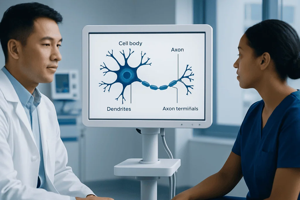
Most neurons have three major structural regions. First are the dendrites, which are branch-like extensions that receive incoming signals from other cells. Second is the cell body, also called the soma, which contains the nucleus and supports the neuron’s daily functions. Third is the axon, a long fiber that carries the electrical message away from the cell body.
At the far end of the axon are axon terminals. These terminals release chemical messengers called neurotransmitters into a tiny gap known as the synapse. The next cell detects these chemicals and converts them into a new signal. In this way, one neuron can influence another neuron, a muscle cell, or a gland.
Many axons are wrapped in myelin, a fatty insulating layer made by support cells. Myelin helps signals travel faster and more smoothly. If myelin is damaged, communication along the nerve can slow down or become unreliable, which may lead to symptoms such as visual changes, tingling, balance problems, or weakness.
- Dendrites: receive information
- Cell body: maintains the neuron and integrates signals
- Axon: carries the electrical impulse
- Axon terminals: release neurotransmitters
- Synapse: connection point between cells
- Myelin: insulation that speeds signal conduction
How neurons send messages

Neurons communicate using both electricity and chemistry. Inside the neuron, a brief electrical change called an action potential travels down the axon. This happens when the neuron is stimulated enough to cross a threshold. The signal then moves in an organized, one-way fashion toward the axon terminals.
When the electrical signal reaches the synapse, it triggers the release of neurotransmitters. These chemicals cross the synaptic gap and bind to receptors on the next cell. Depending on the neurotransmitter and receptor, the message may excite the next cell, calm it, or adjust how strongly it responds.
This constant exchange allows the nervous system to coordinate complex tasks very quickly. For example, touching a hot surface can trigger a rapid reflex, while reading, speaking, and planning involve much larger networks of neurons communicating across many brain regions.
Support cells called glial cells also matter. They do not carry messages in the same way neurons do, but they nourish, protect, and organize the environment around neurons. Healthy nerve function depends on both neurons and their support cells working together.
Different types of neurons and what they do
Neuron anatomy varies depending on function. Sensory neurons carry information from the body to the brain and spinal cord, such as touch, pain, temperature, sound, or visual input. Motor neurons carry commands from the brain or spinal cord to muscles, helping a person move, swallow, breathe, and maintain posture.
Interneurons connect neurons to one another within the brain and spinal cord. They are essential for interpreting information, forming memories, making decisions, and coordinating responses. Many of the brain’s most complex activities rely on dense networks of interneurons.
Neurons can also be grouped by shape. Some are multipolar, with many dendrites and one axon; these are common in the brain and spinal cord. Others are bipolar or pseudounipolar, shapes that are useful in special sensory pathways and peripheral nerves. For patients, the key point is that the structure of each neuron supports the specific job it needs to perform.
When certain neuron types are affected, symptoms often follow recognizable patterns. Damage to sensory pathways may cause numbness or burning pain, while damage to motor pathways may cause weakness, muscle wasting, or stiffness. Conditions such as Parkinson's disease affect specialized neural circuits involved in movement control.
What can damage neurons
Neurons are delicate cells, and many different problems can affect them. Direct injury to the brain, spinal cord, or peripheral nerves can interrupt signaling immediately. Reduced blood flow, as happens in a stroke, can deprive neurons of oxygen and lead to sudden loss of function in the affected area.
Inflammation, infection, autoimmune disease, metabolic disorders, and exposure to toxins may also harm neurons or the myelin around them. Poorly controlled diabetes, vitamin deficiencies, alcohol misuse, and some medications can contribute to nerve damage over time. In the brain, degenerative conditions may gradually affect certain neuron groups more than others.
Age is one factor, but it is not the only one. Genetics, vascular health, sleep, physical activity, blood pressure, and overall nutrition all influence nervous system health. Some symptoms develop suddenly, while others appear slowly and may at first be subtle.
Possible signs of neuron or nerve dysfunction include:
- Numbness, tingling, or burning sensations
- Muscle weakness or cramps
- Tremor, slowness, or poor coordination
- Balance problems or falls
- Vision, speech, or swallowing changes
- Memory, mood, or concentration difficulties
How doctors evaluate neuron-related symptoms
Doctors do not usually diagnose a problem by looking at neuron anatomy alone. Instead, they combine the patient’s symptoms, medical history, and a neurological examination to understand which part of the nervous system may be involved. The pattern of symptoms often provides important clues about whether the issue is in the brain, spinal cord, nerve root, or peripheral nerve.
The neurological exam may assess strength, reflexes, sensation, coordination, balance, eye movements, speech, and mental status. If needed, imaging tests such as MRI or CT can help look for structural problems. In some cases, blood tests are used to check for infections, inflammation, vitamin deficiencies, thyroid disease, or other metabolic causes.
For suspected peripheral nerve or muscle disorders, nerve conduction studies and electromyography may be recommended. For brain-related symptoms, doctors may consider advanced imaging or referral to specialists experienced in neurology care or neurosurgery when structural lesions, pressure on nerves, or certain surgical conditions are being evaluated.
Because many different conditions can affect neurons, diagnosis is often a step-by-step process. Clear communication about when symptoms started, whether they are worsening, and what makes them better or worse can help guide the workup.
Supporting nervous system health
There is no single way to protect every neuron, but healthy daily habits can support the nervous system over time. Good control of blood pressure, cholesterol, and blood sugar helps protect the blood vessels that nourish the brain and nerves. Regular physical activity supports circulation, balance, sleep, and overall brain health.
Nutrition also matters. A balanced diet that provides adequate vitamins, minerals, healthy fats, and protein supports normal nerve function. Avoiding smoking and limiting alcohol can reduce avoidable nerve and vascular damage. Good sleep is another important factor because the brain uses sleep to restore and regulate many processes.
Safety habits help prevent injury-related nerve problems. Wearing seat belts, using helmets when appropriate, and reducing fall risk at home are practical ways to protect the brain and spinal cord. For people with chronic numbness in the feet, regular foot checks and protective footwear can lower the chance of unnoticed injuries.
When a neurological condition is present, treatment depends on the cause. This may include rehabilitation, medicines, surgery, or other targeted care. In selected cases, a doctor may discuss options such as brain tumor treatment if imaging identifies a mass affecting nerve pathways. Near the end of the care pathway, patients seeking international evaluation may wish to know that Acibadem International’s multidisciplinary specialists and JCI-accredited hospitals diagnose and treat neurological conditions for international patients.
When to seek medical care
Medical advice is appropriate if neurological symptoms are new, persistent, worsening, or affecting daily life. Examples include ongoing numbness, repeated falls, unusual weakness, changes in coordination, new tremor, memory decline, or unexplained pain that follows a nerve pattern. Even when symptoms seem mild, early assessment can help identify reversible causes.
Urgent medical care is needed for sudden or severe symptoms. These include sudden weakness on one side, facial drooping, difficulty speaking, sudden confusion, loss of vision, a severe new headache, seizure, or loss of consciousness. These symptoms can signal emergencies such as stroke and should not be ignored.
People with known neurological conditions should also contact a doctor if symptoms change unexpectedly or if treatment no longer seems effective. Prompt follow-up can help reduce complications and support a safer recovery plan.
Frequently asked questions
What is neuron anatomy in simple terms?
Neuron anatomy is the structure of a nerve cell and the way its parts work together. The main parts are dendrites, the cell body, the axon, and the synapse, which allow messages to move through the nervous system.
What is the difference between a neuron and a nerve?
A neuron is a single nerve cell. A nerve is a bundle of many nerve fibers, usually axons, wrapped together in the peripheral nervous system.
Why is myelin important?
Myelin is an insulating covering around many axons. It helps nerve signals travel faster and more efficiently, so damage to myelin can slow or disrupt communication.
Can neurons heal after injury?
Some recovery is possible, but it depends on where the injury occurs and how severe it is. Peripheral nerves may regenerate to some extent, while the brain and spinal cord generally have more limited repair capacity.
What symptoms can happen when neurons are damaged?
Symptoms vary by location and may include numbness, tingling, weakness, pain, tremor, poor balance, speech changes, or memory problems. Sudden symptoms need urgent evaluation because they can indicate a medical emergency.
How do doctors test for nerve cell problems?
Doctors usually begin with a medical history and neurological examination. Depending on the symptoms, they may also use blood tests, MRI, CT, nerve conduction studies, or electromyography.
References
- National Institute of Neurological Disorders and Stroke
- National Library of Medicine
- Merck Manual Consumer Version
- MedlinePlus
- World Health Organization
This article is for general information only and is not a substitute for professional medical advice. Please consult a qualified doctor about your individual situation.
Joint and spine care in Turkey — expert assessment & treatment
JCI-accredited · board-certified surgeons · reply within 24h
Add us as a Preferred Source to see more of our trusted health content across Google Search, AI Overviews and Discover.