Ventricular Septal Defect
Ventricular Septal Defect is a hole between the heart ventricles. Learn symptoms, causes, diagnosis, treatment options and prognosis.

Quick answer
Ventricular septal defect is a congenital heart condition in which an opening in the wall between the heart’s lower chambers allows blood to pass abnormally from one side to the other. Treatment depends on the size of the defect and symptoms, and at Acibadem in Turkey it may include monitoring, medication, or closure with catheter-based techniques or open-heart surgery after…
Ventricular Septal Defect, often called VSD, is a hole in the wall between the heart’s two lower chambers, allowing blood to pass in the wrong direction inside the heart. It is usually present from birth and may be small and harmless, or large enough to require specialist monitoring or treatment.
Overview
Ventricular Septal Defect, or VSD, is a congenital heart defect in which there is an opening in the septum, the wall that separates the right and left ventricles of the heart. The ventricles are the heart’s two lower pumping chambers. When a VSD is present, oxygen-rich blood from the left ventricle can pass through the opening into the right ventricle, instead of flowing normally out to the body.
The effect of a VSD depends mainly on its size, location, and the pressure difference between the two sides of the heart. A small ventricular septal defect may cause little or no disturbance to circulation and may only be noticed because a doctor hears a heart murmur. A larger VSD can allow too much blood to flow toward the lungs, making the heart and lungs work harder.
VSDs are most often diagnosed in babies and children, but some small defects are found later in life. Many children with small VSDs grow and develop normally with regular follow-up. When a VSD is large or causes symptoms, modern pediatric cardiology and cardiac surgery offer effective ways to manage or close the defect when appropriate.
Symptoms
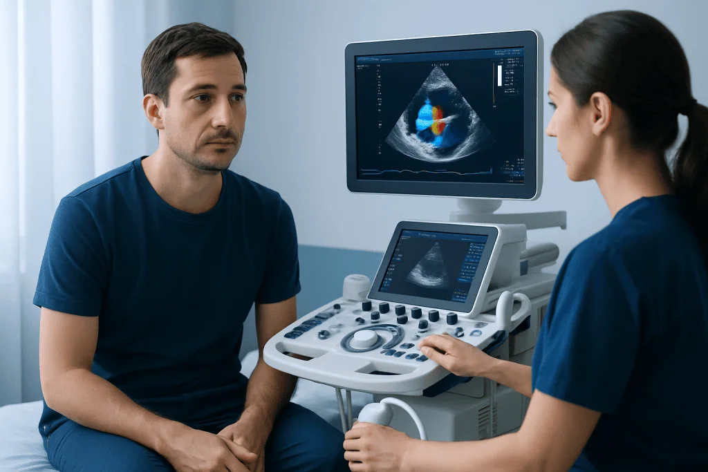
Ventricular Septal Defect symptoms vary widely. Some babies and children with a small VSD have no visible symptoms at all. In these cases, the first sign may be a heart murmur, which is an extra or unusual sound heard through a stethoscope during a routine examination.
When a VSD is moderate or large, symptoms usually relate to extra blood flow to the lungs and increased workload on the heart. Symptoms may appear in the first weeks or months of life, especially as the baby begins feeding more actively. In older children or adults, symptoms may be more subtle and may include reduced exercise tolerance or tiredness.
Possible symptoms and signs include:
- Fast breathing, heavy breathing, or shortness of breath
- Poor feeding, sweating during feeds, or tiring easily while feeding
- Slow weight gain or difficulty growing as expected
- Frequent chest infections or cough
- Fast heartbeat or signs that the heart is working harder
- Fatigue during play or physical activity
- Bluish discoloration in rare advanced cases with significant changes in lung blood pressure
Symptoms do not always match the loudness of the murmur. A small VSD can produce a loud murmur, while a very large defect may have a softer murmur. For this reason, assessment by a qualified doctor and, when needed, a pediatric cardiologist is important.
Causes & Risk Factors
Most ventricular septal defects form during early heart development before birth. The heart begins as a simple tube and gradually develops walls, chambers, valves, and major blood vessels. If part of the ventricular septum does not form completely, an opening can remain between the two ventricles.
In many cases, there is no single identifiable cause. A VSD can occur by itself in an otherwise healthy baby, or it can be part of a more complex congenital heart condition. VSDs may also be associated with some genetic or chromosomal conditions, so a doctor may consider whether additional assessment is needed if there are other physical findings or developmental concerns.
Factors that may be associated with a higher chance of congenital heart defects include a family history of congenital heart disease, certain genetic syndromes, some maternal infections during pregnancy, poorly controlled maternal diabetes, exposure to certain medicines or substances during pregnancy, and other maternal health factors. Having a risk factor does not mean a baby will have a VSD, and many babies with VSD have no known risk factor.
Very rarely, a ventricular septal defect can be acquired later in life, for example after a severe heart attack or heart injury. This is different from the congenital VSD seen in infants and children and requires urgent specialist care. The information on this page focuses mainly on congenital VSD.
Diagnosis
Diagnosis of Ventricular Septal Defect often begins when a healthcare professional hears a heart murmur. A murmur is not a diagnosis by itself; it is a sound caused by blood flow. The doctor will assess the child’s growth, breathing, feeding, oxygen levels, pulses, and overall health, and will decide whether heart imaging is needed.
The main test for diagnosing a VSD is echocardiography, also called a heart ultrasound. This test uses sound waves to show the heart’s chambers, valves, septum, blood flow direction, and the size and location of the defect. It can also help estimate how much extra blood is flowing to the lungs and whether the heart chambers are enlarged.
Other tests may be used depending on the situation. An electrocardiogram can assess heart rhythm and signs of chamber strain. A chest X-ray may show heart size and lung blood flow. In selected cases, cardiac MRI, CT, or cardiac catheterization may be recommended to obtain more detailed information, especially before an intervention or when lung pressure needs careful evaluation.
Some VSDs are suspected before birth during a fetal ultrasound or fetal echocardiogram. Prenatal diagnosis allows parents and doctors to plan delivery and newborn care appropriately. However, some small VSDs are easier to detect after birth, once circulation has changed from fetal to newborn life.
Treatment Options
Ventricular Septal Defect treatment is individualized. The right approach is decided by a pediatric cardiologist, cardiologist, or cardiac surgeon after assessing the person’s age, symptoms, growth, VSD size and location, heart function, lung blood pressure, and whether other heart conditions are present. No single treatment plan is suitable for every patient.
Small VSDs that are not causing symptoms may only need observation and regular follow-up. Some small defects close naturally as a child grows, while others remain open but do not cause important health problems. During follow-up, the specialist monitors growth, heart sounds, echocardiography findings, and any signs that the defect is affecting the heart or lungs.
Medicines may be used when a baby or child has symptoms related to extra blood flow, such as difficulty feeding, poor weight gain, or signs of fluid overload. Medication does not close the hole, but it can help reduce the workload on the heart and support growth while the care team decides whether further treatment is needed. Nutrition support may also be discussed for infants who tire easily during feeding.
If closure is recommended, it may be performed by open-heart surgery or, for selected types of VSD, by a catheter-based procedure. Surgical repair usually involves closing the defect with a patch or stitches. Catheter closure uses thin tubes inserted through blood vessels to place a closure device, but it is not suitable for every VSD location or size. The specialist team explains the benefits, risks, timing, and follow-up needs before any procedure.
Living With / Prognosis
The outlook for many people with Ventricular Septal Defect is very good, especially when the defect is small or when a significant VSD is treated at the right time. Many children with small VSDs live active lives and have normal growth and development. Even after closure, follow-up is important to check heart function, rhythm, valves, and the repair site.
Long-term care depends on the type of VSD and the treatment history. Some people need only occasional cardiology visits, while others require more regular review. Doctors may give guidance about sports, dental care, infection prevention, pregnancy planning, and whether any special precautions are needed before procedures. These recommendations should be personalized rather than assumed.
Parents can support a child with VSD by attending scheduled appointments, tracking feeding and growth in infancy, reporting changes in breathing or activity, and keeping vaccinations and general pediatric care up to date. Adults with a history of VSD, including those who had repair in childhood, should inform healthcare providers about their heart history and should consider care from a cardiologist familiar with congenital heart disease.
For international patients who need evaluation or treatment, Acibadem International provides access to multidisciplinary cardiology, pediatric cardiology, cardiac surgery, imaging, and intensive care teams in JCI-accredited hospitals. Care decisions should always be based on a detailed specialist assessment and the patient’s individual medical needs.
When to See a Doctor
A doctor should assess any baby or child with a newly detected heart murmur, poor feeding, poor weight gain, rapid breathing, unusual sweating during feeds, or reduced activity. Many murmurs are harmless, but a medical examination and appropriate testing help distinguish an innocent murmur from a structural heart condition such as VSD.
Parents should seek prompt medical attention if a baby is breathing very fast, struggling to breathe, feeding much less than usual, becoming unusually sleepy, or showing bluish discoloration of the lips or skin. These signs can have several causes, and timely assessment helps ensure the child receives the right care.
Children and adults with a known VSD should contact their doctor if they develop new shortness of breath, fainting, chest discomfort, palpitations, swelling, reduced exercise capacity, or frequent respiratory infections. People who had VSD repair should also keep planned follow-up visits, even if they feel well.
Before pregnancy, women with an unrepaired VSD or a history of repaired congenital heart disease should consult a cardiologist. Pregnancy is often possible, but heart function, lung pressure, and any residual defect should be reviewed so that safe, coordinated care can be planned.
Frequently asked questions
What is a Ventricular Septal Defect?
A Ventricular Septal Defect is a hole in the wall between the heart’s two lower chambers, called the ventricles. It allows blood to move between the ventricles in an abnormal way. The condition is usually present from birth and can range from very small to large.
Can a VSD close on its own?
Yes, some small ventricular septal defects close naturally during infancy or early childhood. Others may remain open but cause no major symptoms. A cardiologist can monitor the defect with examinations and echocardiography to determine whether observation is safe.
Is Ventricular Septal Defect serious?
It depends on the size and effect of the defect. Small VSDs are often mild and may need only follow-up, while larger VSDs can put extra strain on the heart and lungs. Specialist assessment is important because symptoms alone may not show how significant the defect is.
How is Ventricular Septal Defect diagnosed?
A VSD is often suspected when a doctor hears a heart murmur. The main diagnostic test is echocardiography, which shows the heart structure and blood flow. Additional tests such as an electrocardiogram, chest X-ray, cardiac MRI, or catheterization may be used in selected cases.
What are the treatment options for VSD?
Treatment may include regular monitoring, medicines to manage symptoms, nutritional support for infants, catheter-based closure in selected cases, or surgical repair. The best option depends on the VSD’s size, location, symptoms, heart function, and lung pressure. A specialist team decides the safest approach after a full assessment.
Can a child with VSD play sports?
Many children with a small or successfully treated VSD can be physically active. Activity advice should be individualized, especially if the child has symptoms, high lung pressure, rhythm problems, or other heart conditions. Parents should follow the cardiologist’s guidance about sports and school activities.
Does a repaired VSD need lifelong follow-up?
Some people need only occasional follow-up after successful repair, while others require regular cardiology visits. Follow-up checks for residual leaks, valve changes, heart rhythm issues, and overall heart function. Adults who had VSD as children should tell their doctors about their congenital heart history.
References
- American Heart Association
- Centers for Disease Control and Prevention
- European Society of Cardiology
- Mayo Clinic
- Merck Manual Professional Edition
This article is for general information only and is not a substitute for professional medical advice. Please consult a qualified doctor about your individual situation.
Treatments for This Condition
Doctors Who Treat This Condition
Prof. Dr. Ahmet Tulga Ulus
Cardiovascular Surgery
Prof. Dr. Ahmet Ümit Güllü
Cardiovascular Surgery
Prof. Dr. Bülent Kisacikoğlu
Cardiovascular Surgery
Prof. Dr. Cem Alhan
Cardiovascular Surgery
Prof. Dr. Ersin Erek
Cardiovascular Surgery
Prof. Dr. Eyüp Murat Ökten
Cardiovascular Surgery
Prof. Dr. Fuat Bilgen
Cardiovascular Surgery
Prof. Dr. Hayati Özkan
Cardiovascular Surgery
Prof. Dr. Mehmet Özkan
Cardiovascular Surgery
Prof. Dr. Rıza Türköz
Cardiovascular Surgery
Prof. Dr. Tayyar Sarioğlu
Cardiovascular Surgery
Prof. Dr. Öner Gülcan
Cardiovascular SurgeryRelated Articles

CABG Recovery in Older Adults: What Families Should Expect

Pulmonary Hypertension: What Happens If It Goes Untreated?

Cardiovascular Toxicology: How Drugs and Toxins Can Damage the Heart
Peripheral Artery Disease: Leg Pain, Poor Circulation, and When Angioplasty Helps

Kawasaki Disease in Adults: Can Childhood Heart Changes Appear Later?
Arrhythmia Symptoms That Need Urgent Evaluation Instead of Watchful Waiting

Hyperlipidemia: When High Cholesterol Needs Specialist Cardiology Care

Deep Vein Thrombosis After Travel: Why Long Flights Raise the Risk

Stent Procedure Cost in Turkey: What Can Change the Total Price?

Heart Valve Surgery Recovery: ICU Stay, Walking, and Returning Home
Peripheral Artery Disease vs Deep Vein Thrombosis: How to Tell the Difference