What Is Nuclear Cardiology?
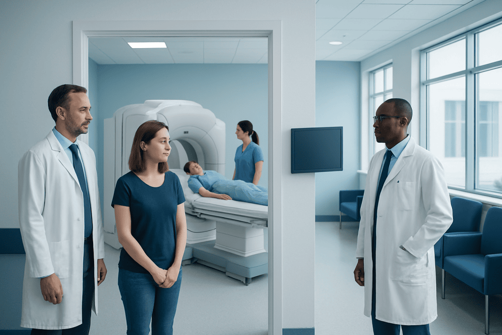
Nuclear cardiology combines a radioactive tracer with special cameras to assess heart blood flow and function. The most common study is a myocardial perfusion scan done at rest and during stress, either with exercise or medication.
Key Takeaways
- Nuclear cardiology combines a radioactive tracer with special cameras to assess heart blood flow and function.
- The most common study is a myocardial perfusion scan done at rest and during stress, either with exercise or medication.
- It can help diagnose coronary artery disease, estimate heart damage after a heart attack, and guide treatment decisions.
- The test is generally safe, and the radiation exposure is usually low and medically justified.
- Preparation may include avoiding caffeine, fasting for a few hours, and temporarily adjusting some medications if advised by a doctor.
Nuclear cardiology is a type of heart imaging that helps doctors see how well blood flows to the heart muscle and how the heart is working. It is commonly used to investigate chest pain, shortness of breath, and possible blocked coronary arteries.
Overview of Nuclear Cardiology
Nuclear cardiology is a specialized branch of heart imaging that uses a very small amount of radioactive tracer and a special camera to create pictures of the heart. These pictures help doctors evaluate blood flow to the heart muscle, heart pumping function, and in some cases the extent of previous heart damage. The information is often different from what can be seen on a standard electrocardiogram, echocardiogram, or routine physical examination.
The most common nuclear cardiology test is myocardial perfusion imaging, often called a nuclear stress test. It usually compares images taken when the heart is at rest with images taken when the heart is under stress. Stress may be created by walking on a treadmill or by using medication that temporarily increases blood flow demands or widens blood vessels in a controlled way.
Nuclear cardiology is widely used to look for reduced blood supply caused by narrowed or blocked arteries, especially coronary artery disease. It may also help assess symptoms such as chest discomfort, unexplained shortness of breath, or reduced exercise tolerance. In some people, it is used after treatment or after a heart event to evaluate recovery and future risk.
How the Test Works
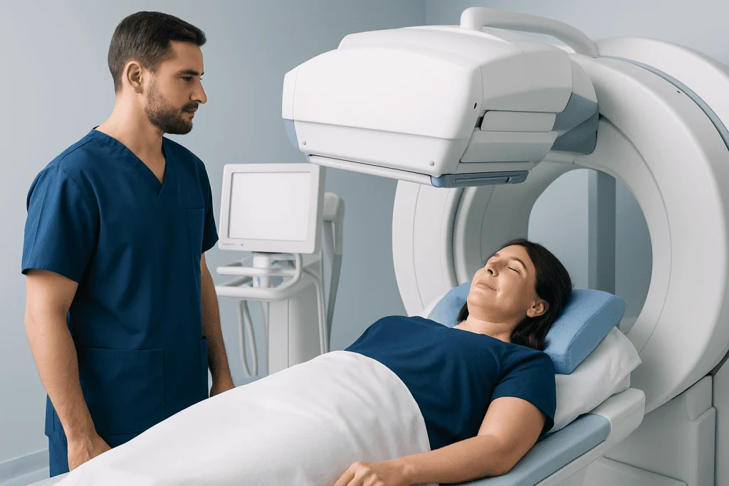
During a nuclear cardiology study, a tracer is injected into a vein. This tracer travels through the bloodstream and is taken up by healthy heart muscle. A gamma camera or similar scanner detects the tracer and creates images showing where blood is flowing well and where flow may be reduced.
If part of the heart receives less blood during stress than at rest, this may suggest a narrowed coronary artery. If a region has poor tracer uptake both at rest and during stress, that can indicate scar tissue from a previous injury, such as a prior heart attack. The test can also provide information about the heart’s pumping strength, often called the ejection fraction.
There are different nuclear techniques, including SPECT and PET imaging. Both can be useful, and the choice depends on the patient’s medical history, body type, local expertise, and the clinical question being asked. The main goal is to obtain a clear and reliable picture of heart blood flow and function so treatment can be tailored appropriately.
When Nuclear Cardiology Is Used

Doctors may recommend nuclear cardiology when a person has symptoms that could suggest heart disease, such as chest pain, pressure, breathlessness with activity, or unusual fatigue. It is often used when the likelihood of a circulation problem in the heart is not fully clear from symptoms, examination, and simpler tests alone.
The test is especially helpful for evaluating suspected blocked or narrowed coronary arteries, estimating the severity of known disease, and deciding whether additional treatment is needed. It can support decisions about medicines, stent procedures, or surgery such as coronary artery bypass surgery. In some people, it is also used to check whether treatment has improved blood flow.
Nuclear cardiology may be used before major surgery in selected patients with cardiac risk factors, or to assess heart function in people with certain forms of heart failure. It can also help identify areas of heart muscle that are still viable and may benefit from restoring blood supply. Not everyone with heart symptoms needs this test, but it can be very informative when used for the right reason.
What to Expect Before, During, and After the Scan
Preparation instructions vary slightly between centers, but patients are often asked not to eat for a few hours before the test. Caffeine may need to be avoided for 12 to 24 hours, especially if a medication-based stress test is planned, because caffeine can interfere with some stress medicines. A doctor may also advise whether certain heart medications should be taken or delayed on the day of testing.
The test commonly has two parts: rest images and stress images. For an exercise stress test, the patient walks on a treadmill or pedals a stationary bicycle while heart rate, blood pressure, and ECG are monitored. If exercise is not suitable, medication can safely mimic the effect of exercise on the heart. The tracer is injected at the right time, and imaging is done after a short wait.
During the scan, the patient usually lies still on a table while the camera moves around the chest. The scan itself is painless, although remaining still can be slightly uncomfortable for some people. Afterward, most patients can return to normal activities unless told otherwise. Drinking fluids may help clear the tracer from the body more quickly.
- Wear comfortable clothing and shoes if exercise testing is planned.
- Bring a list of medications and allergies.
- Tell the team about pregnancy, possible pregnancy, or breastfeeding.
- Report asthma, severe lung disease, or previous reactions to stress medicines.
Understanding the Results
A normal nuclear cardiology study usually means blood flow to the heart muscle appears adequate both at rest and during stress. This makes a major blood flow problem less likely, though no test is perfect. The results are interpreted together with symptoms, examination findings, ECG changes, and other medical information.
An abnormal result may show reduced blood flow during stress, suggesting ischemia, which means the heart muscle is not getting enough oxygen-rich blood during activity. This can occur in people with stable angina or other forms of coronary disease. Some scans show scarred areas from a previous heart attack, while others reveal reduced pumping function or enlargement of the heart chambers.
The results can help guide the next step. For some people, lifestyle changes and medicines are enough. Others may need more testing, such as coronary angiography, or treatment through cardiothoracic surgery or another heart procedure. The aim is to match treatment to the actual degree of risk and the extent of heart muscle affected.
Benefits, Risks, and Limitations
One of the main benefits of nuclear cardiology is that it shows function, not just structure. It can reveal how well blood is reaching the heart muscle under stress, which makes it especially useful for detecting clinically important coronary artery narrowing. It may also help avoid unnecessary invasive procedures in people with low-risk findings.
The test is generally safe. The amount of radiation used is carefully controlled and is typically low enough to be considered acceptable when the test is medically appropriate. Rarely, people may have side effects from stress medicines, such as flushing, headache, chest discomfort, or shortness of breath, but these effects are usually brief and the team is trained to manage them promptly.
Like all tests, nuclear cardiology has limitations. Results can occasionally be affected by body shape, breast tissue, movement during imaging, or certain medical conditions. It also may not be the best first test for every patient. Depending on symptoms and risk factors, a doctor may instead recommend echocardiography, CT coronary imaging, or another form of assessment.
Heart Health, Follow-Up, and When to Seek Medical Care
Nuclear cardiology is only one part of heart care. Whether results are normal or abnormal, long-term heart health still depends on managing risk factors. This often includes controlling blood pressure, stopping smoking, staying physically active, maintaining a healthy weight, improving cholesterol levels, and managing diabetes. In some people, structured cardiac rehabilitation can support recovery and healthier daily habits.
Patients should contact a doctor if they have ongoing chest pain, increasing shortness of breath, dizziness, fainting, or a clear decline in exercise capacity. Immediate emergency care is important for severe chest pain, sudden breathlessness, or symptoms that may suggest a heart attack. A doctor can explain whether nuclear cardiology is appropriate and how the results fit into the overall plan.
For international patients who need advanced heart assessment, Acibadem International’s multidisciplinary specialists in JCI-accredited hospitals diagnose and treat a wide range of cardiac conditions, including the use of modern imaging and tailored treatment plans. Follow-up is important, because the real value of the test lies in using the results to guide safer, more effective care over time.
Frequently asked questions
Is nuclear cardiology the same as a regular stress test?
Not exactly. A regular stress test usually monitors the ECG, heart rate, and blood pressure during exercise, while nuclear cardiology adds imaging to show blood flow to the heart muscle. This can provide more detailed information about possible blocked arteries and heart function.
How long does a nuclear cardiology test take?
The full appointment often takes several hours because it may include preparation, tracer injection, waiting periods, stress testing, and imaging. The exact time depends on the type of test and whether both rest and stress images are done on the same day. The imaging itself is only one part of the visit.
Is the radiation from nuclear cardiology dangerous?
The test uses a small amount of radiation, and the dose is kept as low as reasonably possible. For most patients, the medical benefit of answering an important heart question outweighs the risk. Doctors consider age, symptoms, and other options before recommending the test.
Can someone have a nuclear cardiology test if they cannot exercise?
Yes. If exercise is not possible, medicines can be used to safely create a stress effect on the heart. This allows doctors to evaluate blood flow in a similar way without the patient needing to walk on a treadmill.
What should be avoided before a nuclear stress test?
Many patients are advised to avoid caffeine for a period before the test, and some may need to avoid food for a few hours. Certain medicines may also need special instructions. The imaging center or doctor will provide exact preparation steps based on the type of test.
What happens if the nuclear cardiology scan is abnormal?
An abnormal result does not automatically mean a person needs surgery. It means the doctor will review the pattern of blood flow, symptoms, and overall risk to decide on the next step. This may include medication changes, more testing, or procedures to improve blood supply to the heart.
References
- American Heart Association
- Society of Nuclear Medicine and Molecular Imaging
- American Society of Nuclear Cardiology
- National Heart, Lung, and Blood Institute
- European Society of Cardiology
This article is for general information only and is not a substitute for professional medical advice. Please consult a qualified doctor about your individual situation.
Nuclear Cardiology in Turkey — costs, top hospitals & a free quote
JCI-accredited · board-certified surgeons · reply within 24h
Add us as a Preferred Source to see more of our trusted health content across Google Search, AI Overviews and Discover.
Check your numbers in seconds
BMI, calories, due date, blood pressure and 30+ more clinical calculators — free, instant, doctor-reviewed ranges.

Cardiology Department
Diagnosis and treatment of heart and vascular conditions, from prevention to advanced interventional procedures.
91 specialists in this unit