Dural AV Fistula Treatment: When Endovascular Embolization Is Recommended

A dural AV fistula is an abnormal connection between arteries and veins in the covering of the brain or spinal cord. Endovascular embolization is commonly recommended when the fistula causes symptoms or has high-risk venous drainage.
Key Takeaways
- A dural AV fistula is an abnormal connection between arteries and veins in the covering of the brain or spinal cord.
- Endovascular embolization is commonly recommended when the fistula causes symptoms or has high-risk venous drainage.
- Diagnosis usually involves MRI or CT imaging and is confirmed with cerebral angiography.
- Treatment decisions are individualized and may include embolization alone or combined with surgery or radiosurgery.
- Ongoing follow-up imaging is important because some fistulas can persist or recur after treatment.
Dural AV fistula treatment depends on where the fistula is located, how blood drains from it, and whether it is causing symptoms or bleeding risk. Endovascular embolization is often recommended because it can close or reduce the abnormal connection through a minimally invasive, image-guided approach.
Overview of dural AV fistula treatment
A dural arteriovenous fistula, often shortened to dural AV fistula or dAVF, is an abnormal connection between arteries and veins within the dura, the protective covering around the brain or spinal cord. In a normal circulation pattern, blood moves from arteries into tiny capillaries and then into veins. In a dural AV fistula, blood bypasses the usual pathway and flows directly into veins under higher pressure than they are designed to handle.
This abnormal flow can lead to symptoms such as pulsating noise in the ear, headaches, vision changes, or neurological problems. Some dural AV fistulas are relatively low risk, while others are more serious because they drain into delicate brain veins in a way that increases the chance of bleeding, swelling, or nerve dysfunction. For this reason, treatment is based not only on symptoms but also on the fistula’s drainage pattern and location.
Dural AV fistula treatment may include careful monitoring, endovascular embolization, surgery, radiosurgery, or a combination of these options. Among these, endovascular embolization is often the first-line approach because it is minimally invasive and can often treat the fistula from inside the blood vessels. It is commonly performed by specialists in interventional neuroradiology, neurointervention, or endovascular neurosurgery.
Symptoms and signs that may lead to treatment
The symptoms of a dural AV fistula can vary widely. Some people have a rhythmic whooshing sound in one ear, called pulsatile tinnitus, especially when the fistula is near structures around the ear. Others may develop headaches, eye redness, bulging of the eye, double vision, facial pain, dizziness, or changes in hearing. If the fistula affects veins around the brain or spinal cord, symptoms may include weakness, numbness, difficulty walking, cognitive changes, or seizures.
In some cases, a dural AV fistula is found incidentally during imaging for another reason. Even then, treatment may still be considered if the vessel pattern suggests a higher risk of future problems. Doctors pay especially close attention to signs of cortical venous reflux, which means blood is draining backward into brain veins. This pattern is associated with a greater risk of hemorrhage and neurological injury.
Emergency assessment is important if symptoms begin suddenly or worsen quickly. Warning signs can include severe headache, confusion, weakness on one side, vision loss, trouble speaking, or loss of consciousness. These symptoms do not always mean bleeding has occurred, but they should be evaluated urgently.
When endovascular embolization is recommended

Endovascular embolization is recommended when a dural AV fistula is causing troublesome symptoms, creating strain on nearby veins, or showing imaging features linked to a higher risk of bleeding or neurological damage. In many centers, it is the preferred initial treatment because it can directly target the abnormal vessels through a catheter inserted into an artery or sometimes a vein, usually through the groin or wrist.
The decision depends on several factors, including the fistula’s location, the number and size of feeding arteries, the pattern of venous drainage, and whether there has already been bleeding. Fistulas with cortical venous drainage, rapidly progressive neurological symptoms, eye-related complications, or spinal cord congestion are more likely to need prompt treatment. By contrast, a low-risk fistula with mild symptoms may sometimes be monitored if the expected benefit of treatment is limited.
Embolization may be used as the only treatment when complete closure is feasible, or as part of a combined plan with surgery or radiosurgery if the anatomy is more complex. The aim is to block the abnormal connection while preserving normal blood flow to surrounding tissues. In selected cases, doctors may discuss embolization treatment as the main therapy because it offers a precise, image-guided way to reach the fistula.
People with related vascular conditions may hear comparisons with other vessel abnormalities, but a dural AV fistula is different from a typical brain aneurysm or a brain arteriovenous malformation. These conditions can share certain symptoms or imaging tests, yet their structure and treatment strategy are not the same.
How dural AV fistulas are diagnosed
Diagnosis usually begins with a neurological examination and imaging studies. MRI or CT scans may show indirect signs such as enlarged veins, swelling, prior bleeding, or changes in the brain or spinal cord caused by venous congestion. CT angiography or MR angiography can also suggest the presence of a fistula, but these tests may not provide enough detail to plan treatment fully.
The most important test for confirming a dural AV fistula is catheter angiography, also called cerebral angiography or digital subtraction angiography. During this procedure, contrast dye is injected into blood vessels while X-ray images are taken in real time. This allows doctors to map the exact feeding arteries, the site of the shunt, and the venous drainage pattern. It is essential for deciding whether embolization is possible and how it should be performed.
Because treatment planning is highly anatomical, a multidisciplinary discussion is often helpful. Neurologists, neurosurgeons, and interventional neuroradiology specialists review the imaging together to choose the safest and most effective strategy. If needed, a patient may also be evaluated for broader interventional neuroradiology care when the fistula is complex or located near critical brain structures.
What happens during endovascular embolization
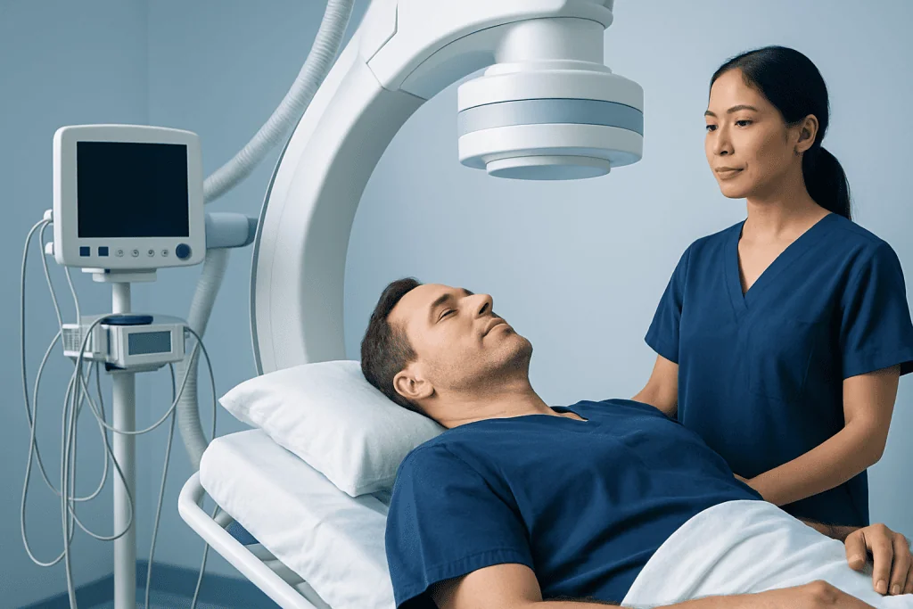
Endovascular embolization is usually performed in a specialized angiography suite. The patient is commonly given anesthesia or sedation, depending on the treatment plan. A doctor guides a thin catheter through the blood vessels to the arteries or veins connected to the fistula. Using live imaging, the team positions the catheter as close as possible to the abnormal connection.
Once the catheter is in place, an embolic agent is delivered to block the fistula. This material may be a liquid embolic substance, coils, or another device chosen for the specific anatomy. The goal is to close the abnormal pathway and reduce the high-pressure blood flow into fragile veins. The specialist then performs repeat angiographic imaging to see whether the fistula has been completely or partially closed.
Some dural AV fistulas can be cured in a single session, while others require staged treatment. The need for more than one procedure does not necessarily mean the first treatment failed; it may reflect a complex vessel network that is safer to treat step by step. In certain cases, embolization is followed by Gamma Knife radiosurgery or surgery if a small residual fistula remains.
Most patients stay in the hospital for observation after the procedure. Recovery time varies, but many people resume light activities within days, depending on their overall condition and whether they had neurological symptoms beforehand. The treating team gives specific advice on activity, medicines, and follow-up appointments.
Benefits, risks, and other treatment options
The main benefit of endovascular embolization is that it is minimally invasive and can often treat the fistula without open surgery. It may quickly improve symptoms caused by abnormal blood flow, reduce the risk of bleeding in high-risk fistulas, and shorten recovery compared with more invasive approaches. It is especially valuable when the fistula is located in an area that is difficult to reach surgically.
Like any procedure involving blood vessels in the brain or spinal region, embolization also carries risks. Potential complications can include stroke, bleeding, damage to a normal blood vessel, reaction to contrast dye, infection, or incomplete closure of the fistula. The exact level of risk depends on the fistula’s anatomy, the embolic material used, and the patient’s general health. These issues are carefully discussed before treatment so the patient can make an informed decision.
Other treatment options may be considered when embolization is not expected to achieve complete closure or when the anatomy is better suited to another method. Microsurgery can directly disconnect the fistula, especially in selected accessible locations. Radiosurgery may be used for smaller residual fistulas, although it works gradually over time rather than immediately. In low-risk cases without dangerous venous drainage, observation with follow-up imaging may be appropriate.
The best plan is individualized. For some people, the safest approach is embolization alone. For others, a staged combination of endovascular treatment and neurosurgery offers the best chance of durable closure while protecting surrounding brain tissue.
Recovery, follow-up, and self-care
After treatment, follow-up is an important part of care. Even when symptoms improve quickly, imaging is usually needed to confirm that the fistula has closed completely. Some patients have a follow-up angiogram or noninvasive vascular imaging after a period determined by their doctor. This is important because a small residual fistula can persist or, less commonly, recur.
During recovery, patients are usually advised to rest, drink enough fluids if appropriate, and follow instructions about wound care at the catheter entry site. They should take medicines only as directed and ask their doctor before restarting strenuous exercise, travel, or blood-thinning medication. If new neurological symptoms appear after the procedure, prompt medical review is needed.
Longer-term outlook depends on the fistula type and whether treatment achieved complete closure. Many people do well, especially when high-risk venous drainage is eliminated early. If symptoms such as pulsatile tinnitus or vision problems were present for a long time, improvement can be gradual rather than immediate. Toward the end of the care journey, some international patients choose evaluation at Acibadem International, where multidisciplinary specialists in JCI-accredited hospitals diagnose and treat complex neurovascular conditions.
When to see a doctor
A person should see a doctor if they notice pulsatile tinnitus, unexplained eye changes, ongoing headaches with new neurological symptoms, or progressive weakness, numbness, or balance problems. While these symptoms can have many causes, a vascular condition such as a dural AV fistula is one possibility that should not be overlooked. Early assessment can help clarify the cause and guide timely treatment if needed.
Urgent medical attention is needed for sudden severe headache, seizure, confusion, fainting, speech difficulty, one-sided weakness, or sudden vision loss. These symptoms may indicate bleeding or another serious neurological event. Fast evaluation can be critical for diagnosis and treatment planning.
People who have already been diagnosed with a dural AV fistula should also contact their care team if symptoms worsen, return after treatment, or new symptoms develop. Regular follow-up matters, even after a successful procedure, because confirming long-term closure is part of safe care.
Frequently asked questions
What is a dural AV fistula?
A dural AV fistula is an abnormal connection between arteries and veins in the dura, the outer covering around the brain or spinal cord. This direct connection can cause high-pressure blood flow into veins and lead to symptoms or complications depending on where it is located and how it drains.
Is endovascular embolization always necessary for a dural AV fistula?
No. Treatment depends on the fistula's symptoms, venous drainage pattern, location, and overall risk. Some low-risk fistulas may be monitored, while higher-risk or symptomatic fistulas are more likely to need treatment.
How do doctors decide if embolization is the right option?
Doctors review detailed imaging, especially catheter angiography, to understand the exact vessel anatomy. Embolization is often chosen when the fistula can be reached safely through the blood vessels and when closing it is expected to reduce symptoms or prevent complications.
Can a dural AV fistula come back after treatment?
Yes, in some cases a small residual fistula can remain or recur later. That is why follow-up imaging is an important part of care, even when symptoms improve after treatment.
What is recovery like after endovascular embolization?
Recovery is often shorter than with open surgery, but it varies from person to person. Many patients stay in the hospital for monitoring and can return to light activity within days, following their doctor's specific advice.
Are dural AV fistulas cancerous?
No, dural AV fistulas are not cancer. They are vascular abnormalities involving blood vessels, and treatment focuses on controlling abnormal blood flow and protecting brain or spinal cord function.
References
- World Health Organization
- National Institute of Neurological Disorders and Stroke
- American Association of Neurological Surgeons
- Society of NeuroInterventional Surgery
- Radiological Society of North America
This article is for general information only and is not a substitute for professional medical advice. Please consult a qualified doctor about your individual situation.
Joint and spine care in Turkey — expert assessment & treatment
JCI-accredited · board-certified surgeons · reply within 24h
Add us as a Preferred Source to see more of our trusted health content across Google Search, AI Overviews and Discover.