

Quick answer
A PET-CT scanner combines metabolic imaging from positron emission tomography with detailed anatomical images from computed tomography, helping specialists identify and assess many conditions, particularly cancer. At Acibadem in Turkey, PET-CT imaging is used to support diagnosis, staging, treatment planning and follow-up after specialist evaluation.
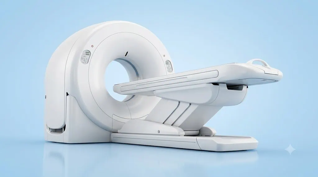
PET-CT Scanner
PET-CT is an advanced imaging technology that combines two complementary scans in one examination: positron emission tomography (PET) and computed tomography (CT). Together, they help doctors see both how tissues are functioning and where those changes are located in the body.
This combined view is especially valuable in cancer care. PET imaging can identify areas with altered metabolic activity, while CT provides detailed anatomical images of organs, bones, lymph nodes and other structures. By matching these images, specialists can assess a condition with greater precision and plan the most appropriate next steps.
At Acibadem International, PET-CT supports the work of Nuclear Medicine, Medical Oncology and multidisciplinary cancer teams. It may be used to clarify a diagnosis, determine the extent of disease, guide treatment planning or monitor response after treatment. Every scan is requested and interpreted according to an individual medical assessment.
What Is PET-CT in Plain Language?
A PET-CT scan is a largely non-invasive imaging examination. It usually begins with the administration of a small amount of a radiotracer, a substance designed to make certain body processes visible on the scan. The tracer is commonly given through a vein and travels through the bloodstream.
Many PET tracers are taken up more actively by tissues that use energy differently from surrounding tissue. This can include some cancer cells, but it can also occur in inflammation, infection or normal body processes. For this reason, PET-CT findings are always assessed carefully alongside your symptoms, laboratory results, previous scans and pathology reports.
The CT part of the examination creates cross-sectional images of the body. It helps the radiology and nuclear medicine teams identify the exact location, size and shape of areas seen on PET imaging. Combining the two methods in a single scan helps create a clearer, more complete picture.
How Does a PET-CT Scanner Work?
PET-CT brings functional imaging and anatomical imaging together. The PET component detects signals released by the radiotracer in the body. These signals are processed into images that show patterns of tracer uptake, sometimes described as metabolic activity.
The CT component uses X-ray technology to produce detailed images of the body’s internal structures. In some cases, a contrast material may be used to improve visibility of blood vessels, organs or tissues. Your clinical team will confirm whether contrast is suitable for you, particularly if you have allergies, kidney concerns or other relevant health conditions.
Specialist software aligns the PET and CT images precisely. This allows doctors to identify whether an area of increased or reduced tracer uptake corresponds to a lymph node, organ, bone, soft tissue structure or another anatomical site. The combined information can support more confident clinical decisions.
PET-CT does not replace every other diagnostic test. Depending on your condition, your doctor may recommend blood tests, ultrasound, MRI, biopsy, endoscopy or additional imaging. PET-CT is one important part of a personalised diagnostic and treatment pathway.
Clinical Applications of PET-CT
PET-CT is most commonly associated with oncology, where it can help specialists understand the location and activity of known or suspected cancer. It may also have selected roles in cardiology, infection assessment and inflammatory disease evaluation when clinically appropriate.
The exact tracer, imaging protocol and area of the body scanned depend on the clinical question. Your physician will determine whether PET-CT is appropriate for your case and which type of examination may provide the most useful information.
| Clinical applications | How PET-CT may help | Conditions commonly assessed |
|---|---|---|
| Initial cancer evaluation | Helps assess suspicious findings and identify areas that may require further investigation. | Selected solid tumours, lymphoma and other cancers, depending on the clinical indication. |
| Cancer staging | Helps show the primary tumour and whether disease may involve lymph nodes or other parts of the body. | Lung cancer, lymphoma, colorectal cancer, head and neck cancers, melanoma and many other oncology conditions. |
| Treatment planning | Supports multidisciplinary planning for surgery, systemic treatment and radiation therapy. | Patients preparing for personalised cancer treatment pathways. |
| Monitoring treatment response | May help evaluate changes in metabolic activity and anatomical appearance during or after treatment. | Cancers treated with chemotherapy, immunotherapy, targeted therapy, radiotherapy or combined approaches. |
| Follow-up after treatment | Can help investigate symptoms, abnormal blood tests or findings that raise concern for recurrence. | Patients under specialist surveillance after cancer treatment. |
| Selected infection or inflammation assessments | May help identify active inflammatory or infectious processes in carefully selected cases. | Complex infections, fever of unknown origin and certain inflammatory conditions, when recommended by a specialist. |
| Selected cardiac assessments | May provide information about tissue viability and metabolic activity in specific cardiac evaluations. | Some patients with coronary artery disease or other complex cardiac indications. |
Why PET-CT Matters in Cancer Staging
Accurate cancer staging is essential because treatment decisions are based not only on the type of cancer, but also on its location, size, biological behaviour and possible spread. PET-CT can help specialists bring these elements together in a single examination.
For patients with a newly diagnosed cancer, PET-CT may help determine whether the disease appears limited to one area or whether lymph nodes or distant organs need further assessment. This information can influence whether surgery, radiotherapy, systemic treatment or a combination of treatments may be considered.
In some situations, PET-CT can also help doctors select the best area for biopsy, evaluate whether a treatment is working or distinguish between active disease and post-treatment changes. Results are reviewed within the context of your full medical history; a scan alone does not determine a diagnosis or treatment plan.
At Acibadem International, PET-CT results can be discussed by relevant specialists within a multidisciplinary approach. This coordinated review helps ensure that imaging findings contribute meaningfully to a personalised care plan.
Benefits and Advantages for Patients
The main advantage of PET-CT is the ability to combine functional and anatomical information in one coordinated imaging examination. Instead of looking only at body structure or only at metabolic activity, the care team can review both together.
For many patients, this can support earlier clarification of important findings and reduce uncertainty when treatment decisions need to be made. It can also help avoid unnecessary procedures when imaging provides information that changes the clinical pathway.
| Patient benefits at a glance | What this can mean for your care |
|---|---|
| Combined imaging in one examination | PET and CT information are reviewed together to give a more complete view of the body. |
| Support for accurate staging | Specialists can assess the known disease site and evaluate areas that may need further investigation. |
| Personalised treatment planning | Imaging results can help guide discussions about surgery, medical oncology treatments or radiation therapy. |
| Largely non-invasive procedure | The scan generally requires an injection and lying still on the scanning table, without surgery. |
| Clearer follow-up assessment | Repeat imaging, when clinically indicated, may help evaluate treatment response or investigate new concerns. |
| Coordinated specialist review | Nuclear medicine physicians, oncologists, radiologists and other specialists can use the findings in shared care planning. |
| International patient support | Care coordinators can help organise appointments, records, language support and travel-related planning. |
What to Expect During a PET-CT Procedure
Your experience begins before the day of the scan. An Acibadem International coordinator can help collect your existing medical records, pathology reports, previous imaging and referral information. These materials help the relevant specialist determine whether PET-CT is appropriate and whether any additional preparation is needed.
Preparation instructions vary according to the type of PET-CT examination and the tracer being used. For many common oncology PET-CT scans, patients may be asked to fast for a period before the appointment. You may also receive guidance about drinking water, exercise, diabetes medications or other regular medicines. It is important to follow the instructions provided by your clinical team.
When you arrive, the team will confirm your identity, review your medical history and ask about allergies, pregnancy, breastfeeding, diabetes, kidney conditions and current medications. Please tell your team if you may be pregnant or are breastfeeding, as this may affect the timing or type of scan.
Before the Scan
A radiotracer is usually administered through a small intravenous injection. The amount is carefully calculated and selected for the diagnostic purpose of the examination. You will then rest quietly while the tracer distributes through your body. The waiting period can vary according to the protocol.
During this time, you may be asked to remain relaxed and avoid significant movement or activity. For certain studies, staff may ask you to keep warm, drink water or follow other specific instructions. Your care team will explain each step clearly.
During the Scan
For the imaging portion, you will lie on a padded table that moves slowly through the PET-CT scanner. The scanner is open at both ends and shaped like a large ring. The examination is generally painless, although you will need to stay still to help produce clear images.
The scan itself often takes a limited period, but the full appointment may be longer because of preparation, tracer uptake time and any additional imaging required. If you are concerned about enclosed spaces, discomfort when lying flat or pain that may make it difficult to remain still, tell your coordinator and medical team in advance.
After the Scan
Most patients can return to normal daily activities after the examination unless their physician gives different advice. You may be encouraged to drink fluids to help your body eliminate the radiotracer. The team will provide specific aftercare guidance, including any advice related to close contact with infants or pregnant individuals if relevant to your scan.
Your images are reviewed by appropriately trained specialists. The final report is then shared with the doctor responsible for your care. For international patients, our coordination team can help arrange follow-up consultations and support the secure transfer of reports and imaging records where appropriate.
Safety, Radiation Awareness and Clinical Standards
PET-CT is a routinely used diagnostic technology when requested for an appropriate medical reason. Like other imaging studies involving radiation, it is performed only after the anticipated clinical benefit has been considered. The amount of radiation depends on the scan protocol, the tracer used and whether CT contrast is required.
Acibadem International teams apply established safety processes in JCI-accredited hospital environments. Nuclear medicine physicians, radiologists, technologists, nurses and other trained professionals work together to ensure that imaging is performed according to clinical standards and patient-specific requirements.
Your care team will consider factors such as your age, medical history, kidney function, diabetes status, allergies and recent imaging. If you have had previous scans, bring the reports and, if available, the image files. Comparing current and prior imaging may be valuable for interpretation.
PET-CT findings must be interpreted by experienced specialists. Increased tracer uptake does not always indicate cancer, and some cancers may not show the same uptake pattern. Inflammation, infection, healing after surgery and certain normal physiological processes can affect results. Your doctor will explain what the findings mean for you and whether any further testing is recommended.
Care Coordination for International Patients
Travelling for diagnostic imaging or cancer care can feel complex, particularly when you are managing new information about your health. Acibadem International provides coordinated support designed to make the process clearer from the first consultation request through follow-up planning.
Our international patient team can help you share reports before travel, coordinate specialist review, arrange appointments and provide language support throughout your visit. If PET-CT is recommended, the team can advise on practical preparation, expected timing and how the scan may fit into a wider diagnostic or treatment schedule.
For patients who need to see several specialists, coordination can help bring consultations, imaging and treatment planning together as efficiently as clinically appropriate. Your team can also help you understand which hospital location is most suitable for your planned examination and related care.
After your visit, we can assist with the continuity of your medical information. This may include supporting access to reports, imaging results and recommendations for ongoing communication with your physician at home. Your care plan remains individual, and any decision to travel should be made after discussing your needs with the relevant medical professionals.
Frequently Asked Questions
Is PET-CT safe?
PET-CT is used routinely under specialist supervision. It involves radiation from the radiotracer and CT component, so it is recommended when the expected diagnostic value is greater than the potential risk. Your team will explain the specific considerations for your health, including pregnancy, breastfeeding, allergies, kidney function and previous imaging exposure.
Will I need contrast material?
Not every PET-CT scan requires CT contrast. Whether contrast is used depends on the clinical question and your individual health profile. If contrast is planned, your team will review relevant medical history and provide preparation guidance.
Can I eat before a PET-CT scan?
Many PET-CT protocols require fasting, but instructions differ depending on the type of scan and your medical condition. Patients with diabetes may need personalised planning. Always follow the preparation instructions given by your clinical team rather than relying on general guidance.
How soon will I receive my results?
Images require careful review by the appropriate specialist before a report is issued. Timing depends on the complexity of the examination and whether the findings need multidisciplinary discussion. Your international patient coordinator can explain the expected process and arrange follow-up with the relevant doctor.
Will my insurance cover PET-CT?
Coverage depends on your policy, insurer and the medical indication for the examination. You may share your insurance details with the international patient team so they can help clarify available options and explain the administrative process before your visit.
How do I get started?
You can submit your available medical reports, pathology results and previous imaging through a consultation request. A relevant specialist can review the information, advise whether PET-CT may be suitable and recommend the next steps for your individual care pathway.
Personalised Imaging for More Informed Decisions
PET-CT is an important technology for evaluating cancer and other selected medical conditions because it brings metabolic and anatomical information together. For patients facing a new diagnosis, planning treatment or monitoring progress, this combined perspective can support timely and informed clinical decisions.
At Acibadem International, PET-CT is integrated into a patient-centred approach that combines advanced imaging, specialist interpretation and coordinated international care. Your suitability for PET-CT will always be determined through an individual medical assessment, with clear communication at every stage of your journey.
Related Medical Units
Available at these Hospitals










Related Treatments
Speak with our medical team
Find out how this technology could support your diagnosis or treatment.