Cardiac Thrombosis: How Blood Clots in the Heart Are Diagnosed and Treated

Cardiac thrombosis is a blood clot that forms in a chamber or on a structure inside the heart. Some people have no symptoms, while others develop chest discomfort, shortness of breath, palpitations, or signs of stroke or poor circulation.
Key Takeaways
- Cardiac thrombosis is a blood clot that forms in a chamber or on a structure inside the heart.
- Some people have no symptoms, while others develop chest discomfort, shortness of breath, palpitations, or signs of stroke or poor circulation.
- Diagnosis usually relies on heart imaging, especially echocardiography, along with blood tests and evaluation of the underlying heart condition.
- Treatment often includes anticoagulant medicines and may also involve procedures or surgery depending on the clot’s size, location, and cause.
- Managing related conditions such as heart failure, arrhythmias, or coronary artery disease helps lower the risk of future clots.
Cardiac thrombosis means a blood clot forms inside the heart. It can be serious because the clot may affect blood flow or travel to other parts of the body, but prompt diagnosis and treatment can reduce complications.
Overview
Cardiac thrombosis refers to a blood clot, also called a thrombus, that develops inside the heart. The clot may form in one of the heart chambers, attach to the heart wall, or involve a heart valve or device. It is not the same as a heart attack, although a heart attack and other cardiac conditions can increase the risk of a clot forming afterward.
Blood normally moves smoothly through the heart. When blood flow becomes sluggish, the heart muscle is damaged, or the inner lining of the heart is abnormal, blood may be more likely to clot. This is why cardiac thrombosis is often linked to other heart problems rather than occurring on its own.
The main concern is that the clot can interfere with normal circulation or break off and travel through the bloodstream. If that happens, it may block blood flow to the brain, lungs, kidneys, or limbs. For this reason, doctors treat cardiac thrombosis as a condition that needs timely medical evaluation and careful follow-up.
Symptoms of cardiac thrombosis
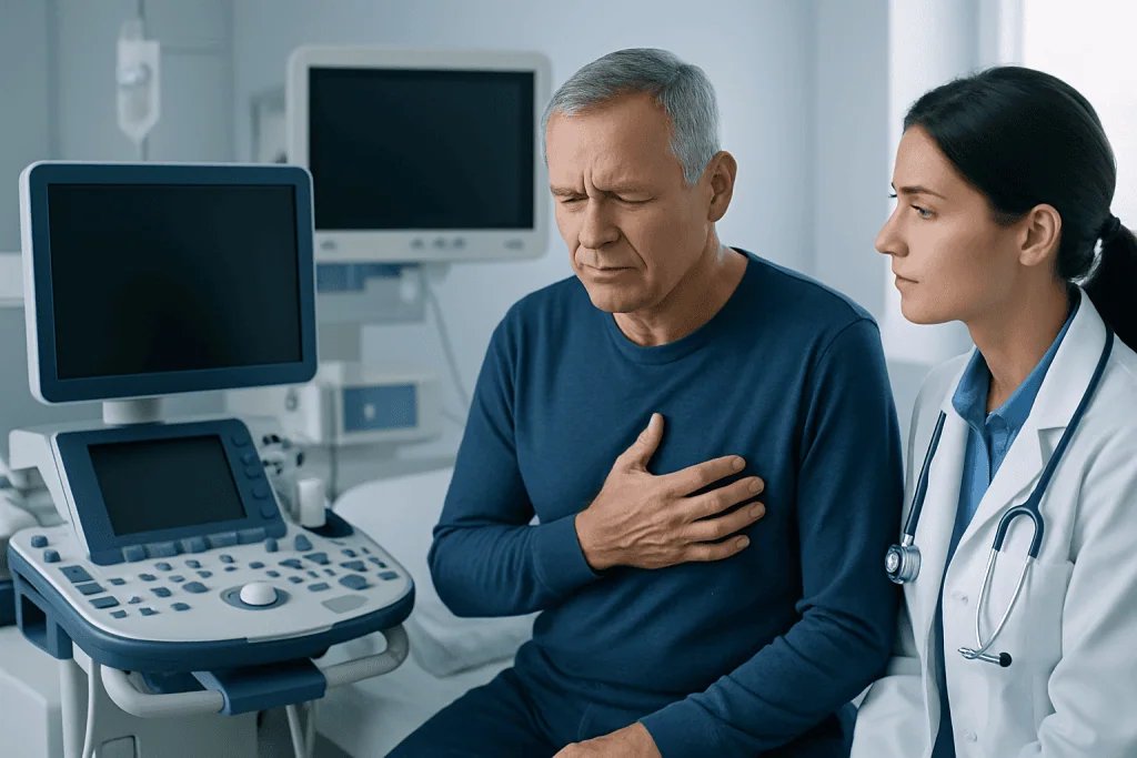
Symptoms vary widely. Some people do not notice any signs, and the clot is discovered during tests done for another reason. Others develop symptoms related to the heart problem that caused the clot or to the clot’s effect on circulation.
Possible symptoms can include chest discomfort, shortness of breath, fatigue, dizziness, fainting, or a racing or irregular heartbeat. In people with rhythm disorders such as atrial flutter or other abnormal heart rhythms, blood may pool in part of the heart and increase the chance of clot formation.
If part of the clot travels to another area of the body, symptoms may appear suddenly. These can include weakness on one side, facial drooping, difficulty speaking, severe limb pain, cold or pale fingers or toes, or sudden shortness of breath. These warning signs need urgent medical attention.
- Chest pain or pressure
- Shortness of breath
- Palpitations or an irregular pulse
- Fatigue or reduced exercise tolerance
- Stroke-like symptoms or sudden limb symptoms if an embolism occurs
Causes and risk factors
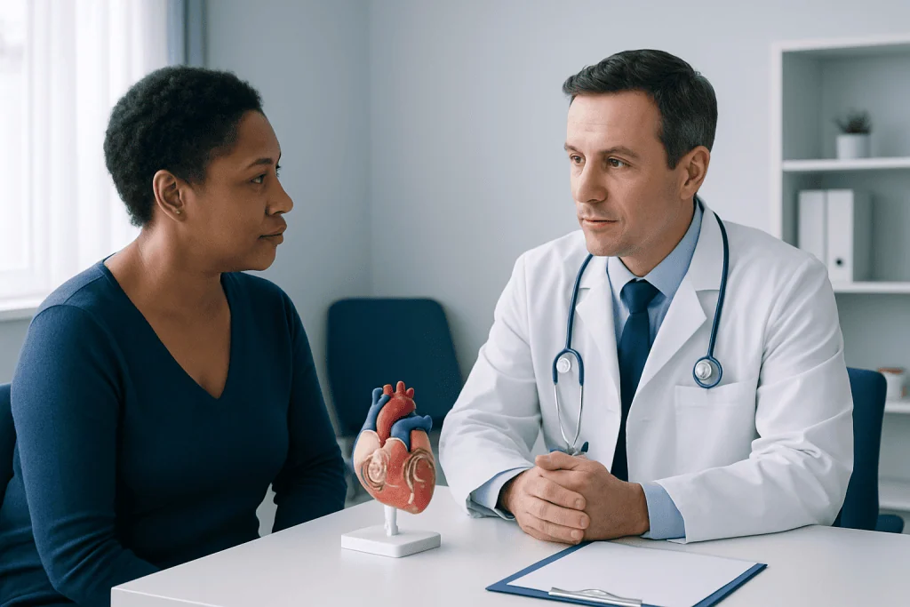
Cardiac thrombosis usually develops when one or more of three factors are present: slow blood flow, damage to the heart tissue or lining, and a tendency for the blood to clot more easily. Doctors often describe this general pattern when evaluating why a clot formed.
Common cardiac causes include recent myocardial infarction, weakened heart pumping from heart failure, enlarged heart chambers, valve disease, inflammation of the heart, and certain rhythm disorders such as atrial fibrillation or atrial flutter. Structural heart problems and some congenital heart conditions can also create areas where blood stagnates.
Other risk factors come from the wider health picture. These may include older age, smoking, prolonged immobility, cancer, inherited clotting disorders, pregnancy or the postpartum period, and some medications. Uncontrolled hypertension, diabetes, and lipid disorders can contribute to heart disease overall and indirectly raise clot risk by damaging the cardiovascular system. People with coronary artery disease or cardiomyopathy may need close monitoring when heart function is reduced.
How cardiac thrombosis is diagnosed
Diagnosis begins with a medical history and physical examination. The doctor asks about symptoms, prior heart disease, rhythm problems, recent heart attack, stroke history, medications, family history, and any factors that may increase clotting. Because symptoms can overlap with many heart conditions, imaging is especially important.
The most commonly used test is echocardiography, an ultrasound of the heart. A standard transthoracic echocardiogram can often detect clots, especially in the ventricles. In some cases, a transesophageal echocardiogram is preferred because it gives a clearer view of structures such as the left atrium and left atrial appendage, where clots may form in people with abnormal rhythms.
Additional tests may include an electrocardiogram to assess the heart rhythm, blood tests, and imaging such as cardiac CT or cardiac MRI when more detail is needed. Doctors also investigate the underlying cause, because treatment is not only about the clot itself but also about preventing it from returning.
- Echocardiography to visualize the clot
- Electrocardiogram to look for arrhythmias
- Blood tests for clotting, inflammation, and organ function
- CT or MRI for selected cases
- Assessment of related conditions such as valve disease or heart muscle weakness
Treatment options
Treatment depends on where the clot is located, how large and mobile it is, whether it has caused complications, and what underlying heart disease is present. In many cases, anticoagulant medicines, often called blood thinners, are the main treatment. These medicines do not dissolve the clot immediately, but they help prevent the clot from growing and reduce the chance of new clots forming while the body gradually breaks the clot down.
The doctor may recommend hospital monitoring at first, especially if the clot is newly discovered or there is concern about embolism. Follow-up imaging is usually needed to see whether the clot is shrinking. The safest medicine and duration of therapy vary from person to person and depend on factors such as bleeding risk, kidney function, valve disease, and the reason the clot developed.
Sometimes the underlying problem also needs direct treatment. If a clot is related to a rhythm problem, managing ablation procedures or other rhythm-focused care may be part of the plan for selected patients. When severe blocked arteries have damaged heart muscle and contributed to clot formation, doctors may evaluate whether stent procedures or coronary artery bypass surgery could help restore blood flow as part of broader cardiac care. Rarely, surgery or catheter-based treatment is considered for a large, highly mobile, or complicated clot.
People recovering from a significant cardiac event may also benefit from supervised cardiac rehabilitation to improve fitness, medication adherence, and risk-factor control. The treatment plan is usually made by a multidisciplinary team, often including cardiologists, imaging specialists, and sometimes cardiac surgeons.
Prevention and self-care
Preventing cardiac thrombosis starts with treating the conditions that make clots more likely. This may include careful management of heart rhythm disorders, heart failure, valve disease, and coronary artery disease. Taking prescribed medicines exactly as directed is important, especially anticoagulants and heart medications.
General cardiovascular self-care also matters. Not smoking, staying physically active within a doctor’s advice, maintaining a healthy weight, and managing blood pressure, cholesterol, and diabetes can all help support heart health. Regular follow-up visits allow doctors to monitor heart function and adjust treatment before complications develop.
People taking anticoagulants should follow safety guidance about missed doses, drug interactions, and signs of bleeding. They should tell all healthcare professionals, including dentists and pharmacists, that they use a blood thinner. Lifestyle measures do not replace medical treatment, but they can lower risk and support recovery.
- Take medicines consistently
- Attend follow-up imaging and cardiology appointments
- Control blood pressure, cholesterol, and blood sugar
- Avoid smoking and long periods of inactivity
- Ask before starting new medicines or supplements
When to see a doctor
Anyone with symptoms that suggest a possible heart problem should seek medical care. Ongoing chest discomfort, new shortness of breath, fainting, or a sudden irregular heartbeat should not be ignored. People who already have heart disease should contact their doctor if symptoms change or worsen.
Emergency care is needed if there are signs that a clot may have traveled elsewhere. These include sudden weakness, numbness, trouble speaking, loss of vision, severe headache, sudden leg pain, or a cold or pale limb. These symptoms can indicate stroke or blocked blood flow and need immediate attention.
Near the end of diagnosis and treatment planning, patients may benefit from evaluation at a center with experience in advanced cardiac imaging and multidisciplinary heart care. Acibadem International’s multidisciplinary specialists and JCI-accredited hospitals diagnose and treat cardiac conditions for international patients when coordinated specialist assessment is needed.
Frequently asked questions
Is cardiac thrombosis the same as a heart attack?
No. Cardiac thrombosis means a blood clot forms inside the heart, while a heart attack usually happens when blood flow to part of the heart muscle is blocked in a coronary artery. A heart attack can increase the risk of a clot forming inside the heart afterward, especially if heart muscle function becomes weak.
Can a blood clot in the heart go away?
It can, especially with appropriate treatment. Anticoagulant medicines help prevent the clot from getting larger and allow the body to gradually break it down over time. Doctors usually repeat imaging to confirm whether the clot is shrinking or has resolved.
What test is most commonly used to find a clot in the heart?
Echocardiography is often the first and most useful test. It uses ultrasound to show the heart’s chambers, valves, and pumping function, and it can often identify a clot. In some cases, a transesophageal echocardiogram, CT scan, or MRI provides more detail.
Is cardiac thrombosis always an emergency?
It can become urgent because of the risk that the clot may block circulation or travel to another part of the body. However, the level of urgency depends on the clot’s location, size, mobility, symptoms, and associated conditions. A doctor should assess it promptly to decide on the safest treatment.
Who is more likely to develop cardiac thrombosis?
People with recent heart attack, heart failure, enlarged heart chambers, valve disease, or abnormal heart rhythms are at higher risk. Other factors such as smoking, cancer, inherited clotting tendencies, and prolonged immobility can also increase the chance of clot formation.
Can cardiac thrombosis be prevented?
It cannot always be prevented, but the risk can often be reduced. Good control of heart disease, blood pressure, cholesterol, and diabetes, along with not smoking and taking prescribed medicines correctly, all help. People with known rhythm problems or weakened heart function may need regular monitoring and individualized prevention strategies.
References
- American Heart Association
- European Society of Cardiology
- National Heart, Lung, and Blood Institute
- Centers for Disease Control and Prevention
- Merck Manual Consumer Version
This article is for general information only and is not a substitute for professional medical advice. Please consult a qualified doctor about your individual situation.
Cardiac Thrombosis in Turkey — costs, top hospitals & a free quote
JCI-accredited · board-certified surgeons · reply within 24h
Add us as a Preferred Source to see more of our trusted health content across Google Search, AI Overviews and Discover.

Cardiology Department
Diagnosis and treatment of heart and vascular conditions, from prevention to advanced interventional procedures.
93 specialists in this unit