Interventional Neurology vs Neurointerventional Radiology: What Is the Difference?

Both specialties use minimally invasive, image-guided techniques to treat blood vessel problems affecting the brain, spine, head, and neck. The biggest difference is typically the doctor’s background training, not the overall purpose of care.
Key Takeaways
- Both specialties use minimally invasive, image-guided techniques to treat blood vessel problems affecting the brain, spine, head, and neck.
- The biggest difference is typically the doctor’s background training, not the overall purpose of care.
- These specialists often work together with stroke neurologists, neurosurgeons, vascular surgeons, and critical care teams.
- Patients are commonly treated for stroke, brain aneurysms, arteriovenous malformations, and carotid artery disease.
- Hospital experience, emergency availability, and multidisciplinary teamwork are often more important to outcomes than the job title alone.
Interventional neurology and neurointerventional radiology are closely related fields that use catheter-based, image-guided procedures to diagnose and treat disorders of the brain, spine, head, and neck. The main difference is usually the specialist’s training pathway and role within the care team, while the procedures and patient goals often overlap.
Overview: How These Specialties Compare
When patients search for the difference between interventional neurology and neurointerventional radiology, they often discover that the two fields are very similar in day-to-day practice. Both focus on diagnosing and treating disorders of the nervous system using minimally invasive, image-guided procedures performed through blood vessels. Instead of large open surgery, a specialist may guide thin catheters through an artery or vein to reach the brain, spinal cord, head, or neck.
These procedures are often called endovascular or catheter-based treatments. They are commonly used in emergencies such as acute ischemic stroke, as well as in planned treatment for conditions like aneurysms, arteriovenous malformations, fistulas, or narrowing of the carotid arteries. Because these diseases can affect blood flow to delicate nervous system structures, rapid diagnosis and expert technique are important.
In many hospitals, the distinction between the two specialties is less about what is treated and more about who trained in which medical discipline first. An interventional neurologist usually begins with neurology training, while a neurointerventional radiologist often begins with radiology training. Both may then complete advanced fellowship training in endovascular treatment of neurological disease.
For patients, the most practical point is that care is usually team-based. A person with stroke symptoms, for example, may be evaluated by emergency physicians, neurologists, neuroradiologists, neurosurgeons, and intensive care specialists. The goal is not to choose between labels, but to receive timely treatment from a qualified team experienced in complex neurological and vascular care.
What Interventional Neurology Is
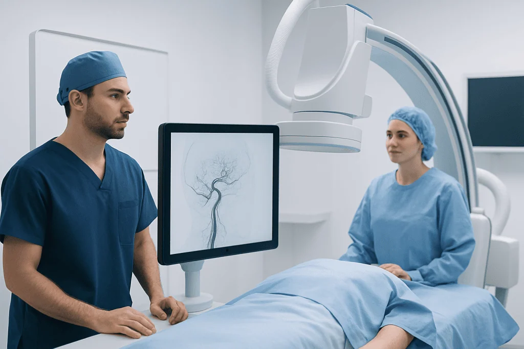
Interventional neurology is a subspecialty focused on catheter-based treatment of neurological and cerebrovascular conditions. Doctors in this field usually come from a neurology background, which means they are trained first in disorders of the brain, spinal cord, nerves, and stroke care before gaining procedural expertise. This background can be especially valuable when a patient’s symptoms, neurological examination, and long-term recovery needs must all be considered together.
Interventional neurologists often help manage acute stroke, transient ischemic attack, aneurysms, vascular malformations, and narrowed blood vessels supplying the brain. They may perform mechanical thrombectomy for selected patients with blocked brain arteries, place stents in certain situations, or use embolization techniques to close abnormal vessels. Their work combines neurological assessment with real-time imaging and endovascular treatment.
Because they are neurologists first, these specialists may be deeply involved in the full stroke pathway, from emergency evaluation to rehabilitation planning. They often work closely with stroke medicine, critical care, and neurorehabilitation teams. In some centers, they also contribute to follow-up care after a procedure, especially when ongoing neurological monitoring is needed.
The exact scope of practice varies by country, hospital, and credentialing system. In some institutions, interventional neurologists and neurointerventional radiologists perform nearly identical procedures. In others, their responsibilities differ somewhat depending on departmental structure, available specialists, and emergency service organization.
What Neurointerventional Radiology Is

Neurointerventional radiology, sometimes called interventional neuroradiology, is a subspecialty that uses advanced imaging to treat vascular disorders of the brain, spine, head, and neck through minimally invasive techniques. Specialists in this area often begin with training in radiology or neuroradiology, building strong expertise in interpreting detailed imaging studies such as CT, MRI, angiography, and fluoroscopy.
These doctors are highly trained in navigating tiny blood vessels under imaging guidance. They may treat brain aneurysms with coiling or flow-diverting devices, remove blood clots in acute stroke, or embolize abnormal connections between arteries and veins. Their imaging-centered training is especially important when planning a procedure, identifying complex anatomy, and monitoring treatment in real time.
Neurointerventional radiology plays a key role in both emergency and elective care. In emergencies, rapid brain and vessel imaging helps determine whether a patient may benefit from thrombectomy or another urgent procedure. In planned care, angiography can clarify the size, shape, and blood flow patterns of abnormalities before treatment begins.
Many patients encounter these specialists when being evaluated for brain aneurysms, vascular malformations, or severe narrowing of neck arteries. Although the specialty name includes radiology, the work is not limited to diagnosis alone. It includes hands-on treatment that can be central to managing serious neurological conditions.
Key Differences: Training, Perspective, and Team Role
The clearest difference between interventional neurology and neurointerventional radiology is the physician’s original specialty training. Interventional neurologists usually complete neurology training first, while neurointerventional radiologists usually train first in radiology and often neuroradiology. Both then receive additional fellowship training in endovascular procedures involving the nervous system.
This difference in training can shape how a specialist approaches a case. A neurologist may naturally emphasize bedside neurological evaluation, stroke syndromes, symptom patterns, and medical management before and after a procedure. A radiology-trained specialist may bring especially deep experience in image interpretation, vascular anatomy, and technical imaging guidance during procedures. In practice, these strengths are often complementary rather than competitive.
Another difference may be where the specialist sits within the hospital system. Interventional neurologists are often based in neurology or stroke services, while neurointerventional radiologists may be based in radiology, neuroradiology, or dedicated endovascular departments. Even so, modern care pathways are usually multidisciplinary, and many decisions are made in joint conferences or emergency stroke teams.
From a patient point of view, the title alone does not reveal overall quality. Useful questions include whether the center offers 24/7 stroke treatment, how often the team performs complex procedures, whether neurosurgery and intensive care are available if needed, and how follow-up care is organized. Experience, coordination, and timely access often matter more than specialty labels.
Conditions and Procedures Both Specialties Commonly Treat
Interventional neurologists and neurointerventional radiologists often treat the same core group of cerebrovascular and neurovascular conditions. The most urgent example is acute ischemic stroke caused by a clot blocking a major brain artery. In selected patients, a catheter-based clot removal procedure called stroke treatment with mechanical thrombectomy may restore blood flow and reduce brain injury.
Another common area is the treatment of aneurysms and other abnormal blood vessel problems. Depending on the anatomy, a team may recommend endovascular coiling, stent-assisted techniques, flow diversion, or embolization. These approaches are often alternatives to open surgery or may be used alongside it. For some patients with carotid artery disease, endovascular methods such as carotid artery disease treatment may also be considered.
Other conditions may include:
- Arteriovenous malformations and dural arteriovenous fistulas
- Intracranial or extracranial arterial narrowing
- Cerebral venous disorders in selected cases
- Tumors or bleeding conditions requiring embolization support
- Vascular causes of pulsatile tinnitus or facial symptoms
Some patients are referred after tests suggest a vascular abnormality but before a definite diagnosis is made. In these situations, cerebral angiography may be used to map the blood vessels in detail. When appropriate, the same team may then proceed to interventional neuroradiology treatment or related therapies designed for the patient’s specific anatomy and symptoms.
How Patients Are Evaluated and Diagnosed
Assessment usually begins with symptoms, medical history, and imaging. In emergencies such as sudden weakness, speech difficulty, vision loss, or severe sudden headache, doctors may quickly order CT, CT angiography, MRI, or MR angiography to look for bleeding, blocked vessels, or aneurysms. Fast imaging helps determine whether an urgent catheter-based procedure may help.
For non-emergency problems, patients may be referred after an incidental finding on a scan or after symptoms such as recurrent transient neurological episodes, dizziness, or unusual headaches prompt further evaluation. The specialist reviews previous images, current symptoms, medications, and overall health. This is important because not every vascular abnormality requires immediate intervention, and some are monitored over time.
Catheter angiography remains an important diagnostic tool in many neurovascular cases. During this test, a thin catheter is guided into the arteries and contrast dye is used to create detailed images of blood vessels. Although it is invasive, it can provide information that other scans cannot fully show, particularly when planning treatment for aneurysms, malformations, or complex narrowing.
Decision-making is individualized. Doctors consider the location and size of the lesion, the risk of stroke or bleeding, the patient’s age and general health, and whether medical therapy, observation, surgery, or endovascular treatment is the safest option. Patients are usually encouraged to ask who will perform the procedure, why a particular approach is being recommended, and what follow-up will involve.
Treatment Planning, Recovery, and Follow-Up
When a procedure is recommended, the care plan depends on the condition being treated. Some interventions are performed in an emergency, while others are scheduled after detailed planning. Before treatment, patients may need blood tests, medication review, and imaging to assess vessel anatomy. The specialist explains the expected benefits, possible alternatives, and potential risks such as bleeding, clotting, vessel injury, or reaction to contrast material.
Most procedures are performed in a specialized angiography suite using X-ray guidance. Access is commonly through an artery in the wrist or groin. The doctor advances tiny catheters to the target area and delivers treatment, such as clot retrieval, coils, stents, or embolic materials. Hospital stay can range from brief observation to several days, depending on the condition and whether it was an emergency.
Recovery also varies. After stroke treatment, follow-up often includes neurological monitoring, rehabilitation, blood pressure management, and prevention of future events. After aneurysm or malformation treatment, repeat imaging may be needed to confirm that the abnormal vessel remains closed or that blood flow has improved. Some patients require ongoing medications, while others mainly need surveillance imaging and regular clinic visits.
Near the end of the treatment journey, multidisciplinary follow-up becomes especially valuable. Patients may continue care with neurology, neuroradiology, neurosurgery, rehabilitation, or vascular medicine depending on the diagnosis. Acibadem International’s multidisciplinary specialists and JCI-accredited hospitals also diagnose and treat complex neurovascular conditions for international patients when advanced evaluation and coordinated follow-up are needed.
How to Choose the Right Specialist and When to Seek Care
For most patients, choosing between interventional neurology and neurointerventional radiology is less important than choosing an experienced center. A high-quality service usually offers around-the-clock emergency stroke capability, advanced brain and vascular imaging, neurocritical care, and access to neurosurgery if needed. Patients can also ask whether treatment decisions are discussed by a multidisciplinary team and how many similar cases the center manages.
It is helpful to seek care promptly for symptoms that may point to a neurological emergency. Warning signs include sudden weakness or numbness on one side, trouble speaking, sudden confusion, abrupt vision loss, severe imbalance, facial drooping, or a sudden unusually intense headache. These symptoms require urgent medical assessment because early treatment may protect brain function.
For non-emergency concerns, specialist review is appropriate if imaging shows an aneurysm, vascular malformation, or artery narrowing, or if symptoms keep returning without a clear explanation. A consultation can clarify whether monitoring, medication, surgery, or catheter-based treatment is the best path. Patients should feel comfortable asking for a clear explanation of the diagnosis and treatment options in everyday language.
In short, both interventional neurologists and neurointerventional radiologists play important roles in modern brain and vascular care. Their expertise often overlaps, and collaboration is common. What matters most is receiving timely, evidence-based treatment from a skilled team that can match the right technique to the patient’s condition.
Frequently asked questions
Is interventional neurology the same as neurointerventional radiology?
They are not exactly the same, but they overlap greatly. The main difference is usually the doctor’s original specialty training, while the procedures and conditions treated are often very similar.
Which specialist treats stroke with catheter-based clot removal?
Both interventional neurologists and neurointerventional radiologists may perform mechanical thrombectomy, depending on the hospital and training pathway. What matters most is that the procedure is done quickly by a qualified team experienced in acute stroke care.
Do these specialists only treat blood vessel problems in the brain?
No. They may also treat vascular conditions involving the spine, head, and neck. Examples include carotid artery narrowing, spinal vascular malformations, and certain abnormal artery-vein connections.
Is neurointerventional treatment considered surgery?
It is usually considered minimally invasive endovascular treatment rather than traditional open surgery. The doctor uses a catheter inserted through a blood vessel, guided by imaging, to reach the treatment area.
How should a patient choose between these specialists?
Patients usually do not need to choose based on the title alone. It is more helpful to look for a center with experienced specialists, 24/7 emergency capability when needed, strong imaging support, and multidisciplinary follow-up care.
Are these procedures always urgent?
No. Some procedures, such as stroke thrombectomy, are emergencies, while others are planned after careful review of scans and symptoms. Many aneurysms or vascular abnormalities are assessed in a non-emergency setting before deciding on treatment.
This article is for general information only and is not a substitute for professional medical advice. Please consult a qualified doctor about your individual situation.
Interventional Neurology in Turkey — costs, top hospitals & a free quote
JCI-accredited · board-certified surgeons · reply within 24h
Add us as a Preferred Source to see more of our trusted health content across Google Search, AI Overviews and Discover.

Neurosurgery
Surgical treatment of the brain, spine and nervous system, including minimally invasive and functional procedures.
70 specialists in this unitMore from the Health Library

Brain Aneurysm Symptoms: When Urgent Endovascular Treatment May Be Needed

Stroke Thrombectomy Recovery: What to Expect After Clot Removal

CSF Leak: Headache, Ear Symptoms, and When Neuroimaging Is Needed

Neurointerventional Radiology Cost: What Affects the Total Price?

Cerebral Angiography Risks and Recovery: What Patients Should Know