Lucency: Symptoms, Causes, and Treatment — Complete Patient Guide

Lucency is a descriptive imaging term, not a final diagnosis. A lucent area may be normal, temporary, or a sign of an underlying condition depending on its location and appearance.
Key Takeaways
- Lucency is a descriptive imaging term, not a final diagnosis.
- A lucent area may be normal, temporary, or a sign of an underlying condition depending on its location and appearance.
- Symptoms, medical history, and follow-up tests help doctors interpret what a lucency means.
- Treatment depends on the cause and may range from observation to medication or a procedure.
- Urgent evaluation is important if lucency is linked with severe pain, breathing trouble, trauma, or neurological symptoms.
Lucency is not a disease itself. It is an imaging description used when an area on an X-ray, CT scan, or other study looks darker than surrounding tissue, usually because it is less dense or contains more air, fat, or bone loss.
Overview: what lucency means on a scan
Lucency is a term radiologists use when an area on an imaging test looks darker than expected. In simple terms, the darker appearance usually means that part of the body is less dense than the tissue around it. This can happen for many reasons, including normal air-filled structures, temporary changes after injury, or an underlying medical condition that needs more evaluation.
The meaning of lucency depends heavily on where it appears. For example, lucency in the lungs may reflect air spaces, while lucency in a bone may suggest bone loss, a cyst, or another structural change. A report may also use related terms such as “lucent lesion,” “radiolucent area,” or “area of decreased density.” These phrases describe how something looks on the image, not what it definitively is.
Because lucency is a finding rather than a diagnosis, the next step is interpretation in context. Doctors look at the patient’s age, symptoms, physical examination, past health history, and the exact pattern seen on imaging. In many cases, a lucency turns out to be benign, but some findings need additional scans, laboratory tests, or specialist review to clarify the cause.
Where lucency may be seen in the body
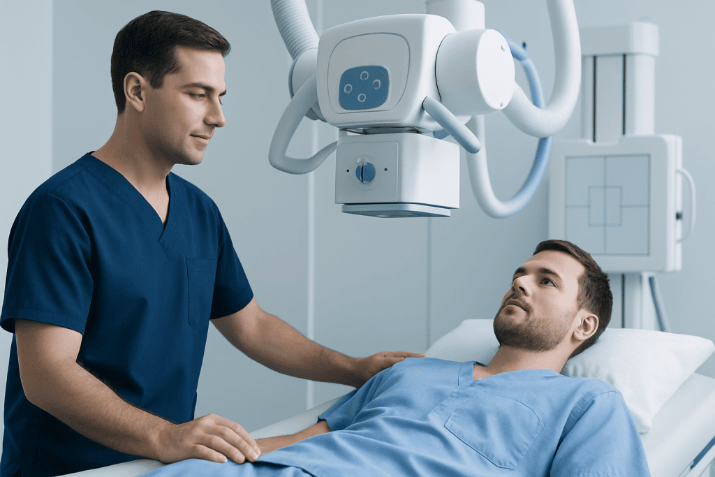
Lucency can appear in many body systems, and its significance changes by location. In chest imaging, lucency may relate to normal air in the lungs or to changes such as overinflation, cavities, or air outside the lung where it should not be. In bones and joints, a lucent area may reflect a benign cyst, healing change, infection, fracture line, or less commonly a tumor-related process.
In dental imaging, lucencies are often used to describe areas around the tooth root or jawbone that may suggest infection, cysts, or bone changes. In the abdomen, a lucent appearance may be connected to gas in the bowel or, more rarely, abnormal air in places that deserve prompt attention. The same word can therefore describe very different situations across specialties.
Radiologists also consider the shape, border, and size of the lucent area. A smooth, well-defined lucency may be less concerning than one with irregular edges, surrounding inflammation, or signs of tissue destruction. Comparison with older images is often very helpful, because a stable lucency over time may be reassuring, whereas a new or enlarging one usually needs closer assessment.
Symptoms that may accompany a lucency

A lucency itself does not cause symptoms; symptoms come from the underlying reason for the imaging finding. Some people have no symptoms at all, and the lucency is discovered incidentally during a scan done for another reason. This is common in routine X-rays, dental imaging, or CT studies.
When symptoms are present, they depend on the body area involved. Bone-related lucencies may be associated with localized pain, swelling, reduced movement, tenderness, or a fracture after minor trauma. Chest-related lucencies may be linked with cough, shortness of breath, chest pain, fever, or wheezing. Dental or jaw lucencies can be associated with tooth pain, gum swelling, drainage, or trouble chewing.
Some symptoms suggest the need for faster medical review. These include sudden severe chest pain, difficulty breathing, new weakness, numbness, fever with significant pain, unexplained weight loss, or persistent night pain. Although many causes of lucency are not dangerous, these accompanying features can point to conditions that should not be ignored.
- Bone pain or tenderness
- Shortness of breath or chest discomfort
- Swelling, redness, or warmth
- Fever or signs of infection
- Tooth or jaw pain
- Symptoms after injury or trauma
Causes and risk factors
There is no single cause of lucency. On imaging, a dark or lucent area may reflect normal anatomy, air, fat, reduced mineral content, or tissue breakdown. In the lungs, lucency may be seen with normal air-containing tissue, but also with conditions such as trapped air, emphysematous change, or a pneumothorax. In the skeleton, lucencies may appear with bone cysts, osteoporosis, infections, stress injuries, healing fractures, and some benign or malignant lesions.
Age and medical history help narrow the possibilities. In children and younger adults, some bone lucencies are benign developmental or cystic findings. In older adults, degenerative changes, lower bone density, and a history of cancer can influence how a lucency is interpreted. A recent injury, surgery, dental problem, smoking history, or chronic steroid use may also change the level of concern and the likely diagnosis.
Risk factors depend on the suspected source. For bone-related lucency, possible factors include osteoporosis, repetitive stress, metabolic bone disease, infection risk, or prior tumors. For chest-related lucency, smoking, chronic lung disease, trauma, and certain infections may matter. In a patient with symptoms such as persistent cough or weight loss, doctors may also evaluate for related conditions such as lung cancer if the overall imaging pattern suggests that possibility.
How doctors diagnose the cause of a lucency
Diagnosis begins with the imaging report but does not end there. A doctor will review the exact wording, location, size, and appearance of the lucency, then connect that information with the patient’s symptoms and examination findings. Sometimes the radiologist can strongly suggest a likely cause, while in other cases the report may recommend correlation with clinical findings or repeat imaging.
Additional tests may be needed depending on the body part involved. These can include targeted X-rays, blood tests, ultrasound, MRI, CT, or occasionally a nuclear medicine scan. MRI is often helpful when doctors need more detail about soft tissues, bone marrow, or the internal structure of a lesion. CT can provide a more precise view of bone and lung anatomy, especially if the original finding was unclear.
If a suspicious or unexplained bone lucency is found, a clinician may consider referral to orthopedics, rheumatology, or oncology depending on the overall picture. In some situations, tissue sampling is required to confirm a diagnosis. Imaging-based assessment is central to this process, and tests such as MRI or CT scan may be used to clarify what a lucent area represents and whether it is stable, benign, or in need of treatment.
Doctors also pay close attention to whether the finding is new, changing, or stable over time. A prior scan can be extremely valuable. If the same lucency has remained unchanged for years and there are no symptoms, watchful follow-up may be enough. If it is growing, causing symptoms, or has aggressive features, the diagnostic process becomes more urgent.
Treatment options depend on the underlying cause
There is no universal treatment for lucency because lucency is only a description of an imaging appearance. Management is directed at the condition causing the finding. In many cases, especially when the area looks benign and the person feels well, treatment may simply be observation with repeat imaging after an appropriate interval.
If the lucency is related to a fracture, bone stress injury, or reduced bone density, treatment may include rest, physical support, rehabilitation, and management of bone health. If infection is suspected, doctors may recommend antimicrobial treatment or a procedure to drain or remove affected tissue. Dental and jaw-related lucencies may need dental care, root canal treatment, or oral surgery depending on the source.
Chest-related lucencies are treated according to the diagnosis. For example, air trapping from chronic lung disease is managed differently from a pneumothorax, which may require urgent monitoring or intervention. Some suspicious lesions need biopsy or surgical removal, while others are followed over time. If a broad evaluation is needed, centers with multidisciplinary imaging and specialty care can help coordinate next steps; Acibadem International’s multidisciplinary specialists at JCI-accredited hospitals diagnose and treat complex imaging findings for international patients.
When a procedure is recommended, the aim is to identify or correct the underlying problem rather than to treat the lucency itself. Depending on the case, this may involve image-guided biopsy, orthopedic care, pulmonary treatment, or surgery. Follow-up is important so doctors can confirm that symptoms improve and that imaging changes are stable or resolving.
Prevention and self-care
Because lucency can arise from many different causes, prevention is mainly about reducing risk factors for the underlying conditions. Good bone health is especially important. Adults can support bone strength by eating a balanced diet, getting appropriate calcium and vitamin D as advised by a clinician, staying physically active, and discussing bone density testing when risk factors are present.
Not smoking is one of the most important steps for lung and overall health. Protecting the body from injury with safe exercise habits, proper equipment, and fall prevention can also reduce some causes of lucent findings in bones and joints. Regular dental care helps lower the chance of tooth and jaw infections that may appear as lucencies on dental imaging.
Self-care should also include attending follow-up appointments and bringing prior imaging studies when possible. People should avoid assuming that any lucency is harmless or, on the other hand, fearing that it always means cancer. The safest approach is to ask what body part is involved, what the doctor thinks the most likely cause is, and whether repeat imaging or specialist review is recommended.
When to seek medical care
Medical care should be sought promptly if a lucency is found after trauma, or if it is associated with severe or worsening symptoms. Examples include chest pain, shortness of breath, high fever, rapidly increasing swelling, inability to bear weight, or significant weakness or numbness. These symptoms may point to an urgent condition that needs timely assessment.
Routine follow-up is still important even when symptoms are mild. A person should contact a doctor if pain persists, a swelling does not improve, a dental problem continues, or an imaging report recommends further evaluation. It is also reasonable to ask for clarification if the report uses technical terms such as “lucent lesion,” “lytic change,” or “indeterminate lucency,” as these often require interpretation in plain language.
People with a history of cancer, immune suppression, osteoporosis, or chronic lung disease should be especially careful not to miss recommended follow-up. In some cases, the next steps may include specialist consultation or advanced testing such as PET-CT if there is concern about a more complex process. Timely evaluation helps distinguish between harmless findings and those that benefit from treatment.
Frequently asked questions
Is lucency always serious?
No. Lucency is a descriptive term on an imaging report, and many lucent findings are benign or even normal for that body part. The level of concern depends on where the lucency is, how it looks, whether symptoms are present, and whether it changes over time.
What is the difference between lucency and a lesion?
Lucency describes how an area appears on imaging, usually darker because it is less dense. A lesion is a broader term for an abnormal area of tissue. A radiologist may describe a “lucent lesion” when both ideas apply.
Can lucency mean cancer?
Sometimes a lucent area can be related to a tumor, but it can also reflect many non-cancerous causes such as cysts, infection, osteoporosis, or normal anatomy. Imaging features, symptoms, and sometimes biopsy are used to determine whether cancer is a concern.
Why would a doctor order more scans after finding lucency?
An additional scan can provide more detail about the exact structure, borders, and surrounding tissues. This helps doctors decide whether the finding is harmless, needs monitoring, or requires treatment. Repeat imaging is also useful for checking whether the area stays the same or changes over time.
Can a lucency go away on its own?
Some lucent findings do resolve, especially if they are related to healing, temporary inflammation, or a treatable infection. Others remain stable for years without causing harm. The expected course depends entirely on the underlying diagnosis.
Should a person worry about a lucency found incidentally?
Incidental findings are common, and many are not dangerous. Still, they should not be ignored. It is best to review the report with a qualified doctor, who can explain whether the finding is likely benign or whether follow-up is needed.
References
- Radiological Society of North America
- American College of Radiology
- National Institutes of Health
- Mayo Clinic
- National Health Service
This article is for general information only and is not a substitute for professional medical advice. Please consult a qualified doctor about your individual situation.
Explore treatments in Turkey — costs, top hospitals & a free quote
JCI-accredited · board-certified surgeons · reply within 24h
Add us as a Preferred Source to see more of our trusted health content across Google Search, AI Overviews and Discover.