Cardiac MRI Before CABG: Assessing Heart Muscle Viability

Cardiac MRI can distinguish permanent scar tissue from viable, under-functioning heart muscle. Viability findings may help the heart team decide whether CABG is likely to improve symptoms or heart function.
Key Takeaways
- Cardiac MRI can distinguish permanent scar tissue from viable, under-functioning heart muscle.
- Viability findings may help the heart team decide whether CABG is likely to improve symptoms or heart function.
- The test is noninvasive and uses magnetic fields; some scans include gadolinium contrast to show scar patterns.
- Results are interpreted together with coronary angiography, echocardiography, symptoms, and overall surgical risk.
- Patients with kidney disease, implanted devices, or severe claustrophobia should discuss MRI safety and alternatives with their doctor.
Cardiac MRI before CABG can help doctors understand whether weakened areas of the heart muscle are scarred or still alive and likely to improve after restored blood flow. This information supports careful planning for patients with coronary artery disease, reduced heart function, or complex bypass decisions.
Overview
Cardiac MRI before CABG is a specialized heart imaging test used to evaluate heart muscle structure, function, blood flow, and scar tissue before coronary artery bypass grafting. CABG is a surgical procedure that creates new pathways for blood to reach areas of the heart affected by narrowed or blocked coronary arteries. For some patients, especially those with reduced pumping function, doctors need to know whether weak heart muscle is permanently damaged or still viable.
Heart muscle viability means that heart cells are alive but may not be contracting normally because they have had limited blood supply over time. This is sometimes called hibernating myocardium. If blood flow is improved through bypass surgery, viable muscle may recover partially or fully, while dense scar tissue usually does not regain normal contraction.
Cardiac MRI, also called cardiovascular magnetic resonance or CMR, is particularly useful because it provides detailed pictures without ionizing radiation. It can show how strongly the heart pumps, which areas are thin or thickened, and where scar tissue is located. In CABG planning, this information is usually considered alongside coronary angiography, echocardiography, blood tests, symptoms, and the patient’s overall health.
Why Heart Muscle Viability Matters Before CABG
When coronary artery disease has been present for a long time, some regions of the heart may become weak because they receive too little oxygen-rich blood. In some patients, this weakness is reversible after revascularization, meaning after blood flow is restored. In others, the affected muscle has been replaced by scar after a previous heart attack and is less likely to recover function.
Knowing the difference can help the heart team balance the potential benefits and risks of coronary artery bypass surgery. If a large amount of dysfunctional heart muscle is still viable, CABG may offer a better chance of improving symptoms, exercise tolerance, or heart pumping function. If most weak areas are scarred, surgery may still be recommended for other reasons, such as symptom relief or prevention of further ischemia, but expectations about recovery of function may be different.
Viability assessment is most important in patients with complex coronary artery disease, previous heart attack, heart failure, or low left ventricular ejection fraction. It can also be helpful when symptoms, angiography findings, and standard ultrasound images do not fully explain the severity of heart dysfunction. The goal is not to make a decision from one test alone, but to add clarity to a broader, personalized treatment plan.
How Cardiac MRI Assesses Viability

Cardiac MRI uses a strong magnetic field and radio waves to create detailed moving and still images of the heart. For viability assessment, doctors commonly use a technique called late gadolinium enhancement. After a gadolinium-based contrast agent is given through a vein, scarred heart tissue tends to hold the contrast differently from healthy muscle, allowing the radiologist or cardiologist to map the extent and depth of scar.
The depth of scarring through the heart wall is important. A thin layer of scar may leave enough living muscle for improvement after restored blood flow, while full-thickness scar is less likely to recover contraction. Cardiac MRI can also measure left and right ventricular size, ejection fraction, wall motion, valve function, and sometimes blood flow or stress-induced ischemia, depending on the scan protocol.
In some cases, additional MRI techniques may be used to evaluate swelling, inflammation, perfusion, or tissue characteristics. The scan may help identify other conditions that can mimic or worsen ischemic heart disease, such as myocarditis, infiltrative disease, or certain cardiomyopathies. This broad tissue assessment is one reason CMR is valued in complex cardiac decision-making.
Who May Need Cardiac MRI Before CABG
Not every patient scheduled for bypass surgery needs a cardiac MRI. Many patients can be evaluated adequately with echocardiography, coronary angiography, computed tomography, and clinical assessment. Cardiac MRI is most often considered when the benefit of CABG is uncertain, when the heart’s pumping function is reduced, or when doctors need more precise information about scar and viable tissue.
Patients who may benefit include those with ischemic cardiomyopathy, previous myocardial infarction, multiple blocked coronary arteries, or unclear findings on other imaging tests. It may also be useful when echocardiogram images are limited by body habitus, lung disease, or other technical factors. For patients being considered for heart bypass surgery, viability imaging can contribute to a more individualized discussion about expected outcomes.
There are situations where MRI may not be suitable or may require special precautions. Patients with some implanted cardiac devices, metal fragments, certain older aneurysm clips, or severe kidney impairment should inform the care team before the scan. Many modern pacemakers and defibrillators are MRI-conditional, but they still require a specific safety protocol and coordination between cardiology, radiology, and device specialists.
What to Expect During the Scan
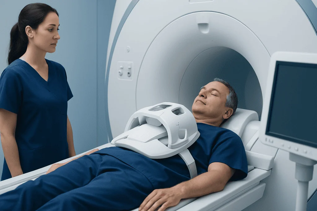
Before the scan, the patient is usually asked about medical history, kidney function, allergies, implanted devices, and previous reactions to contrast agents. If gadolinium contrast is planned, a recent kidney function test may be needed. The patient changes into MRI-safe clothing, removes metal items, and lies on a scanning table with monitoring equipment and a coil placed over the chest.
During the scan, the table moves into the MRI machine, and the patient hears loud tapping or knocking sounds while images are being taken. Ear protection is provided. The technologist may ask the patient to hold their breath for short periods so the heart images are clear. Most cardiac MRI examinations take longer than a simple X-ray or CT scan, but the patient is monitored throughout and can communicate with the team.
If contrast is used, it is injected through an intravenous line during the examination. Some people feel a cool sensation in the arm, but discomfort is usually mild and brief. Patients who feel anxious in enclosed spaces should tell the team in advance, because reassurance, positioning adjustments, or other supportive steps may be available.
Understanding Results and CABG Planning
Cardiac MRI results are interpreted by specialists trained in cardiovascular imaging. The report may describe ejection fraction, areas of reduced movement, the presence and location of scar, and the amount of scar across the thickness of the heart wall. It may also mention ventricular volumes, valve findings, clots inside the heart chambers, or other structural abnormalities relevant to surgery.
The heart team then compares the MRI findings with the coronary artery anatomy. For example, if a blocked artery supplies a region that is mostly viable but under-functioning, bypassing that artery may be expected to help that area recover. If the same region is mainly full-thickness scar, recovery of contraction is less likely, though bypass may still be considered for other territories or for symptom control.
Planning for CABG is multidisciplinary. Cardiologists, cardiac surgeons, anesthesiologists, imaging specialists, and sometimes heart failure specialists review the overall picture, including age, frailty, diabetes, kidney function, lung disease, and patient preferences. When surgery is chosen, related care may include cardiothoracic surgery expertise and structured recovery planning after the operation.
Safety, Alternatives, and Related Tests
Cardiac MRI is generally considered safe when appropriate screening is performed. It does not use ionizing radiation. The main safety considerations involve implanted metal or electronic devices, kidney function when gadolinium contrast is used, and the patient’s ability to lie still for the duration of the scan. Serious reactions to gadolinium contrast are uncommon, but patients should report previous contrast reactions or severe allergies.
Alternative or complementary tests may include echocardiography, stress echocardiography, nuclear perfusion imaging, PET viability imaging, coronary CT angiography, and invasive coronary angiography. Each test provides different information. Echocardiography is widely available and useful for valve and pumping assessment; PET can be highly informative for blood flow and metabolism; angiography remains central for defining coronary blockages.
The best test depends on the clinical question, local expertise, patient safety factors, and timing before surgery. In some patients, one imaging test is enough; in others, combining information from several tests improves confidence. Patients should ask their doctor why a specific test is recommended, what the result may change, and how it fits into the overall treatment plan.
After Testing: Self-Care and When to Seek Medical Advice
After a routine cardiac MRI, most patients can return to normal activities unless their doctor gives different instructions. Drinking fluids may be advised after contrast, especially if there are no fluid restrictions. The imaging report is usually reviewed by the referring cardiologist or heart team, who will explain how the findings affect CABG planning or other treatment options.
Patients with coronary artery disease should continue prescribed medicines unless told otherwise. This may include medications for blood pressure, cholesterol, diabetes, angina, or heart failure. Lifestyle measures such as not smoking, following a heart-healthy eating pattern, staying active within medical limits, and attending follow-up appointments remain important before and after any procedure.
Urgent medical care is needed for chest pain that is severe, new, or not relieved as instructed by a doctor; shortness of breath at rest; fainting; sudden weakness; or symptoms suggesting a heart attack or stroke. For non-urgent concerns, such as questions about MRI safety, contrast, or treatment choices, patients should contact their cardiology team. Acibadem International’s multidisciplinary specialists and JCI-accredited hospitals diagnose and treat coronary artery disease for international patients, including evaluation, bypass surgery planning, and cardiac rehabilitation when appropriate.
Frequently asked questions
Why is cardiac MRI done before CABG?
Cardiac MRI may be done before CABG to determine whether weak areas of the heart muscle are alive or replaced by scar tissue. This helps doctors estimate whether restoring blood flow is likely to improve heart function in those areas. It is especially useful when the heart’s pumping function is reduced or prior heart attacks are suspected.
Does viable heart muscle always recover after bypass surgery?
Viable muscle has a better chance of improving after blood flow is restored, but recovery is not guaranteed. Improvement depends on the amount of viable tissue, the severity of coronary disease, overall heart health, and other medical conditions. Doctors use MRI findings to guide expectations, not to make promises.
Is cardiac MRI painful?
Cardiac MRI is not painful, but the patient must lie still inside the scanner while images are taken. The machine can be noisy, and some people feel uncomfortable in enclosed spaces. The imaging team monitors the patient and can offer support if anxiety or discomfort occurs.
Is gadolinium contrast safe for viability imaging?
Gadolinium contrast is commonly used in cardiac MRI to show scar tissue and is generally safe for appropriately selected patients. Kidney function is usually checked first because severe kidney disease may increase risk from some contrast agents. Patients should tell their doctor about kidney problems, allergies, pregnancy, or previous contrast reactions.
Can a person with a pacemaker have cardiac MRI?
Some people with pacemakers or defibrillators can safely have MRI if the device is MRI-conditional and the correct safety protocol is used. Older or non-compatible devices may make MRI unsuitable. The cardiology and radiology teams must review the device type before scheduling the scan.
How does cardiac MRI compare with echocardiography before CABG?
Echocardiography is often the first test used to assess heart pumping function, valves, and pressures. Cardiac MRI provides more detailed tissue characterization and is particularly strong for detecting scar and measuring ventricular volumes. The two tests often complement each other rather than replace each other.
What questions should patients ask after receiving MRI results?
Patients can ask how much viable heart muscle was found, whether scar tissue is present, and how the findings affect the recommendation for CABG. It is also helpful to ask what benefits are realistic, what risks apply personally, and whether other treatment options should be considered. A clear discussion with the heart team supports shared decision-making.
References
- American Heart Association
- American College of Cardiology
- European Society of Cardiology
- Society for Cardiovascular Magnetic Resonance
- National Heart, Lung, and Blood Institute
This article is for general information only and is not a substitute for professional medical advice. Please consult a qualified doctor about your individual situation.
Heart care in Turkey — expert evaluation and treatment
JCI-accredited · board-certified surgeons · reply within 24h
Add us as a Preferred Source to see more of our trusted health content across Google Search, AI Overviews and Discover.

Cardiology Department
Diagnosis and treatment of heart and vascular conditions, from prevention to advanced interventional procedures.
91 specialists in this unit