Electrophysiology Study: What Happens During EPS and What the Results Mean

An electrophysiology study examines the heart’s electrical signals from inside the heart using thin catheters. EPS can help diagnose the cause of palpitations, fainting, rapid heartbeat, or unexplained rhythm problems.
Key Takeaways
- An electrophysiology study examines the heart’s electrical signals from inside the heart using thin catheters.
- EPS can help diagnose the cause of palpitations, fainting, rapid heartbeat, or unexplained rhythm problems.
- The test may confirm whether a rhythm disorder can be treated with medication, monitoring, or ablation.
- Most people are awake but sedated, and the procedure is usually performed through a vein in the groin or neck.
- Results may be available right away, although the full treatment plan depends on the findings and overall heart health.
An electrophysiology study, or EPS, is a specialized heart test that helps doctors understand why an abnormal heartbeat is happening and where it starts. It is commonly used to diagnose arrhythmias, assess risk, and plan treatments such as medication, a pacemaker, or catheter ablation.
Overview: What is an electrophysiology study?
An electrophysiology study is a procedure that evaluates the heart’s electrical system from the inside. The heart beats because of electrical impulses that travel through specialized pathways. If these signals start in the wrong place, move too quickly, or become blocked, they can cause an arrhythmia, also called an abnormal heart rhythm.
During an EPS, a cardiologist with training in heart rhythm disorders places thin flexible tubes called catheters into a blood vessel and guides them to the heart. These catheters record electrical activity directly from different parts of the heart. They can also deliver carefully controlled impulses to see how the heart responds.
This test is more detailed than a standard electrocardiogram because it can identify where an arrhythmia begins and how it travels. That information helps doctors diagnose conditions such as supraventricular tachycardia, atrial flutter, or ventricular tachycardia. In some cases, treatment may be offered during the same session if it is safe and appropriate.
Why an EPS may be recommended
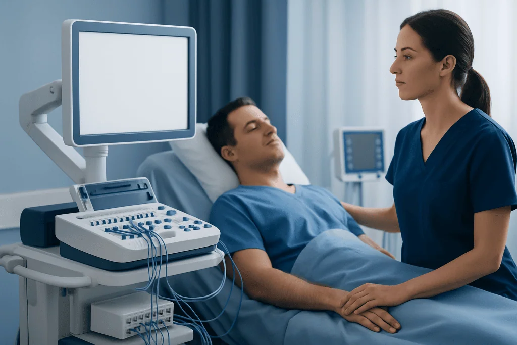
A doctor may recommend an electrophysiology study when a person has symptoms that suggest a rhythm problem but the cause is still unclear. Common reasons include repeated palpitations, episodes of a racing heartbeat, dizziness, fainting, or near-fainting. It may also be used when an abnormal rhythm has already been detected and more precise information is needed.
EPS can help answer important questions. It may show whether symptoms are caused by an arrhythmia, identify the exact source of the abnormal rhythm, and determine how easily the rhythm can be triggered. This can be especially helpful when external tests such as Holter monitoring, event recorders, or a routine ECG have not provided enough detail.
The study is also used to guide treatment planning. For some people, it helps decide whether medicines are likely to work, whether a pacemaker or implantable defibrillator may be needed, or whether ablation procedures could be the best option. People with structural heart disease, including cardiomyopathy or prior heart damage, may also have EPS as part of a broader rhythm evaluation.
What happens during the EPS procedure

An electrophysiology study is usually performed in a hospital electrophysiology laboratory. Before the procedure, the care team reviews medical history, current medicines, allergies, and test results. Some medicines that affect heart rhythm may need to be stopped for a short time beforehand, but this should only be done under medical guidance.
On the day of the procedure, the person changes into a hospital gown and an intravenous line is placed for fluids and medicines. Monitoring devices are attached to check heart rhythm, blood pressure, and oxygen levels throughout the test. Most people receive sedation to help them feel relaxed, although they may remain awake enough to respond to instructions.
After cleaning the skin and numbing the area, the doctor inserts catheters through a vein, most often in the groin and sometimes in the neck or arm. Using X-ray guidance and electrical mapping, the catheters are moved into specific chambers of the heart. The team then records electrical signals and may pace the heart briefly to try to reproduce the abnormal rhythm in a controlled setting.
If the arrhythmia is identified and the person has already agreed to treatment, the same session may include a therapeutic procedure such as catheter ablation. In other cases, the EPS is diagnostic only, and treatment decisions are made afterward. The entire procedure may take a few hours, depending on how complex the rhythm problem is.
How to prepare and what recovery is like
Preparation instructions vary, but fasting for several hours before the procedure is common. Patients are usually asked to bring a list of all medicines and supplements. Because some rhythm medicines, blood thinners, diabetes treatments, or blood pressure drugs may need temporary adjustment, it is important to follow the medical team’s instructions closely rather than making changes independently.
After the EPS, the catheters are removed and pressure is applied to the insertion site to reduce bleeding. The patient is monitored while the sedative wears off and may need to lie flat for a period of time, especially if the groin was used. Some people go home the same day, while others stay overnight depending on the findings, any treatment performed, and their general health.
Mild soreness or bruising at the catheter site can happen and often improves within a few days. The care team usually advises when normal activities, driving, exercise, and work can be resumed. A responsible adult may be needed to take the patient home if sedation was used.
Many people feel reassured to know that recovery is typically straightforward. However, the doctor should be contacted promptly if there is heavy bleeding, increasing swelling, severe pain, fever, chest pain, shortness of breath, or a new fainting episode after going home.
What the results mean
One of the main benefits of an electrophysiology study is that results are often available immediately after the procedure. The doctor can usually explain whether the test reproduced an abnormal rhythm, where it started, and how it traveled through the heart. This information helps turn symptoms such as palpitations or fainting into a clear diagnosis.
A normal or nondiagnostic EPS does not always mean there is no problem. Some arrhythmias happen only under specific conditions and may not be triggered during the study. In that situation, the doctor may recommend additional monitoring, repeat evaluation, or treatment based on the person’s symptoms and overall risk profile.
If the EPS identifies a specific rhythm disorder, the findings help guide the next step. For example, a short electrical circuit in the upper chambers may support a diagnosis such as Wolff-Parkinson-White syndrome or another supraventricular tachycardia. Abnormal signals from the lower chambers may point to a more serious ventricular arrhythmia, especially in people with underlying heart disease.
The results may also show whether the heart’s normal conduction system is too slow or unreliable, which can support the need for a pacemaker. In some patients, the pattern of arrhythmia and the condition of the heart muscle help doctors judge the need for longer-term rhythm protection and follow-up with a specialized cardiac arrhythmia team.
Treatment options after EPS
Treatment after an electrophysiology study depends on the diagnosis, symptoms, and the person’s overall heart health. Some arrhythmias need only observation and follow-up. Others are managed with lifestyle changes and medicines that slow the heart, stabilize the rhythm, or reduce the chance of future episodes.
For many rhythm disorders, catheter ablation is an effective option. During ablation, the doctor uses heat or cold energy delivered through a catheter to treat the small area of heart tissue causing the abnormal electrical pathway. This is often considered when symptoms are troublesome, episodes are recurrent, or medicines are not well tolerated.
Some people may need a pacemaker if the heart’s electrical system is too slow, or an implantable cardioverter-defibrillator if there is concern about dangerous ventricular arrhythmias. In patients who also have underlying heart conditions such as valve disease, congenital heart disease, or heart failure, treatment usually involves a broader plan that addresses both the rhythm problem and the structural issue.
Supportive care may also be important after treatment. Depending on the person’s condition, follow-up may include ECGs, remote rhythm monitoring, medicine review, and risk-factor control such as management of cholesterol, sleep apnea, and hypertension treatment. In selected patients, supervised cardiac rehabilitation can support recovery and confidence after a heart-related procedure.
Risks, safety, and when to seek medical care
An electrophysiology study is generally considered safe when performed by experienced teams, but like any invasive procedure it has some risks. Possible complications include bleeding, bruising, infection at the catheter site, blood vessel injury, temporary rhythm disturbances, blood clots, or reaction to sedation. Rarely, more serious complications can occur, such as damage to the heart or the need for urgent treatment.
The care team works carefully to reduce these risks. Before the procedure, doctors assess other medical conditions, kidney function, medicines, and any previous reactions to contrast dye or anesthesia. Continuous monitoring during the test allows the team to respond quickly if the heart rhythm changes unexpectedly.
People should seek urgent medical attention if they have chest pain, severe shortness of breath, fainting, heavy bleeding from the catheter site, rapid swelling, or signs of a stroke after the procedure. Even if symptoms seem mild, new or concerning symptoms should be discussed with a doctor promptly. It is always safer to ask than to wait.
For international patients who need evaluation for complex rhythm problems, Acibadem International’s multidisciplinary specialists and JCI-accredited hospitals diagnose and treat arrhythmias using advanced electrophysiology techniques and individualized follow-up plans.
Frequently asked questions
Is an electrophysiology study painful?
Most people do not describe EPS as painful, although it can feel uncomfortable at times. The insertion area is numbed, and sedation is usually given to help the patient relax. Some people notice pressure at the catheter site or feel their heart race briefly when the rhythm is being tested.
How long does an EPS take?
The procedure often takes a few hours, but the exact time depends on how complex the rhythm problem is. If a doctor also performs catheter ablation during the same session, it may take longer. Recovery and monitoring time afterward are usually added to the total hospital stay.
Will the doctor know the results right away?
In many cases, yes. Because EPS records the heart’s electrical activity directly, the doctor can often explain the main findings shortly after the procedure. A full treatment plan may still require review of other tests and discussion of the safest next steps.
Can an electrophysiology study treat the problem as well as diagnose it?
Yes, sometimes it can. If the abnormal pathway is identified and the patient has given consent, the doctor may perform catheter ablation during the same procedure. This approach can reduce the need for a separate hospital visit.
What should be avoided after an EPS?
Patients are usually advised to avoid strenuous activity, heavy lifting, and driving for a short period, especially if sedation was used. The exact instructions depend on where the catheter was inserted and whether additional treatment was performed. It is important to follow the discharge guidance from the medical team.
Why would someone need EPS if they already had an ECG or Holter monitor?
An ECG and Holter monitor record heart rhythm from the body surface, which is often enough for many problems. EPS gives much more precise information by measuring electrical signals from inside the heart and testing how the rhythm starts and spreads. This is especially useful when symptoms are unexplained or when treatment decisions depend on exact rhythm mapping.
This article is for general information only and is not a substitute for professional medical advice. Please consult a qualified doctor about your individual situation.
Explore treatments in Turkey — costs, top hospitals & a free quote
JCI-accredited · board-certified surgeons · reply within 24h
Add us as a Preferred Source to see more of our trusted health content across Google Search, AI Overviews and Discover.
Check your numbers in seconds
BMI, calories, due date, blood pressure and 30+ more clinical calculators — free, instant, doctor-reviewed ranges.

Cardiology Department
Diagnosis and treatment of heart and vascular conditions, from prevention to advanced interventional procedures.
93 specialists in this unitRelated Treatments
Related Conditions
More from the Health Library

CABG Before and After: Symptoms, Activity Level, and Realistic Results

Supraventricular Tachycardia: Sudden Fast Heartbeat and Ablation Treatment

Palpitations: Causes, Tests, and When to Worry

Pulmonary Hypertension: Early Signs and How It Is Diagnosed

Coronary Angiography Before Valve Surgery: Why It May Be Needed