

Nuclear Cardiology Scan: What It Shows and When It Is Recommended
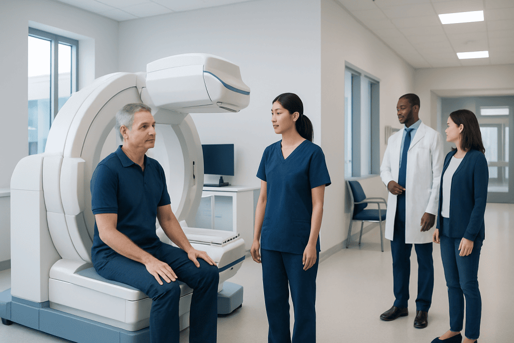
A nuclear cardiology scan assesses blood flow to the heart muscle, usually at rest and during stress. It is commonly used to look for reduced circulation linked to coronary artery disease or prior heart damage.
Key Takeaways
- A nuclear cardiology scan assesses blood flow to the heart muscle, usually at rest and during stress.
- It is commonly used to look for reduced circulation linked to coronary artery disease or prior heart damage.
- The test uses a small amount of radioactive tracer and is generally considered safe when medically appropriate.
- Results can help guide treatment decisions, lifestyle changes, and the need for further cardiac testing or procedures.
- Preparation may include avoiding caffeine and following instructions about food and medications.
A nuclear cardiology scan is an imaging test that shows how well blood reaches the heart muscle at rest and during stress. It can help doctors evaluate chest pain, shortness of breath, suspected blocked arteries, and the effects of some heart conditions or treatments.
Overview
A nuclear cardiology scan is a type of heart imaging test that uses a small amount of radioactive tracer to show blood flow to the heart muscle. It is often called a myocardial perfusion scan. Special cameras detect the tracer and create images that help doctors see whether the heart is receiving enough blood.
In many cases, images are taken both at rest and during stress. Stress may be created through exercise on a treadmill or with medication that makes the heart work harder or widens the heart arteries. Comparing the two sets of images can reveal areas that receive less blood during exertion than at rest.
This test does not usually diagnose every heart problem on its own. Instead, it adds important information to a patient’s symptoms, physical examination, electrocardiogram, and other cardiac tests. It is especially helpful when there is concern for coronary artery disease or when a doctor wants to understand how well the heart muscle is functioning.
What a Nuclear Cardiology Scan Shows
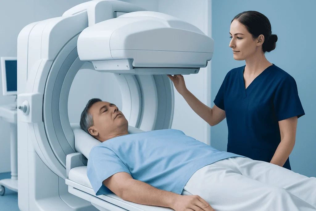
The main purpose of a nuclear cardiology scan is to show how blood moves through the heart muscle. If one area takes up less tracer during stress, it may suggest reduced blood flow caused by a narrowed or blocked coronary artery. If an area has low uptake both at rest and during stress, it may suggest scar tissue from a previous heart attack.
The scan can also provide information about heart pumping function, including ejection fraction in some studies. This helps doctors understand whether the heart is contracting effectively and whether there may be signs of weakened heart muscle, such as in heart failure or certain forms of cardiomyopathy.
Depending on the type of study and the patient’s situation, the scan may help assess:
- Reduced blood flow to parts of the heart
- Evidence of previous heart muscle injury
- How the heart responds to physical or medication-induced stress
- Overall pumping performance
- Whether symptoms may be related to circulation problems in the heart
Although highly useful, the scan has limits. It shows patterns of blood flow and function, but it does not directly treat a problem or always identify the exact cause of symptoms. Some patients may need additional testing, such as echocardiography, CT coronary imaging, or invasive coronary angiography.
When It Is Recommended

Doctors may recommend a nuclear cardiology scan when a patient has symptoms that could be related to reduced blood flow to the heart. Common reasons include chest discomfort, shortness of breath with exertion, unexplained fatigue, or symptoms that raise concern for stable angina or silent ischemia. It may also be considered when an exercise ECG alone is unlikely to give a clear answer.
The test is often used to estimate the likelihood of clinically important coronary artery narrowing and to help decide next steps in care. For example, it may help determine whether symptoms are more likely to be due to a circulation problem in the heart or another cause. It can also be useful after a previous heart attack to evaluate whether damaged or under-supplied heart muscle is present.
A nuclear cardiology scan may be recommended before some surgeries in selected patients with cardiovascular risk factors, or after treatment to see whether blood flow has improved. In people with known heart disease, results may help guide referral for procedures or surgery such as coronary artery bypass surgery or other forms of advanced cardiac care.
Not every patient with chest symptoms needs this test. The decision depends on age, medical history, risk factors, symptoms, ECG findings, and whether another test would be more suitable. A cardiologist or referring physician selects the most appropriate evaluation for each person.
How the Test Is Performed
The test usually takes place in a hospital or imaging center. A small intravenous line is placed, and the tracer is injected into a vein. Images are then taken with a special camera after the tracer has circulated through the body. Depending on the protocol, this may happen once at rest and once after stress.
For the stress portion, some people walk on a treadmill or pedal a stationary bicycle. Others receive medication to simulate stress if exercise is not possible or would not be safe. During this phase, the healthcare team monitors heart rhythm, blood pressure, and symptoms closely.
Patients are usually given preparation instructions in advance. These may include avoiding caffeine for a period before the test, not eating for several hours, and asking whether usual medicines should be taken that day. Because instructions vary, it is important to follow the advice of the testing center exactly.
Most people can go home the same day. The tracer amount is small, and the body gradually clears it. Drinking fluids after the test may be recommended unless a doctor has advised fluid restriction for another medical reason.
Risks, Safety, and Limitations
A nuclear cardiology scan is generally considered safe when ordered for an appropriate medical reason. The tracer exposes the patient to a low dose of radiation, and healthcare teams aim to use the lowest amount needed to obtain useful images. For many patients, the expected benefit of accurate diagnosis outweighs the small radiation risk.
Stress testing can occasionally cause temporary symptoms such as flushing, headache, shortness of breath, chest discomfort, or changes in heart rhythm, especially when stress medications are used. Serious complications are uncommon because the test is supervised by trained professionals who monitor the patient throughout.
Some people should discuss special circumstances with their doctor before the test, including pregnancy, breastfeeding, severe asthma, certain rhythm disorders, or inability to lie still. The team may modify the plan or choose a different test if needed.
Like all tests, nuclear imaging has limitations. Results can sometimes be affected by body shape, breast tissue, the diaphragm, movement during imaging, or other technical factors. In some cases, findings are unclear and need correlation with other cardiac tests or follow-up with general cardiology specialists.
Understanding the Results and What Happens Next
Results are interpreted by a specialist who reviews the images along with the stress data, symptoms during testing, and often the ECG. A normal scan generally suggests that blood flow to the heart muscle is adequate during rest and stress. This can be reassuring and may reduce the need for more invasive testing in some patients.
An abnormal scan may show reduced blood flow during stress, evidence of prior injury, or reduced pumping performance. These findings do not always mean the same thing for every patient. The meaning depends on where the change appears, how extensive it is, and how it fits with the person’s symptoms and overall risk profile.
Next steps may include changes in medication, closer risk-factor control, additional imaging, or referral for procedures to improve blood flow. In some situations, treatment may involve catheter-based care or surgery, followed by cardiac rehabilitation to support recovery and long-term heart health.
If symptoms are related to broader cardiovascular risks such as diabetes, obesity, abnormal cholesterol, or high blood pressure management, doctors usually address those factors as part of the overall care plan. The goal is not only to explain the scan result, but also to reduce future heart risk in a comprehensive way.
Prevention, Self-Care, and When to See a Doctor
A nuclear cardiology scan does not prevent heart disease by itself, but it can help guide preventive care. Many of the conditions it helps identify are linked to modifiable risk factors. Healthy lifestyle habits remain a cornerstone of heart protection, whether scan results are normal or abnormal.
Helpful steps often include not smoking, staying physically active within medical advice, choosing a heart-healthy eating pattern, managing stress, sleeping well, and attending regular checkups. Careful control of blood pressure, cholesterol, blood sugar, and body weight is also important. People with known heart disease should take medicines exactly as prescribed and discuss any side effects with their doctor rather than stopping treatment on their own.
Medical attention is important if a person has chest pressure, pain that spreads to the arm or jaw, sudden severe shortness of breath, fainting, or new symptoms with exertion. These may need urgent assessment. Less urgent but persistent symptoms such as recurring chest discomfort, unexplained exercise intolerance, or worsening fatigue should also be discussed with a healthcare professional.
For patients seeking specialist evaluation, Acibadem International’s multidisciplinary cardiology teams in JCI-accredited hospitals diagnose and treat heart conditions for international patients. A doctor can explain whether a nuclear cardiology scan is the right test and how it fits into a broader plan of diagnosis, treatment, and follow-up.
Frequently asked questions
Is a nuclear cardiology scan the same as a regular stress test?
Not exactly. A regular stress test usually monitors the heart with an ECG during exercise, while a nuclear cardiology scan adds imaging to show blood flow to the heart muscle. This can provide more detailed information, especially when doctors need to look for areas with reduced circulation.
How should a patient prepare for a nuclear cardiology scan?
Preparation instructions vary, but patients are often asked to avoid caffeine and sometimes food for several hours before the test. The doctor may also give instructions about heart medications, diabetes medicines, or inhalers. It is best to follow the testing center’s directions carefully.
Does the test hurt?
Most people tolerate the test well. The main discomfort is usually the brief needle stick for the IV line, and some patients feel temporary effects during the stress portion, such as warmth, flushing, or shortness of breath. Staff monitor the patient throughout and can respond if symptoms occur.
How long does a nuclear cardiology scan take?
The full appointment often takes several hours because it may include resting images, a stress phase, and waiting periods after tracer injection. The exact timing depends on the imaging protocol and whether exercise or medication stress is used. The testing center can provide a more precise estimate beforehand.
Is the radiation from a nuclear cardiology scan dangerous?
The test uses a small amount of radioactive tracer, and the radiation exposure is generally considered low for medically appropriate testing. Doctors order it when the expected benefit of the information gained outweighs the potential risk. If a patient is pregnant, breastfeeding, or concerned about radiation, that should be discussed before the scan.
Can a nuclear cardiology scan miss heart problems?
Yes, no test is perfect. A nuclear cardiology scan is very helpful for many blood flow problems, but some conditions may not appear clearly or may require other tests for confirmation. Doctors interpret the scan together with symptoms, examination findings, and other investigations.
This article is for general information only and is not a substitute for professional medical advice. Please consult a qualified doctor about your individual situation.
Nuclear Cardiology in Turkey — costs, top hospitals & a free quote
JCI-accredited · board-certified surgeons · reply within 24h
Add us as a Preferred Source to see more of our trusted health content across Google Search, AI Overviews and Discover.

Cardiology Department
Diagnosis and treatment of heart and vascular conditions, from prevention to advanced interventional procedures.
93 specialists in this unitRelated Treatments
Related Conditions
More from the Health Library

Coronary Artery Disease: Plaque Buildup, Testing, and Treatment

Coronary Bypass Surgery for Chronic Total Occlusion: When CABG Is Considered

Heart-Lung Machine During CABG: How Cardiopulmonary Bypass Supports Surgery

Coronary Artery Bypass Surgery: Who May Need CABG and Why

Heart Bypass Surgery With Kidney Disease: Risk Planning and Recovery

Valve Surgery Cost in Turkey: What Affects the Total Price?
Cardiology Specialists at Acibadem

Prof. Dr. Ahmet Oytun Baykan
Cardiology
Prof. Dr. Ahmet Tulga Ulus
Cardiovascular Surgery
Prof. Dr. Ahmet Ümit Güllü
Cardiovascular Surgery
Prof. Dr. Aleks Değirmencioğlu
Cardiology