Retinal Tear vs Retinal Detachment: Key Differences and Urgent Signs

A retinal tear is a break in the retina; a retinal detachment happens when the retina lifts away from the tissue beneath it. Retinal detachment is generally more serious because it can quickly threaten permanent vision loss.
Key Takeaways
- A retinal tear is a break in the retina; a retinal detachment happens when the retina lifts away from the tissue beneath it.
- Retinal detachment is generally more serious because it can quickly threaten permanent vision loss.
- Sudden flashes of light, a shower of new floaters, or a dark curtain over vision need urgent eye evaluation.
- Prompt treatment of a retinal tear may help prevent retinal detachment.
- People with high myopia, aging vitreous changes, eye injury, or past eye surgery may have a higher risk.
Retinal tears and retinal detachments affect the light-sensitive tissue at the back of the eye, but they are not the same condition. A retinal tear can sometimes progress to a retinal detachment, which is why sudden flashes, new floaters, or a curtain-like shadow in vision should be assessed urgently.
Overview: What Is the Difference?
The retina is the thin, light-sensitive layer lining the back of the eye. It captures visual information and sends it to the brain through the optic nerve. A retinal tear is a small break or rip in this delicate tissue. A retinal detachment occurs when the retina separates from the layer underneath that supplies it with oxygen and nutrients.
The two conditions are closely related, but they are not identical. A tear may happen first, often when the gel inside the eye, called the vitreous, pulls on the retina. Fluid can then pass through the tear and collect behind the retina, causing it to lift away. In that way, a retinal tear can lead to a retinal detachment.
This difference matters because a tear may sometimes be treated before the retina detaches, helping protect vision. Once a detachment develops, treatment is usually more urgent and often more complex. For patients comparing retinal tear vs retinal detachment, the key point is that both need prompt medical attention, but detachment is generally the more vision-threatening stage.
Symptoms and Urgent Warning Signs
Retinal tears and retinal detachments can cause some of the same symptoms. People may notice sudden flashes of light, especially in the side vision, or a sudden increase in floaters, which can look like dots, threads, cobwebs, or specks drifting through the field of view. Some also describe blurred vision or a change in peripheral vision.
Symptoms can become more concerning if a detachment is developing or has already occurred. A dark shadow, gray curtain, or veil moving across part of the vision is a classic warning sign. Vision may become distorted or reduced, and the effect often does not clear by blinking or resting the eyes.
Not everyone with flashes or floaters has a retinal tear, and many floaters are harmless age-related changes. Still, new or sudden symptoms should never be ignored because it is not possible to tell the difference without an eye examination. Urgent assessment through a dilated eye examination is important.
- Sudden flashes of light
- A shower or sudden increase in floaters
- A curtain, shadow, or dark area in vision
- Sudden loss of side vision
- Unexplained blurred or distorted vision
Causes and Risk Factors

The most common cause of a retinal tear is traction from the vitreous. With age, the vitreous gel naturally shrinks and becomes more liquid. As it pulls away from the retina, called posterior vitreous detachment, it may tug strongly enough to create a tear. In many people this process happens without serious harm, but in others it can cause a break in the retina.
Several factors can increase risk. These include high myopia, previous retinal tear or detachment, a family history of retinal detachment, eye injury, and certain previous eye operations. People who have had cataract surgery may have a somewhat higher risk than those who have not, especially if other risk factors are also present. Some retinal diseases and severe inflammation can also contribute.
Diabetes can affect the retina in different ways and may lead to tractional detachment in advanced cases, although this is not the usual pathway for a simple retinal tear. Eye trauma, including sports injuries or accidents, can also trigger a tear or detachment at any age. Patients already being followed in retina and vitreous care may be advised about their individual risk based on the structure of their retina and vitreous.
How Doctors Diagnose These Conditions
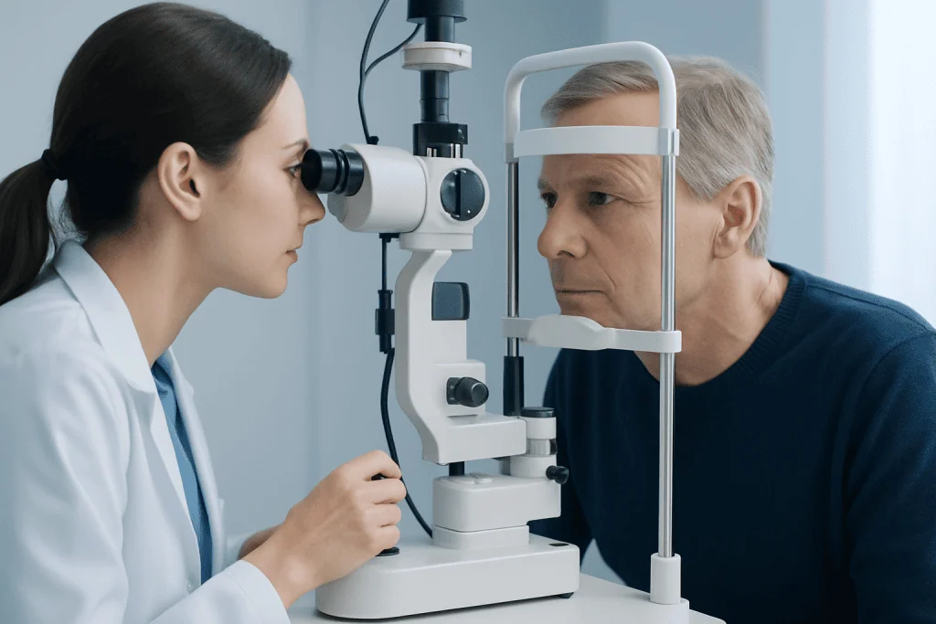
Diagnosis usually begins with a careful discussion of symptoms, when they started, and whether they are worsening. An ophthalmologist will typically check visual acuity, eye pressure, and the front part of the eye. The most important part is a dilated retinal examination, which allows a detailed view of the back of the eye.
Using special lenses and bright light, the doctor looks for tears, bleeding, weak retinal areas, and signs that the retina has lifted. If the view is blocked by blood or a dense cataract, ultrasound imaging of the eye may help show whether a detachment is present. In some cases, additional evaluation in general ophthalmology or retinal subspecialty care is needed.
Fast diagnosis is important because timing can affect treatment choices and visual outcome. A small tear found early may be sealed before fluid tracks underneath it. A detachment, especially if the central retina is affected, often requires more urgent intervention to preserve as much vision as possible.
Treatment Options for Retinal Tears and Detachments
Treatment depends on whether there is only a tear, whether fluid has already gone under the retina, and how much of the retina is involved. A retinal tear without detachment is often treated with laser therapy or freezing treatment, called cryopexy, to create a scar that seals the retina in place. These treatments are intended to reduce the chance that the tear will progress.
Retinal detachment usually requires a procedure or surgery rather than simple observation. Options may include pneumatic retinopexy, where a gas bubble helps press the retina back into position, scleral buckle surgery, or vitrectomy, which removes the vitreous gel and relieves traction. The choice depends on the location and size of the detachment, the number of tears, the condition of the vitreous, and the patient’s overall eye health.
After treatment, follow-up is important because new tears can sometimes appear, and vision may take time to stabilize. Recovery instructions vary by procedure and may include positioning the head in a certain way, avoiding some activities, and attending repeated retinal checks. In complex cases, care may involve specialists in eye surgery and retinal disease.
Prevention, Self-Care, and What Not to Ignore
There is no guaranteed way to prevent every retinal tear or detachment, especially when age-related vitreous changes are the cause. However, regular eye examinations can help identify weak retinal areas or other eye problems before symptoms become severe. People with high myopia, previous tears, or a family history of retinal detachment may benefit from regular ophthalmic follow-up.
Protecting the eyes from injury is also important. Wearing appropriate protective eyewear during sports, home repairs, and high-risk work can reduce the chance of trauma-related retinal damage. Managing other eye conditions and attending follow-up after eye surgery may also lower the chance that warning signs will be missed.
Self-care at home should never replace urgent evaluation when symptoms are sudden. Resting the eyes, using lubricating drops, or waiting to see if floaters improve is not enough if there are new flashes, many floaters, or a curtain-like shadow. Quick care can make a meaningful difference, especially when the problem is caught before full detachment occurs.
When to See a Doctor
Anyone with sudden flashes, a rapid increase in floaters, or a dark shadow in vision should seek urgent same-day or emergency eye care. These symptoms do not always mean detachment, but they can signal a retinal tear that needs prompt treatment. Waiting can allow fluid to move under the retina and make the condition more serious.
Urgent care is especially important if vision suddenly becomes blurry, side vision narrows, or a curtain seems to move over part of the visual field. Symptoms in one eye should still be treated as important, even if the other eye seems normal. People who recently had eye surgery or eye trauma should be particularly cautious.
Ongoing or recurrent floaters without sudden change should still be discussed at a routine eye visit, especially in people at higher risk. Near the end of the care pathway, it may be reassuring to know that Acibadem International’s multidisciplinary specialists and JCI-accredited hospitals diagnose and treat retinal conditions for international patients, including advanced retinal disorders and related neuro-ophthalmology support when needed.
Frequently asked questions
Is a retinal tear the same as a retinal detachment?
No. A retinal tear is a break in the retina, while a retinal detachment means the retina has lifted away from the tissue beneath it. A tear can lead to a detachment, which is why early assessment is important.
Are flashes and floaters always an emergency?
Not always, because many floaters are related to normal aging changes in the vitreous. However, sudden new flashes, a large increase in floaters, or any shadow in vision should be treated as urgent until a doctor examines the eye.
Can a retinal tear heal on its own?
A retinal tear usually does not reliably seal itself in a way that prevents detachment. Doctors often recommend prompt treatment, such as laser or freezing therapy, to reduce the risk of fluid passing under the retina.
How quickly should someone get help for possible retinal detachment?
Possible retinal detachment should be assessed as soon as possible, ideally the same day. Early diagnosis can affect treatment options and may improve the chance of preserving vision.
Who is most at risk for retinal tears or detachments?
People with high myopia, older age-related vitreous changes, previous retinal tears or detachments, eye trauma, and certain prior eye surgeries may be at higher risk. A family history can also matter.
Can vision return after treatment?
Many people keep or regain useful vision after treatment, but recovery depends on how severe the problem was and how quickly it was treated. If the central retina has been detached, vision may not fully return to its previous level.
References
- American Academy of Ophthalmology
- National Eye Institute
- Royal College of Ophthalmologists
- Mayo Clinic
- National Health Service
This article is for general information only and is not a substitute for professional medical advice. Please consult a qualified doctor about your individual situation.
Explore treatments in Turkey — costs, top hospitals & a free quote
JCI-accredited · board-certified surgeons · reply within 24h
Add us as a Preferred Source to see more of our trusted health content across Google Search, AI Overviews and Discover.