

Quick answer
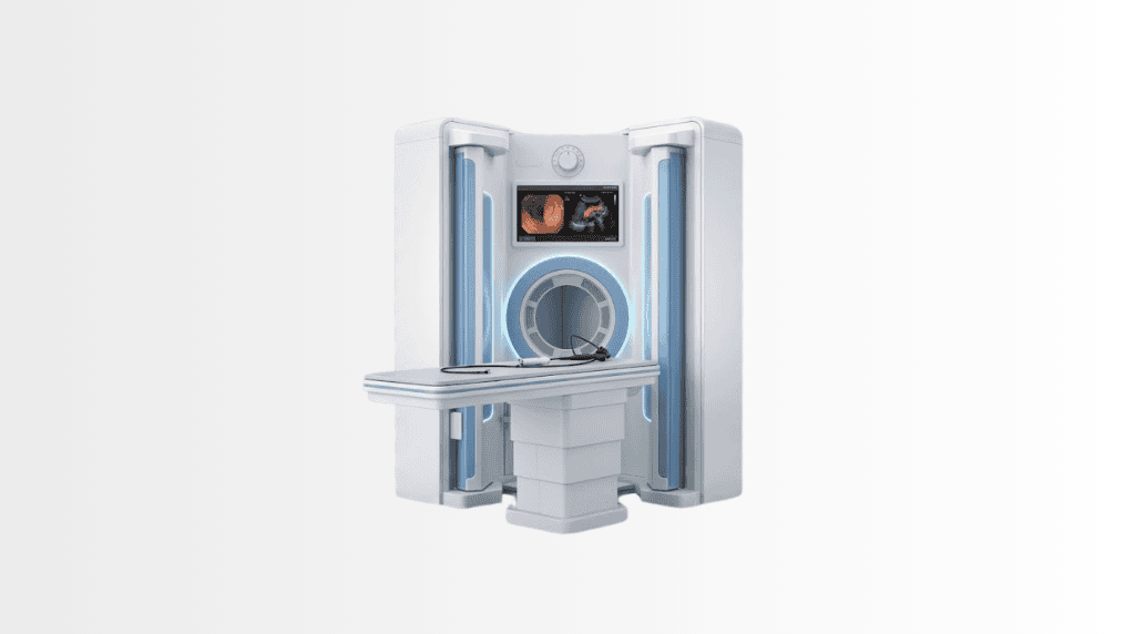
Endoscopic ultrasound (EUS), or endoultrasonography, combines a flexible endoscope with ultrasound imaging to examine the digestive tract and nearby organs in detail. At Acibadem in Turkey, EUS is used to assess areas including the oesophagus, stomach, pancreas, bile ducts and lymph nodes, and can guide tissue sampling when needed.
Endoscopic ultrasound (EUS) solves a geographic problem in diagnosis: the pancreas, bile ducts and the layers of the gut wall sit deep behind gas and bone, where surface ultrasound squints and CT sees structure but not always subtlety. EUS mounts a miniature ultrasound probe on the tip of an endoscope and images those organs from millimetres away, and, in the same session, can pass a fine needle through the gut wall to sample what it sees. One examination: high-resolution imaging plus tissue diagnosis, no incision.
What is an endoscopic ultrasound?
A flexible endoscope carries an ultrasound transducer through the mouth into the stomach and duodenum (or, for rectal indications, from below). From inside, the probe images adjacent anatomy with a clarity distance cannot match: the pancreas in its entirety, the bile duct and gallbladder, the gut wall’s five distinct layers, nearby lymph nodes and vessels. When something needs proof rather than description, EUS-guided fine-needle aspiration or biopsy (FNA/FNB) samples it under continuous ultrasound vision: the standard route to a tissue diagnosis of pancreatic masses without surgery.
What EUS is used for
The classic indications: evaluating pancreatic masses and cysts, where EUS both characterises and biopsies; hunting small bile-duct stones that other imaging equivocates on; staging how deeply an oesophageal, gastric or rectal tumour has grown through the wall and whether nearby nodes are suspicious, depth-staging is EUS home ground; clarifying submucosal bumps seen at routine endoscopy, which layer they arise from and what they likely are; and assessing chronic pancreatitis. On the therapeutic side, EUS increasingly acts as well as looks: draining pancreatic fluid collections and guiding nerve-block procedures such as coeliac plexus block for pancreatic pain.
The procedure: what to expect
EUS is performed under sedation, most patients sleep through it, after a fasting period your instructions specify. The examination itself typically takes twenty to sixty minutes depending on whether sampling or a therapeutic step is included. Afterwards: an hour or two of recovery while sedation wears off, a companion to accompany you home, no driving that day, and at most a mild sore throat or bloating. If biopsy was taken, pathology results follow within days and are read into the plan, at Acibadem, into a tumour board where the finding warrants one.
EUS vs CT, MRI and ERCP
CT and MRI/MRCP map the region and remain the first pictures in most pathways; EUS adds near-field resolution, the small lesion, the wall layer, the equivocal node, plus the needle that turns an image into a diagnosis. Against ERCP, the division of labour is clean: ERCP is a treatment tool for the bile duct (removing stones, placing stents) with real procedural risks; EUS is the low-risk way to look and decide whether ERCP is needed at all. Modern units run them as a tandem, sometimes in one sedation.
Honest risks
Diagnostic EUS is a low-risk procedure; sampling adds small risks, bleeding, infection, and uncommonly pancreatitis after pancreatic puncture, quantified for your case beforehand. Perforation is rare. The sedation itself is screened and monitored by anaesthesia protocols; tell the team about heart or lung conditions and blood thinners early, since they shape the plan rather than cancel it.
EUS at Acibadem
EUS runs within the group’s gastroenterology practice, with corporate-verified endosonography capability at campuses including City Clinic Tokuda in Sofia, Mladost’s diagnostic floors and the Varna medical centrewith pathology through the group’s ISO-accredited laboratories and findings that need oncology read directly into tumour boards. Records-first applies: prior imaging travels ahead, so the examination answers a defined question rather than starting from zero.
Pancreatic cysts: EUS’s most consequential clinic
Modern scanning finds pancreatic cysts constantly, most incidental, most harmless, a minority carrying real malignant potential, and sorting them is where EUS earns its keep. Near-field imaging reads the features that matter: septations, wall thickening, mural nodules, duct communication and calibre. Fluid aspirated through the needle speaks its own dialect: CEA levels and molecular markers separating mucinous cysts (the surveillance-or-surgery family) from serous ones (the reassurance family), amylase hinting at pseudocysts after pancreatitis. The output is a disposition, not a shrug: discharge with reassurance, a defined surveillance interval by size and features, or referral to surgery when worrisome features stack up. For a patient carrying an “incidental cyst” sentence on a scan report, this examination is usually the difference between years of vague worry and a plan with dates on it.
Therapeutic EUS: when the needle becomes the treatment
The same access that samples can treat, and interventional EUS has grown into a discipline of its own. Established territory: draining pancreatic fluid collections and walled-off necrosis through the stomach wall with stents, replacing operations that once dominated severe pancreatitis care; coeliac plexus block and neurolysis, delivering pain control to the nerve bundle behind the pancreas for cancer pain that medication ceilings cannot reach; and pseudocyst management on the same logic. The expanding frontier: EUS-guided bile-duct and gallbladder drainage where ERCP’s route is blocked, belongs to high-volume centres and honest case selection. Each of these carries its own risk-benefit conversation, held with the team that also offers the alternatives; interventional EUS at its best is exactly that: an alternative offered, not an inevitability sold.
Frequently Asked Questions
My scan found a pancreatic cyst, should I worry?
Most are harmless, a minority matter: EUS with fluid analysis sorts which family yours belongs to and converts worry into a plan: reassurance, dated surveillance, or referral.
What does cyst fluid analysis show?
Chemistry and markers: CEA and molecular tests separating mucinous from serous cysts, amylase flagging pseudocysts, read together with the imaging features.
Can EUS treat pain from pancreatic cancer?
Yes, coeliac plexus neurolysis delivers targeted pain control to the nerves behind the pancreas, one honest tool where medication alone falls short.
Is an endoscopic ultrasound painful?
No: the procedure is performed under sedation and most patients sleep through it; a mild sore throat afterwards is the common complaint.
How long does an EUS take?
Typically twenty to sixty minutes, longer when needle sampling or a therapeutic step is included.
Do I need to fast?
Yes: a fasting period before sedation is required; your appointment instructions state the exact hours.
Will tissue be taken during my EUS?
Only if the finding warrants it: EUS-guided fine-needle sampling is done in the same session when a lesion needs a tissue answer, and your consent covers it beforehand.
Is EUS the best test for the pancreas?
For characterising and biopsying pancreatic lesions it is the reference tool, working alongside CT and MRI rather than replacing them.
Can EUS find bile duct stones that other scans missed?
Yes, small stones and sludge are a classic EUS catch, often deciding whether an ERCP is genuinely needed.
What is the difference between EUS and ERCP?
EUS looks and samples with low risk; ERCP treats the bile duct, stones, stents, with higher procedural risk. EUS often decides whether ERCP is warranted.
How safe is the procedure?
Diagnostic EUS is low-risk; sampling adds small, quantified risks, bleeding, infection, uncommonly pancreatitis, discussed for your specific case before consent.
When do biopsy results come?
Within days, through the group’s accredited pathology, and where the answer changes a cancer plan, it is read into the tumour board directly.
Can I fly home after an EUS?
Usually the next day is comfortable; the same day depends on sedation recovery and your team’s advice, planned visits account for it.
What is EUS-guided coeliac plexus block?
A pain procedure: medication delivered under EUS vision to the nerve plexus behind the pancreas, one of the tools for hard-to-control pancreatic pain.
Do I need someone with me?
Yes, sedation means no driving that day and a companion for the journey home.
Can my previous scans replace an EUS?
They complement it, send them ahead; EUS exists precisely for the questions CT and MRI leave open, and records-first review confirms whether you need it at all.
Related Medical Units
Available at these Hospitals


















Related Treatments
Speak with our medical team
Find out how this technology could support your diagnosis or treatment.