

Chronic Occlusion Treatment
Chronic occlusion treatment restores blood flow in a completely blocked artery, most often using advanced catheter-based techniques. It may relieve angina, improve heart function, and reduce cardiovascular risk.
Quick answer
A chronic occlusion, in cardiology, a chronic total occlusion or CTO, is a coronary artery that has been completely blocked for around three months or longer. Treatment may involve medication, a specialised catheter procedure (CTO PCI) that crosses the blockage and places stents, or bypass surgery. The right approach depends on symptoms, heart function, the amount of muscle at risk and the anatomy of the blocked vessel.
Chronic Occlusion and the CTO Meaning in Cardiology
A chronic occlusion is an artery that has been completely blocked for a prolonged period, in the coronary arteries, conventionally for three months or longer. The blockage stops blood flowing through that vessel to the heart muscle it supplies, and it is usually built from cholesterol-rich plaque that has hardened over years into fibrous, scar-like and often calcified tissue. Treatment ranges from medication alone to a specialised catheter procedure or bypass surgery, and the right choice depends on your symptoms, your heart function and the anatomy of the blocked vessel.
If you have looked up the CTO meaning after reading an angiogram report, you are in good company. It is one of the most searched cardiology abbreviations, and the reports that contain it rarely explain it. This page sets out what the term means, how the condition is evaluated, what the treatment options genuinely involve, what can go wrong, and what a realistic recovery looks like.
A completely blocked artery does not always mean the heart muscle beyond it has died. Over months and years, the heart often grows small natural bypass channels called collateral arteries, which carry blood around the blockage from neighbouring vessels. These collaterals can keep the muscle alive, but they are usually narrow and inefficient. They may deliver enough blood at rest yet fall short during exercise, emotional stress or illness, which is why many people with a chronic occlusion feel reasonably well sitting down but become breathless or develop chest tightness on a hill or a staircase.
What does CTO mean?
In cardiology, CTO means chronic total occlusion: a coronary artery that is completely blocked, with no blood flowing through the blocked segment, and has been that way for an extended period. Much of the confusion around the CTO meaning comes from the business world, where the same three letters stand for chief technology officer: a senior executive role that typically reports to the chief executive and has nothing to do with medicine. If the abbreviation appears on a cardiac catheterisation report, a stress test summary or a scan result, it refers to the artery, not the job title.
What is a CTO in medical terms?
The CTO medical abbreviation stands for chronic total occlusion, and each word carries weight. “Total” means the artery is completely blocked, not merely narrowed. “Chronic” means the blockage is longstanding, which distinguishes it from the sudden occlusion of an acute heart attack: a different clinical situation that is managed as an emergency. “Occlusion” is simply the medical word for blockage. Because a CTO develops slowly, decisions about it are made in a planned, considered way, with time to weigh medication, catheter treatment and surgery against each other rather than acting within minutes.
What is an occluded artery?
An occluded artery is an artery through which blood can no longer pass at all. This is different from a stenosis, where the vessel is narrowed but still open, and different again from a partial occlusion, where a trickle of flow remains. On an angiogram, an occluded coronary vessel appears to stop abruptly, with contrast dye unable to travel further down its course. Whether that occlusion matters clinically depends on how much heart muscle sits beyond it, whether that muscle is still alive, and how well collateral channels are compensating.
What is CTO in healthcare?
In healthcare documents about the heart, CTO almost always denotes chronic total occlusion. The same letters occasionally carry a second meaning in a completely different field: in UK mental health law, a CTO is a community treatment order. If you are checking the CTO acronym against medical paperwork, context settles it quickly, on an angiography, cardiology or imaging report, it means the blocked artery. Understanding the CTO meaning is the first step; the harder and more important questions are whether the blockage explains your symptoms and whether opening it is likely to help you.
What Chronic Occlusion Treatment Is
Chronic occlusion treatment is a set of therapies designed to restore or improve blood flow in an artery that has been completely blocked for a long time. In the heart, this usually means treating a chronic total occlusion in a coronary artery. The most common modern procedural approach is a catheter-based technique called percutaneous coronary intervention, or PCI. When PCI is applied to a chronic total occlusion, it is usually called CTO PCI, and it sits at the demanding end of the spectrum of coronary stent applications.
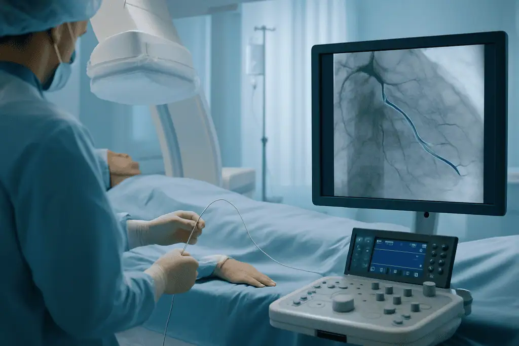
During CTO PCI, an interventional cardiologist guides very thin tubes called catheters through a blood vessel, usually from the wrist or the groin, up to the blocked coronary artery. Using live X-ray imaging and contrast dye, the physician navigates specialised guidewires and small supporting devices through or around the blocked segment. Once a channel has been created, a balloon is inflated to open the passage, and one or more stents are usually placed to scaffold the artery and help keep it open.
A coronary occlusion that has been in place for months or years behaves differently from a fresh narrowing. The blocked segment is often firm, calcified, long, or ambiguous in its course, which makes crossing it the central technical challenge of the procedure. In some cases the physician works forwards through the blockage in the usual direction: the antegrade approach. In others, the physician travels through collateral vessels and approaches the blockage from the far side: the retrograde approach. The choice depends on anatomy, prior procedures, kidney function, bleeding risk and the experience of the treating team.
Medication remains a cornerstone of treatment whether or not a procedure is performed. Anti-anginal medicines, cholesterol-lowering therapy, blood pressure control, diabetes management, antiplatelet treatment, nutrition, structured exercise and smoking cessation all reduce cardiovascular risk, and all of them continue after a successful procedure. For some patients, coronary artery bypass surgery is the better option, particularly when several arteries are severely diseased, diabetes is present, or the heart team judges that surgery offers a more complete way to restore blood flow.
In other words, chronic occlusion treatment is not a single technique applied to every patient. It is a personalised decision-making process that may involve medical therapy, catheter-based reopening of the artery, bypass surgery, or a combination of strategies over time. A blockage is never treated simply because it exists; it is treated when doing so is likely to improve how you feel, how your heart functions, or both.
Who May Need Chronic Occlusion Treatment
Patients are most often evaluated for chronic occlusion treatment after symptoms, stress testing or coronary imaging suggest that part of the heart muscle is not receiving enough blood. Some have already had a heart attack in the past, and the occlusion is found in the artery responsible. Others have never had a recognised heart attack but develop symptoms gradually as coronary disease progresses and the artery closes slowly enough for collaterals to form.
Typical symptoms include chest pressure, tightness, burning or heaviness, especially with physical activity. Some people feel discomfort in the arm, shoulder, back, neck or jaw. Shortness of breath, reduced stamina, unusual fatigue, palpitations, dizziness or nausea may also occur. Symptoms can be more subtle in older adults, in women, and in people with diabetes. A common pattern is quieter than any of these: patients simply stop doing the things that bring symptoms on. They take the lift instead of the stairs, walk more slowly, avoid hills, and only in retrospect realise how much their daily range has narrowed.
How is a chronic total occlusion diagnosed?
A chronic total occlusion is usually diagnosed during a coronary angiogram, a test that uses contrast dye and X-ray imaging to show the inside of the heart arteries. Before angiography, patients often undergo an electrocardiogram, echocardiography, stress testing, myocardial perfusion imaging or cardiac MRI. A coronary CT angiography scan can be particularly useful in planning, because it shows the length of the blockage, the calcium burden and the course of the vessel beyond it. Together, these tests answer two questions: are your symptoms likely caused by reduced blood flow, and is the heart muscle beyond the blockage still viable?
The viability question matters more than almost anything else. If the muscle beyond the occlusion has been permanently scarred by a previous heart attack, opening the artery may not improve function in that region, and the risk of the procedure may not be justified. If the muscle is alive but chronically under-supplied, restoring circulation may relieve symptoms and, in selected patients, support pumping performance. This is why a thorough diagnostic pathway comes before any recommendation to intervene: a principle that applies across the spectrum of coronary artery diseases.
Patients may be considered for treatment when they have persistent angina despite medication, significant ischaemia on testing, reduced quality of life because of symptoms, or impaired heart function plausibly linked to the blocked artery. Treatment may also come up when another heart procedure is being planned and the overall strategy for restoring blood flow needs review. Not everyone is a candidate for CTO PCI: severe kidney disease, high bleeding risk, very complex anatomy, frailty, extensive calcification, absence of viable muscle or other medical conditions may tip the balance towards optimised medical therapy or surgery instead.
Conditions and Indications This Treatment Addresses
Chronic occlusion treatment is primarily used for coronary chronic total occlusion: a complete blockage in one of the arteries supplying the heart muscle. The condition is one expression of coronary artery disease, which develops as plaque accumulates inside the arteries over many years. The risk factors are the familiar ones: raised cholesterol, high blood pressure, smoking, diabetes, excess weight, chronic kidney disease, family history and advancing age.
What is chronic occlusive arterial disease?
Chronic occlusive arterial disease is the broader term for long-standing narrowing or complete blockage of arteries anywhere in the body caused by atherosclerosis. When it affects the coronary arteries, it produces angina and, in its complete form, chronic total occlusion. When it affects the leg arteries, it is called peripheral arterial disease and typically causes pain in the calves or thighs while walking, and in advanced cases non-healing wounds. The disease process is the same; the treatment pathways differ. Leg artery occlusions are managed by vascular surgery and interventional radiology teams, while coronary occlusions are managed by cardiologists and cardiac surgeons. This page concerns the coronary form.
The most common indication for treating a coronary CTO is stable angina that continues despite appropriate medication. Stable angina occurs when the heart needs more oxygen than the blocked artery and its collaterals can deliver. It tends to appear predictably with exertion and to ease with rest or medication. Even when the pattern is stable, it can restrict daily life considerably and create a constant background anxiety about activity.
Another indication is documented ischaemiatest evidence that a significant area of heart muscle is receiving insufficient blood flow. Ischaemia can be silent, producing no symptoms the patient recognises, yet it still weighs on treatment planning, particularly when it involves a large region of the heart or coexists with reduced heart function.
Some patients are evaluated because of left ventricular dysfunction, meaning the heart’s main pumping chamber is not contracting as strongly as expected. If the weakened territory belongs to the occluded vessel and the muscle there remains viable, restoring blood flow may help support recovery or slow further deterioration in selected cases. How much improvement is realistic varies widely, and depends on the duration of the blockage, the amount of scar tissue and any other heart conditions present. An honest heart team will say so plainly rather than raise expectations it cannot justify.
Treatment may also be reconsidered after a previous unsuccessful attempt to open the artery. A failed attempt does not automatically close the door, but it demands a careful review: which approach was used, how much contrast and radiation the earlier procedure required, what the angiogram actually shows, and how the patient’s condition has changed. Sometimes a second attempt at a centre experienced in complex CTO work is reasonable; sometimes the risk-benefit balance now favours surgery or medication. The point is that the decision is remade on evidence, not repeated by default.
What does a blocked RCA artery mean?
A blocked RCA artery means a complete occlusion of the right coronary artery, the vessel that typically supplies the bottom (inferior) wall of the heart and, in most people, part of its electrical conduction system. The right coronary artery is among the vessels in which chronic total occlusions are most often found. When it closes slowly, collaterals frequently develop from the left-sided arteries, which is one reason a fully blocked RCA can be discovered in a patient who has never knowingly had a heart attack. Whether a blocked RCA should be opened follows exactly the same logic as any other CTO: symptoms, ischaemia, viability and anatomy decide, not the mere presence of the blockage.
How Chronic Occlusion Treatment Is Performed
Preparation and Evaluation Before the Procedure
The process begins with a detailed review of your medical history: symptoms and how they limit you, prior angiograms, any previous stents or bypass grafts, current medications, allergies, kidney function and bleeding risk. Prior imaging and reports are typically reviewed in advance so the team can judge whether further testing is likely to be needed and whether the case is technically suitable for a catheter approach at all.
Pre-procedure evaluation may include blood tests, electrocardiography, echocardiography and functional imaging to assess ischaemia or viability. A coronary CT scan helps in selected patients by mapping the length of the occlusion, its calcium content, the vessel’s course and the most promising crossing strategy. The team also reviews all current medicines, including blood thinners, diabetes drugs and kidney-related medication, because timing around the procedure matters; any adjustment is decided and directed by the treating doctors, never improvised by the patient.
Before CTO PCI, patients receive guidance on fasting, hydration and timing. Antiplatelet therapy is normally required before and after stent placement, and understanding this commitment is part of consenting to the procedure. Kidney protection is considered carefully, especially in patients with diabetes, reduced kidney function or previous contrast-related problems. The physician explains the planned approach, the realistic alternatives, and the specific risks: bleeding, vessel injury, heart rhythm disturbance, contrast effects on the kidneys, and the possibility that the artery cannot be crossed safely and the attempt is stopped. Stopping is not a failure of nerve: it is a designed safety decision, agreed in advance.
The Procedure, Step by Step
CTO PCI takes place in a cardiac catheterisation laboratory with continuous monitoring. Sedation is commonly used so that patients remain comfortable while breathing on their own; deeper anaesthesia is considered in selected complex cases. The procedure then follows a broadly consistent sequence:
- Step 1: Access. A small sheath is placed in an artery at the wrist or groin. In many CTO cases, two access points are used at once, so the team can image the artery from both sides of the blockage or work through collateral channels if needed.
- Step 2: Mapping. Contrast dye is injected to outline the coronary arteries, the occluded segment, the vessel beyond it and the collateral pathways. Dual injections from both access points often reveal anatomy a single injection would miss.
- Step 3: Crossing. The cardiologist advances specialised guidewires with different tip stiffnesses and shapes through the blockage, supported by fine microcatheters. If the central channel cannot be crossed, the wire may be steered deliberately through a layer of the vessel wall and back into the true channel beyond the blockage: a controlled dissection and re-entry technique. In selected cases, the occlusion is approached from the far side through collaterals: the retrograde approach.
- Step 4: Opening. Once a wire sits securely beyond the blockage, balloons are inflated to create and widen a channel through the occluded segment. Heavily calcified segments may need additional preparation before the vessel will yield.
- Step 5: Stenting. If the vessel is suitable, one or more stents are placed to scaffold the artery open along the treated length.
- Step 6: Verification. Imaging from inside the artery, such as intravascular ultrasound, may be used to confirm vessel size, plaque characteristics, stent expansion and final positioning, allowing the operator to optimise the result before finishing.
The technology serves the judgement, not the other way around. High-resolution angiographic imaging shows the vessel path and collateral circulation; physiologic and intravascular tools show how the artery is functioning and how well a stent is deployed; radiation-management systems and contrast-sparing strategies protect the patient during longer cases. Which of these tools is used depends on the individual anatomy and the equipment of the treating hospital, and a capable operator will also know when the safest tool is patience, or a planned second session.
What about perforations during chronic total occlusion angioplasty?
Perforation, a tear or puncture in the vessel wall, is a recognised complication of CTO angioplasty, and it occurs more readily here than in routine stenting because the wires used are stiffer, the vessel course can be ambiguous, and techniques deliberately work within the layers of the artery wall. This is precisely why detailed analyses of perforations during chronic total occlusion angioplasty exist in the medical literature: specialist teams study where and how perforations happen in order to prevent them and to respond quickly when they occur.
Experienced CTO operators prepare for this possibility before it arises. Most perforations are small and are managed during the same procedure, with prolonged balloon inflation, covered stents that seal the tear from inside, or tiny coils that close a leaking collateral channel. The team monitors continuously for bleeding around the heart, and drainage of that space is available immediately if ever needed. None of this makes the risk disappear, and no honest centre will claim it does. What it means is that the risk is anticipated, rehearsed for, and weighed openly against the expected benefit before anyone agrees to proceed.
How long does the procedure take, and what happens immediately afterwards?
CTO PCI usually takes longer than a standard angioplasty because crossing the blockage is the hard part. Some procedures are completed within a few hours; complex cases take longer. If the anatomy is especially difficult, or if limiting contrast and radiation exposure is the safer course, the physician may recommend staging treatment across more than one session rather than pushing on in a single sitting.
Afterwards, patients are monitored in a recovery area or hospital room. The access site is checked for bleeding or swelling, heart rhythm is observed, and blood pressure, kidney function and symptoms are assessed. Many patients stay in hospital overnight; the length of stay depends on the complexity of the procedure, other medical conditions and whether anything unexpected occurred.
Before discharge, patients receive clear instructions on antiplatelet medication, activity limits, care of the access site and hydration. Because stents depend on consistent medication adherence for a defined period, understanding this plan before discharge is essential. Ongoing follow-up with a cardiologist then monitors symptoms, fine-tunes medication and guides cardiac rehabilitation or a structured return to exercise.
Can Vitamins or Chelation Therapy Clear a Chronic Total Occlusion?
No. There is no reliable evidence that vitamins, supplements or chelation products marketed under names such as “cardio chelate” can open a chronic total occlusion. A CTO is a physical structure, organised plaque, fibrous tissue and calcium occupying the full channel of the artery, and no oral product has been shown to dissolve it. Searches asking whether a CTO can be reversed with vitamins or chelation are common, and the honest answer is that the only established ways to restore flow through a fully occluded coronary artery are catheter-based intervention and bypass surgery.
Chelation therapy, in which a binding agent such as EDTA is infused to remove metals from the blood, has been studied in cardiovascular disease, but it is not an established treatment for opening occluded coronary arteries, and unregulated over-the-counter “chelation” supplements have no demonstrated ability to do so at all. Some such products can interact with prescribed heart medication, which is a further reason any supplement should be raised with the treating cardiologist rather than added quietly alongside prescriptions.
What genuinely does change the course of the disease is unglamorous: cholesterol-lowering therapy, blood pressure control, diabetes management, smoking cessation, nutrition and exercise. These do not reopen a blocked segment, but they slow the disease everywhere else, support collateral circulation and protect the result of any procedure. That is where the real long-term leverage lies.
Why Acting Early Matters and the Risks of Delay
A chronic total occlusion is, by definition, not a sudden blockage, but that is not a reason to shelve it. If symptoms are present, or tests show a significant territory of reduced blood flow, delaying evaluation prolongs the strain on the heart and quietly shrinks what you can do. Patients often adapt around their symptoms so gradually that neither they nor their families notice how much has been given up until it is pointed out.
Persistent ischaemia can sustain angina, limit exercise tolerance and erode quality of life. In selected patients, chronically inadequate blood supply may worsen heart function or leave the heart more vulnerable during future illness. Delay also gives disease in the other coronary arteries time to progress if risk factors are not being treated aggressively: the occlusion you know about is rarely the whole picture.
There is also a practical argument for early specialist review: the decision is usually more nuanced than open-or-don’t-open. A thorough evaluation may identify medication changes that improve symptoms, imaging that clarifies viability, or reasons why bypass surgery would serve better than a catheter procedure. Assessing the situation early gives you time to compare options calmly, rather than deciding under pressure after symptoms have escalated. It is worth noting that new, rapidly worsening or rest symptoms belong to a different clinical picture, acute coronary syndrome, which is assessed as an emergency and follows entirely different pathways from the planned care described here.
Benefits of Chronic Occlusion Treatment
The potential benefits depend on the individual: on how much heart muscle the occluded vessel serves, on whether that muscle is viable, and on the success and durability of the strategy chosen. The table below summarises what appropriately selected patients can realistically hope for, none of it is promised, all of it is possible.
| Benefit | What It Means for You |
|---|---|
| Improved blood flow to heart muscle | Opening the blocked artery may increase oxygen delivery to a region of the heart that has been under-supplied, especially during activity. |
| Relief of angina symptoms | Many appropriately selected patients experience less chest discomfort, breathlessness and exertional limitation after successful revascularisation. |
| Better exercise capacity | With improved circulation and a supervised recovery, patients may walk, climb stairs and manage daily activities with fewer symptoms. |
| Support for heart function | In selected patients with viable heart muscle, restoring blood flow may help stabilise or improve pumping performance over time. |
| A clearer long-term care plan | A comprehensive evaluation defines whether medication, PCI, surgery or continued monitoring is the most appropriate strategy, and why. |
Recovery Timeline After Chronic Occlusion Treatment
Recovery varies with the complexity of the procedure, the access site used, your overall health, and whether cardiac rehabilitation or a staged second procedure is planned. The pattern below describes a typical, uncomplicated course.
| Time Period | What Patients Can Expect |
|---|---|
| Day 1 | Monitoring of the access site, heart rhythm, blood pressure, kidney function and symptoms. Some patients walk with assistance the same day or the next morning. |
| First Week | Heavy lifting and strenuous activity are limited while the access site heals. Medication adherence, hydration and follow-up instructions matter most in this window. |
| First Month | Activity increases gradually. Your cardiologist may adjust medication and recommend cardiac rehabilitation or a structured exercise plan. |
| Longer Term | Ongoing prevention continues indefinitely: cholesterol management, blood pressure control, diabetes care, smoking cessation, healthy nutrition and regular cardiology follow-up. |
Cardiac rehabilitation deserves particular emphasis. A structured programme typically combines supervised, gradually progressing exercise with education on medication, nutrition, stress and warning symptoms, and it gives many patients the confidence to become active again after years of unconsciously adapting around angina. Where a formal programme is not available, a cardiologist-guided walking plan with scheduled reviews serves a similar purpose. Either way, the goal is the same: to convert a reopened artery into a genuinely wider daily life, safely and progressively.
Factors That Influence Outcomes
The first factor is patient selection. The patients most likely to benefit are those whose symptoms or test results clearly trace back to the blocked artery and whose heart muscle remains viable. When symptoms actually stem from another cause, lung disease, anaemia, valve disease, rhythm problems or simple deconditioning, opening a chronic occlusion will not deliver the improvement everyone hoped for. Good centres invest as much effort in this question as in the procedure itself.
The second is anatomy. Shorter blockages, lighter calcification, a clearly visible vessel beyond the occlusion and favourable collateral pathways make treatment more straightforward. Long occlusions, heavy calcium, twisting vessel courses, ambiguous entry points, prior bypass grafts and previous failed attempts all increase complexity. Complexity does not necessarily rule treatment out, but it changes the honest conversation about risk and about how likely technical success really is.
The third is the condition of the heart muscle itself. Viable muscle has the potential to work better once blood flow returns. Scarred muscle generally will not, although symptom relief may still be possible if the surrounding regions are ischaemic. Advanced imaging exists precisely to draw this distinction before the procedure, so that expectations are set on evidence rather than optimism.
Overall health shapes everything around the procedure. Diabetes, kidney disease, anaemia, frailty, chronic lung disease, peripheral arterial disease, bleeding tendency and prior stroke all influence planning and recovery. So does medication tolerance: stents require antiplatelet therapy for a defined period, and patients who cannot safely take these medicines may need a different strategy from the outset.
The experience and coordination of the team matter as well. CTO procedures demand detailed pre-procedure planning, fluency in several crossing strategies, disciplined radiation and contrast management, and the readiness to change approach, or stop, when the anatomy dictates. For complex cases, collaboration among interventional cardiologists, non-invasive cardiologists, cardiac surgeons, anaesthesiology teams, imaging specialists and intensive care physicians is part of what safety looks like in practice.
Finally, long-term results depend heavily on prevention. Reopening an artery does not remove coronary artery disease; the disease that built one occlusion can build another. Continued treatment of cholesterol, blood pressure, diabetes, weight and sleep apnoea where present, together with not smoking, protects the result and reduces future cardiovascular events. Patients who engage actively with follow-up and prevention are consistently the ones best placed to keep what the procedure gave them.
How Acibadem Approaches Chronic Occlusion Care
Patients weighing up chronic occlusion treatment usually want more than a technically capable procedure. They want a clear diagnosis, a straight explanation of the options, including the option of not intervening, and care that stays coordinated from first consultation to long-term follow-up. Acibadem’s cardiovascular centres are organised around exactly that sequence: evaluation first, recommendation second, procedure only where it is justified.
Chronic occlusion cases are assessed through a structured cardiology pathway. Interventional cardiologists review the angiographic anatomy, symptoms, prior procedures, medication history and non-invasive test results. Where the decision is genuinely contested: CTO PCI versus bypass surgery, complex multivessel disease, reduced heart function, significant coexisting conditions, cases are discussed within multidisciplinary heart teams, so that the recommendation reflects the whole clinical picture rather than a single procedural viewpoint. The way records and imaging are examined before any plan is issued is described in how Acibadem reviews medical records before giving a treatment plan.
Diagnostic work-up follows evidence-based protocols and may include echocardiography, stress imaging, cardiac CT, cardiac MRI, coronary angiography, intravascular imaging and laboratory assessment, depending on the individual case. The purpose is always the same: establish whether the chronic occlusion truly explains the symptoms, whether the muscle beyond it can benefit from restored flow, and which route offers a reasonable balance of benefit and risk for this particular patient.
For catheter-based treatment itself, Acibadem’s teams work in modern catheterisation laboratories equipped for complex coronary intervention: detailed visualisation of coronary anatomy, intravascular imaging, specialised wires and microcatheters, contrast-management strategies and continuous monitoring through longer procedures. Just as important is the judgement to recognise when intervention is not the right answer: a thoughtful recommendation may instead involve medication optimisation, staged PCI, surgical consultation or further testing before any final decision.
Discharge planning is treated as part of the treatment itself. The team provides medication instructions, discharge summaries, procedure reports and follow-up recommendations written for the cardiologist who will continue care afterwards. That documentation is especially important after stent placement, when antiplatelet therapy and any future procedures must be coordinated between different physicians without gaps.
Weighing the Decision
A chronic occlusion is a demanding diagnosis, but it comes with well-established evaluation pathways and several genuine options. The pivotal question is not whether the artery can be opened, but whether opening it is likely to help you, whether the blockage is causing your symptoms, whether the muscle beyond it is alive, and whether the risks of the specific procedure are justified by what it can realistically deliver.
Patients researching this condition often hold conflicting opinions from different doctors: one says the artery cannot be opened, another recommends bypass, a third advises medication alone. Such differences are usually explained by anatomy, symptom burden, viability findings, procedural risk or local expertise rather than by anyone being simply wrong. Understanding why the recommendations differ is what turns a confusing situation into a decision you can actually own.
For some patients, catheter-based treatment brings meaningful relief from angina and a wider daily life. For others, surgery, medication optimisation or watchful monitoring is the better path. The strongest position from which to choose is an informed one: a reviewed angiogram, a clear viability assessment, an honest account of the risks, including the possibility that an attempt is stopped for safety, and a written plan for what happens long after you leave the hospital.
Preparation
- Patients usually need cardiology evaluation, ECG, blood tests, and coronary imaging to assess the blocked vessel. Blood-thinning medications may be adjusted before the procedure. Fasting is typically required for several hours, and patients should inform the team about allergies, kidney disease, or current medications.
Aftercare
- After the procedure, patients are monitored for heart rhythm, blood pressure, and the catheter entry site. Antiplatelet medication and lifestyle changes are important to keep the artery open. Follow-up visits help assess recovery, symptoms, and long-term heart health.
Frequently Asked Questions
What affects the cost of chronic occlusion treatment?
The main factors are the chosen treatment method, complexity of the blocked artery, required imaging, stents or other devices, hospital stay, specialist fees and follow-up needs. Travel, accommodation and interpreter support may also affect the total cost for international patients.
How can I get a personalised quote for chronic occlusion treatment in Turkey?
You can request a free consultation by sharing your medical records, recent angiography images, test results and current medication list. A cardiology team can review your case and provide a personalised treatment plan and cost estimate.
Is catheter-based treatment always the right option for a chronic occlusion?
No. Some patients are best managed with medicines, while others may benefit from catheter-based intervention, bypass surgery or a staged approach. Suitability is decided by a specialist after assessing symptoms, heart function and artery anatomy.
What is usually included in an international patient package?
Packages may include specialist consultation, medical record review, diagnostic planning, the procedure, hospital stay and care coordination. Some hospitals also help with interpreter services, airport transfers and appointment scheduling, but inclusions should be confirmed before travel.
Will I need to stay in Turkey after the procedure?
A short recovery and observation period is commonly recommended after chronic occlusion treatment, but the length of stay depends on the procedure type, recovery, medications and the cardiologist’s advice. Your care team will explain follow-up needs before you travel.
Medically reviewed by the Acıbadem International Medical Board August 30, 2026
See our medical review board →
Update history
- PublishedJune 8, 2026
- Medical review approvedAugust 30, 2026
- Last content updateSeptember 8, 2026
Trusted care for international patients
From the Acibadem Blog

Chronic Total Occlusion (CTO) of a Coronary Artery: How Treatment Is Chosen and Why
Chronic total occlusion treatment options fall into three groups: medicines with risk-factor control, a specialized catheter procedure (CTO PCI) that reopens the artery and…

After CTO Treatment: The Medicines and Habits That Help Keep the Reopened Artery Open
After CTO PCI, keeping the reopened artery open usually rests on three things: taking the antiplatelet medicines exactly as prescribed for the full period…



































