Eye Angiography
Eye angiography is a diagnostic retinal imaging test that uses an injected dye to visualize blood flow in the retina and choroid, helping detect vascular eye disease.
Quick answer
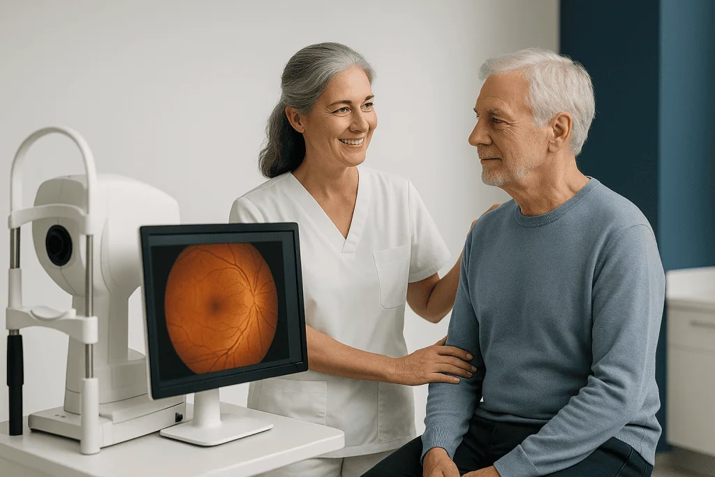
Eye angiography is a diagnostic imaging test that shows blood flow through the retina and choroid. A dye, usually fluorescein or indocyanine green, is injected into a vein in the arm, and a specialised camera photographs the back of the eye in rapid sequence. The images reveal leaking, blocked or abnormal blood vessels, helping ophthalmologists diagnose and plan treatment for retinal conditions.
What Is Eye Angiography?
Eye angiography is a diagnostic imaging test that shows how blood flows through the retina, the light-sensitive tissue at the back of the eye, and through the choroid, the deeper vascular layer beneath it. A dye is injected into a vein, usually in your arm or hand, and a specialised camera photographs the back of your eye in a rapid sequence as the dye circulates. The images reveal leaking blood vessels, blocked circulation, abnormal new vessel growth, inflammation, swelling or scarring that may not be fully visible during a standard eye examination. It is used when an ophthalmologist needs dynamic information about the eye’s blood supply, not just a static picture of its structure.
Most people arrive at this test because something has changed. Blurred central vision, straight lines that look wavy, dark or empty spots in the visual field, flashes, new floaters, or unexplained loss of sight can all point to a problem in the retina. Some people have no symptoms at all: early diabetic eye disease, in particular, can progress quietly and is often picked up during screening or routine follow-up. In either case, the concern is usually the same, not only what is happening now, but whether the condition could progress and affect long-term vision. Eye angiography exists to answer that question with evidence rather than guesswork.
It is worth being clear about what the test is and is not. Eye angiography is diagnostic; it does not treat anything by itself. What it does is guide treatment decisions. Depending on what the images show, your ophthalmologist may recommend intravitreal injections, laser therapy, retinal surgery, medication for inflammation, or simply continued monitoring. In many retinal diseases, the right plan depends not only on what the retina looks like, but on whether its blood vessels are leaking, closing off, or growing where they should not.
What is fluorescein angiography for the eyes?
Fluorescein angiography is the most common form of eye angiography: a fluorescent yellow dye is injected into a vein and highlights the blood vessels of the retina as a camera captures its passage through the eye. Because the dye glows under a specific light filter, the camera can record exactly where blood is flowing normally, where it is leaking out of damaged vessels, and where circulation has stopped altogether.
Fluorescein angiography is particularly useful for detecting diabetic retinopathy, macular oedema, retinal vein occlusion, retinal artery problems, inflammatory conditions and many disorders affecting the macula: the central part of the retina responsible for fine, detailed vision such as reading and recognising faces. When your doctor refers to an “eye angiography” without further detail, this is usually the test they mean.
What is indocyanine green (ICG) angiography?
Indocyanine green angiography, usually shortened to ICG angiography, is the second main form of eye angiography and uses a different dye that is better suited to imaging the choroid, the layer of blood vessels beneath the retina. Because ICG dye fluoresces at a wavelength that passes through the retinal pigment layer, it can show deeper vascular structures that fluorescein cannot reach clearly.
ICG angiography may be recommended when doctors need more information about conditions such as certain forms of age-related macular degeneration, polypoidal choroidal vasculopathy, central serous chorioretinopathy, or inflammatory choroidal disease. In some cases both dyes are used at the same appointment, because the two tests answer different questions about the same eye.
How is eye angiography different from coronary angiography?
Eye angiography and coronary angiography share a name but are very different procedures. Coronary angiography examines the arteries of the heart and involves threading a thin catheter through a blood vessel, usually from the wrist or groin, while X-ray images are taken. Eye angiography requires no catheter at all: the dye is given through an ordinary intravenous line in the arm, and the images are taken with a camera pointed at the back of the eye rather than with X-rays. There is also a non-invasive scan of the heart’s arteries, coronary CT angiography, which again serves an entirely different purpose. If you have been researching angiography of the heart’s vessels and their treatment, that is a cardiology pathway with its own preparation, procedure and recovery; this page deals only with imaging of the eye.
Which other tests are used alongside eye angiography?
Eye angiography is rarely interpreted in isolation; it is usually combined with other retinal imaging tests that provide complementary information. Optical coherence tomography (OCT) is a non-invasive scan that shows the retina in cross-section: its layers, any swelling, fluid pockets or structural damage. Angiography shows blood flow and leakage patterns; OCT shows the physical consequences of that leakage inside the retinal tissue. Depending on the suspected condition, your assessment may also include fundus photography, autofluorescence imaging, ocular ultrasound, or OCT angiography: a newer, dye-free way of visualising retinal blood flow. Each test answers a different question, and the retina specialist builds the diagnosis from the combined picture rather than from any single image.
Who May Need Eye Angiography?
Your ophthalmologist may recommend eye angiography when symptoms, examination findings or previous imaging suggest a vascular or inflammatory retinal condition. Some patients are referred after a routine eye examination detects bleeding, swelling, abnormal vessels or pigment changes at the back of the eye. Others seek care because they have noticed a change in vision that affects reading, driving, face recognition, work or daily activities, and want to know what is causing it before deciding what to do.
The diagnostic pathway usually begins with a detailed history and vision testing. The ophthalmologist asks about the onset and progression of symptoms, medical conditions such as diabetes or high blood pressure, previous eye treatments, current medications, allergies, pregnancy status, kidney or liver disease, and any prior reactions to dyes or contrast materials. A dilated eye examination then allows the retina and optic nerve to be evaluated in detail. Only after this assessment are imaging tests selected, including whether dye angiography is actually the right test for your situation.
Which symptoms can lead to eye angiography?
The symptoms that most often lead to eye angiography are those suggesting a problem with the retina’s blood supply or its central macular region. They include:
- Blurred vision, especially in the centre of sight, where fine detail is processed;
- Distorted vision, where straight lines, door frames, tiles, text, appear wavy or bent;
- Dark or empty spots in the visual field that do not move with the eye;
- Reduced colour perception, with colours appearing washed out or dim;
- Sudden vision loss, whether partial or affecting the whole field of one eye;
- New floaters, particularly when associated with retinal bleeding or inflammation.
Some patients have none of these symptoms. In early diabetic eye disease, damage to the small retinal vessels often develops before vision changes are noticeable, which is why the need for angiography is sometimes identified during screening or scheduled follow-up rather than because of a complaint.
Does everyone with a retinal condition need dye angiography?
No, not every patient with a retinal condition needs dye angiography. In many cases, non-invasive imaging such as OCT or OCT angiography provides sufficient information to diagnose and monitor the condition without an injection. Eye angiography is most valuable when the physician needs dynamic information about blood flow: whether vessels are leaking, how quickly the dye fills the circulation, and where perfusion is reduced. Your physician weighs the potential benefit of the test against your medical history, the urgency of diagnosis, and whether the results are genuinely likely to change management. This individualised approach matters particularly for international patients coordinating diagnosis, treatment and travel within a limited timeframe: a test is only worth scheduling if its answer will shape what happens next.
Conditions Eye Angiography Can Help Evaluate
Eye angiography is used across a broad range of retinal and choroidal conditions. Its value lies in making hidden circulation problems visible and in distinguishing between conditions that can look similar during a standard examination. The same symptom, blurred central vision, for instance, can have many causes, and angiography helps narrow the diagnosis so that treatment addresses the actual problem.
One of the most common indications is diabetic retinopathy. Diabetes can damage the small blood vessels of the retina, leading to leakage, swelling of the macula, bleeding, and eventually the growth of fragile new vessels that bleed easily. Angiography can show whether macular oedema is being driven by leaking microaneurysms, and whether there are areas of reduced perfusion, retina that is no longer receiving adequate blood supply, which increase the risk of progression to the proliferative stage of the disease.
Another major indication is age-related macular degeneration. In some patients, abnormal blood vessels grow beneath the retina and leak fluid or blood into the macula. Angiography can help confirm the presence, location and activity of these vessels, particularly when combined with OCT and other macular imaging. In selected cases, ICG angiography adds information about deeper choroidal vessel abnormalities that fluorescein alone cannot resolve: a distinction that can change the treatment approach.
Eye angiography is also important in retinal vein occlusion, in which one of the veins draining blood from the retina becomes blocked. The blockage causes haemorrhages, swelling and reduced vision. Angiography helps assess the remaining circulation, the extent of leakage, and the degree of ischaemia, factors that influence how intensively the eye needs to be followed and which treatments are considered. Less commonly, angiography is used in retinal artery occlusion or other vascular disorders to evaluate perfusion patterns.
In inflammatory eye diseases such as uveitis, retinal vasculitis or choroiditis, angiography can reveal leakage from inflamed vessel walls, leakage at the optic disc, or choroidal inflammatory lesions that are not obvious on examination. This information helps determine whether the disease is active, how well it is responding to treatment, and whether collaboration with rheumatology, infectious disease or other specialties is warranted when a systemic cause is suspected.
Other recognised indications include central serous chorioretinopathy, macular telangiectasia, inherited retinal disorders, ocular tumours with vascular features, unexplained retinal bleeding, optic nerve head abnormalities, and monitoring after certain retinal treatments. In every one of these situations, angiography is one component of a broader clinical assessment rather than a standalone answer: the images are only as useful as the clinical reasoning around them.
How Is Eye Angiography Done?
Eye angiography is done as an outpatient test: dilating drops widen your pupils, a small intravenous line is placed in your arm or hand, dye is injected, and a specialised camera photographs the back of your eye in timed sequence over several minutes. Most patients do not stay in hospital. A typical appointment includes registration, consultation, pupil dilation, imaging, a short period of observation after the dye injection, and, where appropriate, a discussion of preliminary findings. The exact sequence depends on your clinical situation and on whether additional tests are scheduled for the same day.
Before the Test
Before angiography, the care team reviews your medical history in detail. Tell the ophthalmologist if you have ever reacted to fluorescein, indocyanine green, iodine-containing substances, shellfish, medications or other contrast materials. A shellfish allergy alone does not necessarily rule out the test, but every part of your allergy history is relevant to planning it safely. Asthma, severe heart disease, kidney disease, liver disease, pregnancy and breastfeeding should also be mentioned, along with a complete list of your current medications, decisions about any of them belong to your treating doctor, and the team can only plan properly if the list is complete.
Your pupils are usually dilated with eye drops so the camera can capture clear views of the retina. Dilation blurs your vision and increases light sensitivity for several hours afterwards, so you should not plan to drive after the appointment. If you are travelling for the test, it is sensible to arrange transport back to your hotel or residence in advance, and to bring sunglasses for the journey.
A small intravenous line is then placed, typically in a vein of the arm or hand. Baseline photographs are taken before any dye is given. These pre-dye images document the appearance of the retina and give the physician a reference point against which the dye-phase images can be compared.
Step by Step: What Happens During the Test
Although the details vary between clinics and conditions, the sequence of an eye angiography session generally follows the same steps:
- Positioning. You sit at the imaging camera with your chin on a rest and your forehead against a bar, much like at a standard eye examination.
- Dye injection. The dye is injected through the intravenous line already in place. No further needles are needed during the imaging itself.
- Rapid early imaging. Within seconds, the dye reaches the eye. The camera captures a fast series of photographs recording how the dye fills the retinal and choroidal vessels: the phase where circulation problems first become visible.
- Directed views. The photographer asks you to look in different directions so both the central retina and the periphery can be imaged. Holding a steady gaze on the fixation target helps produce sharp images.
- Later-phase images. Additional photographs are taken minutes later to detect slow leakage, staining or pooling of dye, patterns that distinguish one condition from another.
- Line removal and observation. Once imaging is complete, the intravenous line is removed and you are observed briefly before leaving.
Is eye angiography painful?
For most people, eye angiography involves little discomfort: the main sensation is the brief sting of the needle used to place the intravenous line, and the imaging itself does not involve any contact with the eye. The camera flashes can feel bright and repetitive, and some patients find the sequence of flashes tiring. After the dye injection, you may notice a brief warm sensation, mild nausea or an unusual metallic taste; these effects are usually short-lived and settle on their own. Nothing touches the surface of your eye during the photography, and no anaesthetic is needed beyond the routine placement of the intravenous line.
How much time does it take for eye angiography?
The active imaging portion of eye angiography usually takes only several minutes, but you should plan for a longer appointment overall. Pupil dilation takes time to work, baseline images are taken before the dye, and a brief observation period follows the injection. If additional scans such as OCT are scheduled on the same visit, or if both fluorescein and ICG angiography are needed, the appointment is extended accordingly. Because dilation blurs vision for several hours afterwards, it is realistic to set aside a good part of the day rather than a single tightly scheduled hour. The team will tell you what to expect in your specific case, including whether the physician plans to discuss preliminary findings on the same day.
Technology Used in Eye Angiography
Modern eye angiography uses high-resolution retinal imaging systems designed to capture timed images of dye movement through the eye. These systems may include wide-field imaging, which visualises the peripheral retina where diseases such as diabetic retinopathy and vasculitis often show early changes, and macular-focused imaging, which provides fine detail of the central retina. Digital image processing allows the physician to compare the phases of dye circulation, early filling, peak fluorescence and late leakage, and to correlate the findings with other scans of the same eye. Many patients also undergo OCT at the same visit: where angiography identifies leakage, the cross-sectional scan shows whether fluid is actually accumulating within or beneath the retinal layers. The value lies not in any single image, but in how the findings are interpreted together by an experienced retina specialist.
After the Test
After the images are completed and the intravenous line removed, some patients are observed briefly, particularly those with a history of allergies, asthma or sensitivity to medications. You can usually eat and drink as normal afterwards unless your physician gives different instructions. Drinking fluids helps the body eliminate the dye, provided you have no medical reason to restrict fluid intake.
Your vision will remain blurry and light-sensitive from the dilating drops for several hours, so sunglasses are useful when leaving the clinic and you should not drive until the effect has fully worn off. Most patients resume routine non-driving activities the same day. If treatment is recommended on the basis of the findings, it can be scheduled promptly or coordinated with other tests, depending on urgency and, for travelling patients, the practicalities of the itinerary.
Side Effects and Safety of Eye Angiography
What are the possible side effects of an eye angiogram?
The most common side effects of an eye angiogram are mild and temporary: a brief warm sensation or mild nausea as the dye is injected, an unusual taste, temporary yellowish discolouration of the skin after fluorescein dye, and bright yellow or orange urine for a period as the dye leaves the body through the kidneys. With ICG dye, visible colour changes are less common. Blurred, light-sensitive vision after the appointment is caused by the dilating drops rather than the dye itself and wears off over several hours. Occasionally, dye can leak from the vein at the injection site, causing local irritation; the team monitors the line during injection to minimise this.
Serious reactions to the dye are uncommon, but they are recognised: they can include rash, swelling, faintness, breathing difficulty or, rarely, a severe allergic reaction. This is one reason the test is performed in settings where staff are trained and equipped to recognise and manage adverse reactions, why your allergy history is reviewed carefully beforehand, and why patients with relevant risk factors are observed after the injection. Your physician balances these small risks against the diagnostic value of the information the test provides, and will suggest a different imaging strategy if dye angiography is not appropriate for you.
Recovery After Eye Angiography
How long does it take to recover from fluorescein angiography?
Recovery from fluorescein angiography is quick for most people: the main after-effects, blurred, light-sensitive vision from the dilating drops, settle within hours, and the visible traces of the dye clear as the body eliminates it over the following day or so. There is no wound to heal and no activity restriction beyond not driving while your pupils remain dilated. The table below summarises what patients can typically expect.
| Time Period | What Patients Can Expect |
|---|---|
| Day 1 | Vision may be blurry and light-sensitive because of the dilating drops. Some patients notice temporary skin discolouration or bright-coloured urine after fluorescein dye. Driving should wait until dilation wears off. |
| First 24 Hours | Most dye-related changes fade as the body clears the dye through the kidneys. Mild nausea, if it occurred, usually resolves quickly. Drinking fluids supports elimination unless a medical condition restricts intake. |
| First Week | No special recovery is usually needed. Your physician may discuss results, request additional tests, or recommend retinal treatment depending on the findings. |
| First Month | If a condition is diagnosed, follow-up may focus on treatment response, additional imaging, or monitoring for changes in swelling, bleeding or circulation. |
| Longer Term | Some retinal diseases require periodic imaging and ongoing care. The timing of any repeat angiography depends on the diagnosis, symptoms and treatment plan. |
Why Acting Early Matters
Many retinal vascular diseases are time-sensitive. The retina is delicate neural tissue, and prolonged swelling, poor circulation, bleeding or abnormal vessel activity can lead to lasting structural damage. Eye angiography helps identify active disease before the full extent of that damage becomes apparent in daily life. In some conditions, early detection allows treatment to begin at a stage when more vision can potentially be preserved than would be the case after months of untreated activity.
Delaying evaluation carries specific risks that vary by condition. In diabetic retinopathy, untreated leakage or proliferative changes can progress towards vitreous haemorrhage, tractional retinal detachment and significant vision loss. In wet age-related macular degeneration, active abnormal vessels can scar the macula if not managed in a timely manner, and scarring, unlike fluid, does not resolve. In retinal vein occlusion, delayed assessment may postpone treatment for macular oedema and delay recognition of the complications that follow poor perfusion. None of this means every retinal finding is urgent; it means the decision about urgency should rest on evidence, which is precisely what angiography provides.
There are also practical reasons to establish the diagnosis early. For international patients in particular, an accurate diagnosis at the start of the care journey helps avoid unnecessary travel delays, repeated testing in different systems, or treatment that is not well matched to the actual condition. When angiography is performed as part of a coordinated retinal assessment, the results can determine whether care is needed promptly, whether treatment can be safely scheduled around other commitments, or whether careful monitoring is the more appropriate course.
Benefits of Eye Angiography
The main benefits of eye angiography relate to diagnostic clarity, treatment planning and the timely monitoring of retinal disease. The table below summarises what each benefit means in practice.
| Benefit | What It Means for You |
|---|---|
| Detailed view of retinal blood flow | Shows how blood moves through the eye and reveals leakage, blockage or abnormal vessels that may not be visible on routine examination. |
| More precise diagnosis | Helps distinguish between conditions with similar symptoms, such as macular degeneration, diabetic macular oedema, inflammation or vascular occlusion. |
| Guidance for treatment planning | Supports decisions about injections, laser treatment, medication, surgery or observation based on the location and activity of disease. |
| Monitoring disease activity | Allows physicians to compare findings over time and assess whether a retinal condition is stable, improving or progressing. |
| Support for second opinions | Provides objective imaging that can be reviewed by retina specialists when confirming a diagnosis or evaluating previous recommendations. |
Factors That Influence Outcomes and a Good Result
A good result from eye angiography means more than obtaining clear images. It means the test answers the clinical question, helps explain your symptoms, and supports the next step in care. Several factors influence how useful the angiography will be and how well the overall pathway proceeds.
The underlying condition is the most important factor. Some retinal diseases are chronic and require ongoing management; others may resolve or stabilise after targeted treatment. Angiography can reveal how active the disease is right now, but the long-term visual outlook depends on the diagnosis itself, its severity, how long it has been present, and how it responds to therapy. The test informs the plan: it does not change the underlying biology.
Timing of diagnosis also matters. Retinal swelling or abnormal vessel growth identified early tends to be more manageable than disease that has already caused scarring, extensive ischaemia or structural damage. This is why people with diabetes, high blood pressure, previous retinal disease or new visual symptoms are generally advised not to postpone specialist assessment once it has been recommended.
Image quality affects interpretation. Clear angiography depends on adequate pupil dilation, patient positioning, steady fixation on the target, and the transparency of the eye’s optical media. Cataracts, corneal opacity, vitreous haemorrhage or difficulty holding the eye still can all reduce image clarity. When that happens, the physician does not simply accept an incomplete answer, complementary tests such as OCT or ultrasound can be used to complete the assessment.
Medical history and safety considerations shape how the test is planned. Patients with previous dye reactions, significant allergies, pregnancy, severe systemic disease or complex medication histories may require modified planning: a different imaging strategy, additional precautions, or a decision that dye angiography is not needed at all. Honest, complete communication before the test is what allows the team to select the safest and most informative approach for your specific circumstances.
Expert interpretation is central to the value of the test. Angiography produces a timed sequence of images that must be read in context: how quickly the dye fills the vessels, the pattern of any leakage, areas of staining, blocked fluorescence, zones of non-perfusion, and how all of this correlates with the clinical examination and the other scans. Subtle differences between patterns can change the diagnosis and, with it, the treatment plan. This is why retinal imaging is most valuable when reviewed by physicians experienced in medical retina and its related conditions.
Integration with treatment ultimately determines your course. If angiography shows active disease, the next step may involve medication injections into the eye, laser therapy, systemic evaluation, management of an inflammatory disease, or a surgical consultation. A clear plan, realistic expectations and timely follow-up are essential, particularly for patients who travel for care and later need coordination between the treating hospital and their local ophthalmologist at home.
How Much Does an Eye Angiography Test Cost?
There is no single standard price for an eye angiography test, and any figure quoted without context is unlikely to reflect what you would actually pay. The cost depends on several concrete variables: which dye is used and whether fluorescein and ICG angiography are performed together, which additional imaging tests such as OCT or wide-field photography accompany it, whether a specialist consultation is included in the same visit, and the type of institution and healthcare system in which the test is performed. Because eye angiography is a diagnostic test rather than a treatment, it is also often priced as part of a broader retinal assessment rather than as an isolated item. The reliable way to understand the cost of your own test is to ask for an itemised quotation based on the specific imaging your ophthalmologist has recommended, rather than relying on generic figures found online.
Eye Angiography at Acibadem
For an international patient, eye angiography is rarely just a single test. It sits inside a larger set of decisions: where to obtain a reliable diagnosis, whether treatment is needed at all, how quickly care can be organised, and how to navigate a healthcare system in an unfamiliar country. Acibadem’s approach is built around clinical accuracy and coordinated support, particularly for patients who arrive with previous reports, imaging or treatment recommendations from another healthcare system.
In ophthalmology, patients are evaluated by experienced physicians using evidence-based diagnostic pathways. When retinal findings are complex or connected to systemic disease, care may involve collaboration with other specialties such as endocrinology, cardiology, rheumatology, neurology, infectious disease, oncology or internal medicine. This multidisciplinary structure matters because many retinal vascular problems, diabetic retinopathy above all, are windows onto broader health conditions rather than isolated eye diseases.
Advanced diagnostic imaging supports the retinal assessment. Eye angiography may be combined with high-resolution retinal scans, wide-field imaging, macular analysis and other ocular tests as clinically appropriate. The purpose of the technology is practical rather than decorative: to identify disease activity, localise leakage or poor circulation, document findings objectively, and give treatment decisions a firmer footing. Patients benefit most when images are not only captured well but interpreted within a careful medical framework.
International patient services support the practical side of the visit: appointment coordination, medical document transfer, language support in the patient’s preferred language, hospital navigation and communication with the care teams. Personalised planning is especially relevant in retinal care, because needs differ so widely. Some patients require only diagnostic clarification and a written recommendation for follow-up at home; others need treatment during the same trip, such as intravitreal injections, laser procedures or additional systemic evaluation. The care plan depends on the diagnosis, the urgency of treatment, medical history, visual goals and travel schedule, and it is designed around safety and appropriateness rather than around the calendar.
Second opinions are a common reason patients undergo this test. Many arrive uncertain whether they have wet macular degeneration, diabetic macular oedema, retinal vein occlusion, an inflammatory disease or something else entirely. Eye angiography provides objective information that can confirm or refine a prior recommendation. Sometimes the test supports a decision to treat; sometimes it shows that close observation or further testing is the more appropriate path. Both answers are valuable, because both replace uncertainty with evidence.
For patients who feel anxious about the dye injection, the imaging process or the implications of a retinal diagnosis, clear communication is part of good care. The ophthalmology team explains why the test is recommended, what sensations to expect, which side effects are normal and which are not. When these conversations happen in the patient’s preferred language, discussions about vision, risk and treatment choices become genuinely informed rather than merely translated.
Understanding What Comes After the Results
Eye angiography can be an important step towards understanding a retinal or choroidal condition that may affect vision. By showing how blood flows through the eye, it helps physicians identify leakage, blockage, abnormal vessels, inflammation and other changes that guide diagnosis and treatment planning. For many patients, the real value of the test is clarity: what is happening, how active the condition is, and what should be done next, whether that is treatment, further testing or structured monitoring.
If angiography has been recommended for you, a little preparation makes the appointment more productive. Previous eye scans, imaging reports, a current medication list and relevant medical history all help the retina specialist read your images in context, compare them against earlier findings, and judge whether the condition is new, stable or progressing. The same records matter after the test as well: angiography images become part of your ongoing record, and future physicians, at home or abroad, can use them as a baseline for comparison. In retinal disease, where change over time often matters as much as any single finding, that continuity is one of the quiet advantages of having the test done thoroughly and documented well.
Preparation
- Tell your ophthalmologist about allergies, pregnancy, kidney disease, and all medicines you take. Your pupils are usually dilated before the test, so bring sunglasses and arrange someone to drive you home if needed.
Aftercare
- Mild nausea, light sensitivity, blurred near vision, or yellow discoloration of the skin and urine may occur briefly after dye injection. Drink water unless advised otherwise, and contact the clinic urgently for rash, breathing difficulty, severe dizziness, or worsening vision.
Frequently Asked Questions
What affects the cost of eye angiography?
The final cost depends on the type of angiography, whether extra retinal imaging is required, the specialist consultation, hospital or clinic policies, reporting, follow-up needs and any international patient services such as translation or travel coordination.
How can I get a personalised quote?
You can request a free consultation and share your eye reports, diagnosis, symptoms, previous imaging and medication history. A retinal specialist can then advise which test is appropriate and provide a personalised quote.
Is eye angiography usually included in a package?
Some providers may bundle consultation, imaging, specialist interpretation and written reporting, while others may bill each part separately. It is important to confirm exactly what is included before travel or booking.
Will I need other tests in addition to eye angiography?
Possibly. A specialist may recommend OCT, OCT angiography, fundus photography or other retinal tests to understand the cause of symptoms and plan treatment. This can affect the total cost.
Is travelling to Turkey for eye angiography practical?
For many international patients, it can be practical when appointments, imaging, reporting and language support are coordinated by an international patient team. Travel plans should be made only after medical review and confirmation of suitability.
Medically reviewed by the Acıbadem International Medical Board September 1, 2026
See our medical review board →
Update history
- PublishedJune 8, 2026
- Medical review approvedSeptember 1, 2026
- Last content updateSeptember 1, 2026
References1
- Fluorescein angiography medlineplus.gov
Trusted care for international patients
From the Acibadem Blog

Yellow Skin, Nausea and Blurry Hours After Eye Angiography: Expected Effects and Red Flags
After fluorescein eye angiography, a yellowish tint to the skin for a few hours, dark orange urine for about a day, brief nausea during…